- "Cervical Radiculopathy (Pinched Nerve)". OrthoInfo by American Academy of Orthopaedic Surgeons. June 2015. Retrieved 22 September 2017.
- "Pinched Nerve Symptoms & Treatment | Advanced Neurosurgery". Advanced Neurosurgery Associates. Retrieved 2020-12-14.
- Tarulli AW, Raynor EM (May 2007). "Lumbosacral radiculopathy" (PDF). Neurologic Clinics. 25 (2): 387–405. doi:10.1016/j.ncl.2007.01.008. PMID 17445735. S2CID 15518713. Archived from the original (PDF) on 2019-02-20.
- Iyer S, Kim HJ (September 2016). "Cervical radiculopathy". Current Reviews in Musculoskeletal Medicine. 9 (3): 272–80. doi:10.1007/s12178-016-9349-4. PMC 4958381. PMID 27250042.
- Kuijer PP, Verbeek JH, Seidler A, Ellegast R, Hulshof CT, Frings-Dresen MH, Van der Molen HF (September 2018). "Work-relatedness of lumbosacral radiculopathy syndrome: Review and dose-response meta-analysis". Neurology. 91 (12): 558–564. doi:10.1212/01.wnl.0000544322.26939.09. PMC 6161552. PMID 30120136.
- Childress MA, Becker BA (May 2016). "Nonoperative Management of Cervical Radiculopathy". American Family Physician. 93 (9): 746–54. PMID 27175952.
- Tawa N, Rhoda A, Diener I (February 2017). "Accuracy of clinical neurological examination in diagnosing lumbo-sacral radiculopathy: a systematic literature review". BMC Musculoskeletal Disorders. 18 (1): 93. doi:10.1186/s12891-016-1383-2. PMC 5324296. PMID 28231784.
- Dworkin RH, Johnson RW, Breuer J, Gnann JW, Levin MJ, Backonja M, et al. (January 2007). "Recommendations for the management of herpes zoster". Clinical Infectious Diseases. 44 Suppl 1: S1-26. doi:10.1086/510206. PMID 17143845. S2CID 10894629.
- Shapiro ED (May 2014). "Clinical practice. Lyme disease" (PDF). The New England Journal of Medicine. 370 (18): 1724–31. doi:10.1056/NEJMcp1314325. PMC 4487875. PMID 24785207. Archived from the original (PDF) on 19 October 2016.
- "Lyme Disease Data and surveillance". Lyme Disease. Centers for Disease Control and Prevention. 2019-02-05. Retrieved April 12, 2019.
What is Radiculopathy, or Pinched Nerve? Manifestations and Treatment
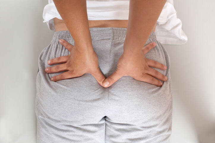
Photo source: Getty images
Radiculopathy, radicular syndrome, or pinched nerve. These terms describe pain caused by nerve compression. It is reported that up to 90% of the population will have experienced back pain at least once in their lifetime.
Most common symptoms
- Shoulder Blade Pain
- Muscle Pain
- Chest pain
- Flank Pain
- Right Flank Pain
- Headache
- Limb pain
- Nerve pain
- Leg Pain
- Shooting pain in fingers and toes
- Pain that Radiates into the Shoulder
- Groin Pain
- Shooting Pain in the Ears
- Back Pain
- Spirituality
- Muscle stiffness
- Defence
- Tingling
- Erectile dysfunction
- Muscle weakness
- Pressure on the chest
- Head spinning
- Fatigue
- Anxiety
Show more symptoms ᐯ
Characteristics
Radiculopathy, radicular syndrome - root stimulus syndrome indicate problems associated with the compression of the root of the nerve, and thus the nerve that emanates from the spinal cord. The most commonly affected areas are the lumbar and the cervical spine.
Damage and displacement, i.e. disc herniations, are a major problem in radiculopathy.
In addition to neck and back pain, there are a number of other problems associated with the syndrome.
It is reported that 60-90% of the population will have experienced back pain and difficulties associated with it at least once in their lifetime.
In most cases, these are less severe difficulties indicating spinal blockage. The most common difficulties occur in the lumbar and cervical spine.
The problem associated with spinal cord compression is referred to as nerve root syndrome, radicular syndrome, or radiculopathy. It is a state in which the nerve is compressed, or more precisely, the nerve root exiting the spinal cord.
This problem is associated with pain, radiating pain, but also various other neurological problems. They reduce the quality of life and restrict the affected person intensely and acutely, but also in the long term.
According to reports, up to 90% of cases with the first case of radiculopathy are cured by conservative treatment. In other words, there is no need for a surgery.
About the Spinal Cord and Spinal Nerves - A Brief Overview
The spinal cord, also called the medulla spinalis, is located in the spinal canal (canalis vertebralis). It is formed by the vertebrae with their openings. The connected vertebrae form the spine.
The spine is...
The spine - columna vertebralis, is the axis of the human body, which has a supporting, locomotor but also protective function, ie it protects the spinal cord.
The inward curvature of the lumbar (lower back) and cervical (the neck) spine is called lordosis. The curvature in the lumbar region is called kyphosis, i.e. curvature towards the back. It is found in the thoracic and sacral areas.
This curvature is physiological, i.e. natural.
Conversely, scoliosis is unnatural as it is a pathological sideways curve of the spine. There is a slight physiological sideways curve in every person.
A temporary sideways curve of the spine can be seen when standing on one leg, transferring the weight to one limb, or when carrying a heavier load in one hand.
The spine consists of 33 or 34 vertebrae.
Table: The Spine is Divided into Vertebral Segments by Their Location
| Part - segment | Latin name | Description |
| Cervical spine | vertebrae cervicales |
|
| Thoracic spine | vertebrae thoracicae |
|
| Lumbar spine | vertebrae lumbales |
|
| Sacral spine | vertebrae sacrales |
|
| Coccygeal spine | vertebrae coccygeae |
|
The spinal cord is...
The spinal cord is located inside the spine and is enclosed by it.
The spinal cord passes through the spinal canal from the first cervical vertebra - C1. As it grows more slowly, it reaches about the level of the second lumbar vertebra, L2. The rest of the spine contains bundles and tangles of nerve fibres.
Cauda equina
The role of the spinal cord is to carry reflexes and transmit signals, ie it mediates nerve impulses to the brain. Spinal nerves go towards the periphery, to the body and its end parts, such as the limbs.
A fuller description of the pinal cord
It is approximately 40-50 centimetres in length.
It weighs between 30 and 50 grams.
The spinal cord, like the brain, is enclosed and protected by membranes called meninges. The contain white and gray matter. White matter is found on the surface. Gray matter is found on the inside and has the shape of the letter H.
Gray matter neurons form the anterior, posterior spinous processes and transverse processes.
In the area of the dorsal root, there is a nerve nodule, which is called the spinal ganglion.
It is divided into individual segments.
The spinal cord is made of 31 segments from which branch one pair of sensory nerve roots and one pair of motor nerve roots
- 8 cervical segments
- 12 thoracic segments
- 5 lumbar segments
- 5 sacral segments
- 1 coccygeal segments
The spinal nerve roots are formed by the connection of individual spinal nerves.
Spinal nerves...
...connect the spinal cord and the brain with other parts of the human body. The Latin name is nervi spinales.
The spinal nerves exit the spinal cord. Their initial part is called fila radicularia. These join into the anterior and posterior spinal nerve roots.
Anterior spinal nerve roots - radices anteriores. These exit the anterior processes of the spinal cord.
Lateral to the spinal cord, the entry and exit points of the spinal nerve roots are located, i.e. the posterior spinal nerve roots - radices anteriores.
Anterior efferent roots - leading stimuli from the centre, brain and spinal cord to the periphery.
Posterior afferent roots - leading stimuli from the periphery, body, spinal cord or brain.
Depending on their function, they are also referred to as motor or sensory fibres.
Anterior (front) - motor.
Posterior (back) - sensory.
In addition, the anterior roots have sympathetic fibres in a certain section, i.e. C8 to L2 and in the part S2-S4 parasympathetic fibres.
Both of these roots join together to form a spinal nerve. It passes through an opening in the vertebra - foramen intervertebrale.
And here we encounter the issue of compression of the nerve or its root in the vertebrae.
Radicular Syndrome
This syndrome is a set of symptoms that stems from compression, i.e. pressing on the nerve root. Compression can occur on one side, i.e. unilaterally, or on both sides, i.e. bilaterally.
Radicular syndrome = pain, which radiates into the relevant innervation area of the damaged segment.
Besides pain, there are other neurological problems radiating towards the limb or other areas of the body.
The most frequently affected areas in radiculopathy are in the cervical and lumbar segments of the spine. According to rough estimates:
- cervical segment - cervical C
- C7 - 60 %
- C6 - 20 %
- C5 a C8 10 %
- lumbar segment - lumbar L
- L5 and S1 up to 90 %
- L4 - 10 %
- other segments rarely affected
- thoracic segment - thoracic Th
- less frequently affected
Radiculopathy is one of the most common causes of chronic back pain.
There are several concepts in use when it comes to spinal problems. They are not always about radiculopathy.
Back pain is usually local, i.e. with no serious radiating pain, for example in low back pain.
Radical syndrome, as we describe it, manifests itself in radiating pain and other neurological pain.
Yet another type is pseudoradicular pain. It is similar to radiculopathy. However, there is pain that spreads but in an open-ended space and does not have the accompanying neurological difficulties of nerve oppression. In most cases, they don't reach below the knee.
Learn more:
Lower back pain - a less serious problem with the spine
Sciatic nerve, it is not always about inflammation as such
Spondylosis - degenerative disease
Spondylarthrosis - degenerative disease of the articular surfaces of the vertebrae
Causes
Radiculopathy can be caused by a variety of conditions. It is often a case of the disc compressing the nerve root. This happens in intervertebral disc herniation. However, there are other situations.
Radiculopathies can be more generally divided into compressive and non-compressive. That is, forms that cause compression, i.e. pressure, and those without compression of the nerve root.
Table: Various Causes of Radiculopathies
| Main category | Subcategory | Description |
| Compressive | Degenerative | Discogenic - in spinal disc herniation, often at a young age, up to 50 years of age |
| Non-discogenic - spondylosis - degenerative spinal injury, even in young people, up to 70 years of age, narrow spinal canal | ||
| Non-degenerative | Injury | |
| Spondylolisthesis | ||
| Inflammation - spondylitis, spondylodiscitis, rheumatic disease, abscess | ||
| Epidural hematoma, at disc herniation | ||
| Tumour, cancer, metastases | ||
| Non-compressive | Metabolic |
|
| Infectious |
| |
| Other |
| |
Several factors are involved in the compression form, i.e. spinal disc herniation with disc degeneration and reduction of its overall height.
However, a problem with congestion is associated, ischemia - non-congestion and ischemic changes occur. It is an inflammatory process with swelling - edema.
Dislocations of the intervertebral disc is the most common reason, i.e. a section of the lumbar spine called L5 and S1.
Apart from platelet herniation, these may be degenerative changes in terms of osteophyte formation. Osteophytes are bony spurs, projections or appositions. These reduce the space in an already tight arrangement of the spine and cause compression.
Compression may also occur due to the displacement of a vertebra. It is called spondylolisthesis. It often occurs at a young age.
Learn more:
The displacement of a spinal vertebra - spondylolisthesis
Spinal disc herniation
Vertebrogenic algic syndrome
Facet syndrome
Sacroiliac joint blockage
Some risk factors that contribute to problems are also mentioned in connection with the radicular syndrome.
Risk factors:
- work-related factors
- vibrations
- long-term sitting
- inappropriate workplace ergonomics
- lifting heavy loads
- hereditary
- frequent colds, hypothermia
- bad biomechanics affecting the spine
- flat feet
- lower limbs of various lengths
- spinal deformities, scoliosis, pelvic problems
- being overweight and obese
- unsuitable footwear
- vitamin deficiency and malnutrition
Cervical radiculopathy
This is about pain radiating from the spinal segments of the neck. In most cases, the cause is intervertebral disc degeneration.
Let us illustrate:
Cervical disc height reduction - one or more - reduced space = nerve compression.
The highest frequency of pain is reported in the cervical vertebrae sections C6 and C7.
Another problem is the degenerative process on the articular surfaces and bones of the vertebra. Osteoarthritis affecting the intervertebral joints and the development of osteophytes, i.e. bone spurs.
Cervical disc herniation. The cause is direct damage to the disc. Nerve compression is caised by the curved part.
Lumbar radiculopathy
The pain occurs in the lower back area - the lumbar region.
It is very common, with 60-90% of the population facing this problem at least once in their lives. The vast majority of cases is radiculopathy caused by disc herniation.
More precisely:
It is mainly due to herniation between vertebrae L5-S1, L4-L5, and less often L3-L4.
Another common cause is degenerative changes across the entire vertebral segment. This pathological process is then referred to as spondylosis. Another cause, even in adolescence and adolescence, is spondylolisthesis, i.e. displacement of a spinal vertebra.
Symptoms
The disease presents with a awide range of symptoms. They are characterized by the site of compression and damage.
Typically, radiculopathy symptoms can be classified into three groups:
- back pain
- pain and other problems radiating into the innervation area - towards the upper or lower limbs
- muscle weakness + hypotension, hypotrophy and reduction of associated reflexes
Radiculopathy is manifested by neurological issues. The first group presentws with irritative symptoms and the second one with paretic symptoms.
What does this mean?
Irritative issues are those manifested by pain, paresthesias (e.g., tingling). So it's an irritation.
On the other hand...
Paretic symptoms and these indicate disorders of sensitivity or reflexes. Less muscle strength and lower muscle tone are also associated - hypotension and hypotrophy.
Table: Several Irritative and Paretic Symptoms
| Irritative symptoms | Paretic symptoms |
| Rest pain | Hypoesthesia
|
| Pain shooting into the innervation part | Anesthesia
|
| Increased response to painful stimulation | Hypoalgesia
|
Paresthesia - abnormal sensation of the skin
| Analgesia
|
The pain is localized in the spine and thus at the site of injury. Impaired mobility may occur along with pain.
However, pain radiating to another part of the body is also a symptom of nerve root irritation.
So:
Sensitive dermatoma symptoms that are localized at the site of nerve innervation. Sensitive sensation means pain and, for example, tingling, numbness or other abnormal sensation.
Unlike in pseudoradiculopathy, the issues are well demarcated.
The second group is segmental motor symptoms. Examples include muscle weakness, reduction of muscle tension or changes in reflexes.
Table: Some Radiculapthies Based on the Most Common Area of Damage
| Spinal segment | Nerve root | Symptoms that may include pain, paraesthesia, or movement disorder |
| C2 - C3 | C3 | Sensory - radiation to the head, namely the head, the sleeping area, the ear and the eye Motor - they cannot be observed |
| C3 - C4 | C4 | Sensory - neck, its front and back area, sometimes on the upper chest Motor - they cannot be observed |
| C4 - C5 | C5 | Sensory - neck, shoulder, upper side of the arm Motor - on the m. deltoideus and biceps brachii muscles, weakening of abduction in the arm and rotators of the arm |
| C5 - C6 | C6 | Sensory - side of the limb - to the thumb and forefinger, sometimes to the 3rd finger of the hand, with reduced sensitivity Motor - disorder, weakening of flexion and extension in the wrist occurs |
| C6 - C7 | C7 | Sensory - dorsal side of the limb Motor - at the level of the m. triceps brachii muscles |
| C7 and Th1 | C8 | Sensory - back of shoulder, little finger of limb, up to 4th and 5th finger Motor - finger flexors and small hand flexors |
| L3 - L4 | L4 | Sensory - front thigh to the knee, inner side of the calf, to the inside of the foot, to the 1st toe Motor - muscles - quadriceps femoris, m. tibialis anterior, weakening of the dorsal flexion of the leg and extension of the knee The limb is weakened - poorer lifting from the squat, a problem with walking, especially on the stairs |
| L4 - L5 | L5 | Sensory - outer thigh, calves up to the back of the foot to the toe up to the 3rd toe Motor - the m. extensor and hallucis longus muscles, weakening of the dorsal flexion of the foot |
| L5 and S1 | S1 | Sensory - back of the seat area (buttocks), thighs, calves, up to the outer edge of the foot and the 4th and 5th toes Motor - the m. triceps surae, mm. fibulares muscles, weakening of plantar leg flexion |
Diagnostics
Diagnosis is based on a medical history and clinical signs. She is followed by a physical examination. Postural stability, posture and gait are evaluated as well.
This is done by an expert, i.e. a neurologist. The GP, neurologist, orthopedist, neurosurgeon, physiotherapist all cooperate in case issues arise.
Imaging methods are used for detection:
- X-ray
- CT
- MRI
- EMG - electromyography
Differential diagnosis is important, as problems in the back can indicate another problem.
For example:
- lumbago - general term for lower back pain, without sciatica
- coxarthrosis - arthrosis of the hip
- abdominal diseases - ulcer disease, gallbladder colic, diverticulitis, tumour
- retroperitoneal kidney disease, abdominal aortic aneurysm
- gynecological diseases - in women, ectopic pregnancy, inflammation, tumour
- shingles - neuralgia after herpetic inflammation
- vascular diseases
- tumourous diseases
- spinal inflammation
- injury
- psychological causes
The presence of three symptoms can help with the differentiation, as described in the section on symptoms. Issues with mobility that worsens the other issues are also indicative.
Course
Symptoms vary according to the site of the dysfunction. Usually, there is local pain in the spine. However, the pain radiates to the relevant area of innervation = the spread is within the associated dermatoma.
When the cervical spine is affected, it can be located in the head, shoulders or in different areas of the upper limb. Of course, other neurological difficulties may be associated. An example is a disorder of sensitivity, but also momentum or muscle weakness.
If the problem occurs in the lumbar region, it radiates to the pelvis, sciatic muscle and lower limb, up to the fingers.
Both local spinal pain and radiating pain to lower levels are present. It is associated with aforementioned neurological symptoms.
The pain is usually intense, sharp, burning, jerky or even convulsive. It is worsened by certain movements, but also by the constricting the abdominal muscles, such as during bowel movement, coughing and sneezing. The mobility of the spine is limited.
Pain may occur unilaterally (on one side) or bilaterally (on both sides). It depends on the extent of the damage.
Caudy Equina Syndrome is a Serious Medical Condition
It is a serious condition that arises as a result of nerve entanglement - that nerve bundle is cauda equina. The spinal cord runs through the spinal canal to approximately the level of the lumbar vertebrae L1 to L2. Subsequently, this entanglement of nerves recedes from it.
Learn more about cauda equina.
The syndrome is characterized by significant impairment of both motor and sensory functions. There is a disorder at the level of the organs of the pelvis and pelvic floor, as well as the lower limbs.
The reason is the disc herniation in the area of the lumbar spine lower than vertebra L2.
The symptoms are variable and depend on the site and extent of the compression. There is sensitivity disorder in the genital and rectal areas. Lower back pain radiating to both or one side of the body after dermatoma.
An example of motor dysfunction is the weakening of the muscles in the lower limbs, but also a dysfunction of sphincter muscles (opening/closing) and associated incontinence, i.e. involuntary excretion of bodily fluids.
There is a risk of sexual dysfunction.
How it is treated: Radiculopathy
Treatment of radiculopathy: Medication, lifestyle changes, rest, or surgery
Show moreRadiculopathy is treated by
Other names
radicular syndrome, , cervical radiculopathy, nerve root syndrome
Interesting resources

Bc. Lukáš Tóth
Healthcare worker
The secondary medical school in Nitra gave me the basis for my career in the field of health and diseases. Thanks to it, I worked for 2 years in the traumatology clinic and outpatient clinic at the Nitra Hospital. Since 2006 I was employed in the emergency medical service, where I stayed until 2017.
I completed my bachelor's degree at the University of Constantine the Philosopher in Nitra in the field of emergency health care. The bachelor's degree allowed me to continue my mission as a paramedic. In the meantime, I got a job at the emergency line 155. I have been working in pre-hospital health care until today.
I had an interest in people, health and even diseases in my childhood, which gave me the prerequisite to pursue this topic in adulthood. Studying and acquiring new information in practice provided me with a great basis for writing professional texts, in the form of articles that can be understood by ordinary people. Thus, my interest in the Health Portal has a solid foundation in years of practice and personal interest. Similarly, I am also interested in healthy eating, nutrition and overall healthy lifestyle. I fill my free time with family and sports.
View all articles by the same author


