SI joint block: What is it and why does the blockade of the sacroiliac joint occur?

SI joint block: What is it and why does the blockade of the sacroiliac joint occur?
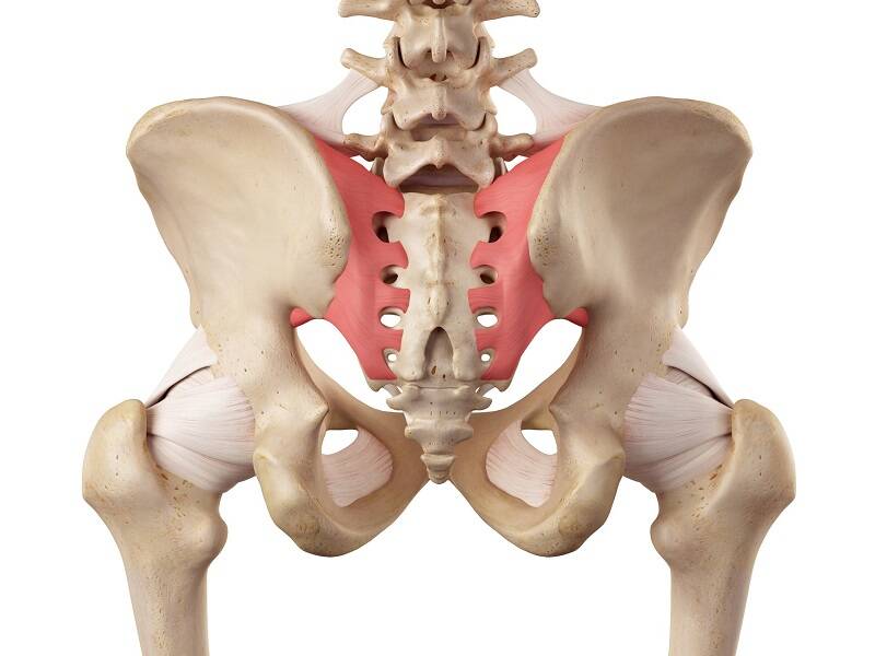
Photo source: Getty images

Blockade of the sacroiliac joint, which is also referred to as the SI joint, is characterized by unpleasant pain in the back, crosses with transfer to the sciatic muscles, ie the buttocks, but also to the groin. Difficulty is aggravated by movement.

Most common symptoms

Show more symptoms

Characteristics

Sacroiliac joint blockage is a relatively common cause of back pain. Many times it is mistaken for another problem associated with the spine and is therefore treated inefficiently.

The sacroiliac joint is also called SI joint.
SI joint block = sacroiliac block or displacement.

It is a joint that connects the sacrum and the hip bones. Of this also the professional designation sacro - iliac joint.

Sacrum = sacrum axis + lumbar bone = ilium axis.

Difficulties with this joint cause unpleasant pain, which occurs in the area of ​​the back, lower back, with the transfer to the sitting area or groin. It is aggravated by walking, changing position, getting up from sitting. It wakes you up.

It frequently affects pregnant women.

In short about SI joints

The lumbar bones are fed to the sacrum on both sides. They form the connection of the spine with the pelvis. This connection is referred to as the sacro-iliac (cruciate-hip) joints. 

Sacroiliac joints are not joints as we usually imagine. They do not have a joint head and a sheath, such as a hip joint.

Their shape is uneven, it contains many edges and depressions. They are said to resemble an ear. Due to the complexity of the articular surface, they are almost immobile.

The movement occurs only in the minimum range of a few millimeters - 2 to 4 millimeters. Slightly more during the last trimester of pregnancy and during childbirth.

In the past, this joint was thought to be rigid and immobile.

However, despite slight movement, SI joints have an important function

The SI joint for the body does not have as much a movement function as a stabilizing function. This joint does not have its own muscles.
The surrounding muscles and ligaments, such as the muscles of the back, pelvis, pelvic floor, sciatic and thigh muscles, and last but not least, the abdominal muscles, are involved in the stabilization and balance of the body.

The main function of SI joints is...

Especially while maintaining the statics and balance of the body. They participate in the stabilization between the spine, pelvis and lower limbs, during movement, walking or running.

They then withstand heavy loads , shock absorption and stress. As the whole weight of the organism is transmitted in this place towards the hip joints and lower limbs.

Causes

What is the cause of the difficulty in this joint?

SI joints can be affected by a functional disorder, such as blockage or displacement. But also, for example, another type of disease, such as inflammation , which is technically referred to as sacroileitis.

Pain in the SI joint can be caused by:

  1. SI block of the joint - is the primary disability of the joint, ie the difficulties stem directly from the joint
  2. SI shift - secondary difficulties outside the joint, faulty movement mechanism and the like
  3. inflammation that can be
    • infectious, in case of spread of bacterial infection, specific inflammation in TB, brucellosis
    • non-infectious as in rheumatoid arthritis or in addition to Bechtereva disease
  4. degenerative changes and osteoporosis
  5. pregnancy, last trimester and childbirth
  6. injury

Difficulties are also conditioned by certain risk factors and conditions, such as:

  • excessive overload
  • one-sided load
  • sedentary lifestyle, sedentary work and long sitting
  • metabolic disease
    • obesity
    • diabetes, diabetes mellitus
  • autoimmune diseases
    • systemic lupus
    • Crohn's disease
    • psoriasis
  • tumor

Symptoms

The main symptom is pain. It can affect one or both joints.

The pain may be pseudoradiculopathy.

The pain is located in the area of ​​the back, crosses with a transfer to the upper part of the sitting region, ie the buttocks, but also to the hip joint and lower along the back surface of the lower limb. May radiate into the groin.

It is aggravated by movement, change of position.Walking and positioning in bed are
problematic. The reason for this is also night pain, which can wake the affected person from sleep.

It is stated that the SI joint will cause low back pain to every 4 people.

Long-term sitting or standing is the reason for the escalation of difficulties. Getting up from a chair is a problem, for example getting out of a car or walking up stairs.

In some cases, agitation and movement alleviate the problem. On the contrary, other times the affected person cannot travel the distance without difficulty. 

Displacement or SI block of the joint causes a malfunction, which is further manifested by a change in movement patterns. What can be seen in posture and walking. Walking can typically naširoko that the present limp and rocking walking.

Diagnostics

Diagnosis is made with the help of anamnesis. The anamnesis also includes, for example, a work history, whether it is a sedentary type of employment, or employment with long-term overload of the spine and pelvis in general.

Are important data about sports or previous trauma. For example, in the case of a painful coccyx, difficulties may manifest themselves over time.

Equally important is information on associated diseases, such as rheumatic problems or metabolic and autoimmune diseases.

Subsequently, a professional examination is important.
Here, the overall evaluation of the figure plays a role, especially weight  and other parameters.
Examination by sight (aspect) begins, when the attitude, movement habits, movement pattern are evaluated. Plus the added palpation examination, ie palpation

For example, the following are investigated:

  • spinal motility
  • dynamics
  • stability
  • hypermobility
  • examination of attitude and movement stereotypes
  • muscle strength
  • muscle shortening
  • slope of the pelvis
  • different length of lower limbs

Various provocative maneuvers and tests are helpful, such as the Lewit test, Patrick test, gapping test, posterior pressure. Reflex examinations are also added.

For the diagnosis of SI joint block, local injection of an anesthetic may be supplemented.
Which has a sedative but also diagnostic significance.

Among the imaging methods that can be used in differential diagnosis are X-ray, CT, MRI or SONO and EMG. However, in not every case can the causes of SI joint problems be identified on their basis.

X-rays can detect bone fractures, CT inflammatory or degenerative changes, and MRI will complete a more detailed overview of all soft structures.

It is necessary to distinguish other causes, such as lumbago or arch of the intervertebral disc, as well as other and more serious diseases of the digestive, excretory or genital system. To eliminate inflammation and laboratory blood tests.

Read also: 
Painful coccyx syndrome
Hernia disc
Lumbago
Radiculopathy

Course

The course and intensity of the difficulties are variable and, of course, depend on the exact cause and extent of the damage. There will be slight differences between SI joint blockade or a degenerative or rheumatic process.

The inflammatory process is also about fatigue, an increase in body temperature.

There is back pain in the lower back, buttocks, hips and groin, with transfer to the lower limbs.

It worsens when changing position, especially when getting out of bed, sitting or getting out of the car. An example is the pain aggravated by walking up stairs.

It is also enhanced by long-term sitting or standing in one position. There are nocturnal pains that wake you up. Someone may have painful gait, but it has also been described to reduce the difficulty of moving and moving. 

When tracking movement, it is possible to notice a change in movement patterns.

How it is treated: SI joint block

Treatment of SI joint block: Medication, physiotherapy, rehabilitation and exercises

Show more
fshare on Facebook

Interesting resources

  • Cibulka MT; Delitto A & Erhard RE (1992). "Pain patterns in patients with and without sacroiliac joint dysfunction". In Vleeming A; Mooney V; Snijders CJ & Dorman T (eds.). First Interdisciplinary World Conference on Low Back Pain and its Relation to the Sacroiliac Joint. pp. 363–70. 
  • Fortin, J. D.; Falco, F. J. (1997). "The Fortin finger test: An indicator of sacroiliac pain". American Journal of Orthopedics26 (7): 477–80. 
  • Sturesson, B; Selvik, G; Udén, A (1989). "Movements of the sacroiliac joints. A roentgen stereophotogrammetric analysis". Spine14 (2): 162–5. 
  • Sturesson, B; Uden, A; Vleeming, A (2000). "A radiostereometric analysis of movements of the sacroiliac joints during the standing hip flexion test". Spine25 (3): 364–8. 
  • Schwarzer, A. C.; Aprill, C. N.; Bogduk, N (1995). "The sacroiliac joint in chronic low back pain". Spine20 (1): 31–7. 
  • Maigne, J. Y.; Boulahdour, H.; Chatellier, G. (1998). "Value of quantitative radionuclide bone scanning in the diagnosis of sacroiliac joint syndrome in 32 patients with low back pain". European Spine Journal7 (4): 328–31. 
  • Maigne, J. Y.; Aivaliklis, A; Pfefer, F (1996). "Results of sacroiliac joint double block and value of sacroiliac pain provocation tests in 54 patients with low back pain". Spine21 (16): 1889–92.