- Mosby's Medical Dictionary, 8th edition. © 2009
- Howe HS, Zhao L, Song YW, et al. (February 2007). "Seronegative spondyloarthropathy--studies from the Asia Pacific region" (PDF). Ann. Acad. Med. Singap. 36 (2): 135–41.
- "Seronegative Spondyloarthropathies: Joint Disorders: Merck Manual Professional".
- Luong AA, Salonen DC (August 2000). "Imaging of the seronegative spondyloarthropathies". Curr Rheumatol Rep. 2 (4): 288–96.
- Elizabeth D Agabegi; Agabegi, Steven S. (2008). Step-Up to Medicine (Step-Up Series). Hagerstwon, MD: Lippincott Williams & Wilkins. ISBN 978-0-7817-7153-5.
- Ankylosing Spondylitis and Undifferentiated Spondyloarthropathy Workup Author: Lawrence H Brent. Chief Editor: Herbert S Diamond.
- Várvölgyi C, Bubán T, Szakáll S, et al. (April 2002). "Fever of unknown origin with seronegative spondyloarthropathy: an atypical manifestation of Whipple's disease". Ann. Rheum. Dis. 61 (4): 377–8.
- Shankarkumar U, Devraj JP, Ghosh K, Mohanty D (2002). "Seronegative spondarthritis and human leucocyte antigen association". Br. J. Biomed. Sci. 59 (1): 38–41.
- Maria Antonietta D'Agostino, MD; Ignazio Olivieri, MD (June 2006). "Enthesitis". Best Practice. 20 (3): 473–486.
Spondylarthrosis: Facet Arthrosis and Uncovertebral Arthrosis of the Spine
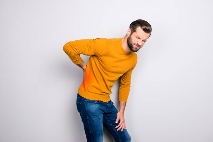
Photo source: Getty images
Spondylarthrosis is a degenerative disease of the intervertebral joints. It affects the adult population and mainly men. It is manifested by both pain and stiffness of the spine.
Most common symptoms
- Shoulder Blade Pain
- Chest pain
- Flank Pain
- Headache
- Limb pain
- Nerve pain
- Leg Pain
- Shooting pain in fingers and toes
- Pain that Radiates into the Shoulder
- Groin Pain
- Back Pain
- Tinnitus
- Muscle stiffness
- Defence
- Tingling
- Muscle weakness
- Head spinning
Show more symptoms ᐯ
Characteristics
Spondylarthrosis is a degenerative disease affecting the intervertebral joints.
The name is a compound of two words, namely vertebra, i.e. spondylos, and arthrosis. Osteoarthritis is a degenerative disease affecting the joints.
During this process, it is possible to notice pathological (morbid) changes on:
- joint cartilage
- joint capsule
- joint cavity
- bones
- tendons
- and muscles
In general, arthrosis can take a primary or secondary form. Primary has no clear cause and secondary arises from ageing, injury, developmental defects or other disease (arthritis, diabetes, gout).
For more general information, see the article on arthrosis and the magazine article Stop arthrosis.
Back pain is a significant contributor to incapacity for work. It does not only affect the elderly, but, on the contrary, the productive and young part of the population.
The causes of disorders and diseases affecting this part of the musculoskeletal system are manifold.
Many times their reason is unknown. However, to a large extent, it is attributed to improper lifestyle, sedentary lifestyle, overweight and incorrect or excessive overloading of the spine.
Problems are acute = sudden, not lasting long. The affected person is fit in three or four days.
The second group consists of chronic cases = when they persist for a long time or recur continuously (returning, relapsing). These have a negative impact on physical but also mental fitness = reduce quality of life.
If the arthrosis affects the joints of the spine, i.e. the intervertebral joints, it is therefore referred to as spondylarthrosis.
For the most part, it mainly affects men, starting as early as the age of 40.
Until then, it may proceed completely asymptomatically or the difficulties are of a mild nature. However, it is the neglect of this initial stage that can lead to irreversible - irreversible damage to the spine.
We briefly describe the spine and intervertebral joints, then return to the issue of spondylarthrosis.
The spine and intervertebral joints
The spine is the supporting axis of the human body. It has both a locomotor and protective function.
Across the spine runs the spinal cord, which is an important transmission pathway of nerve reflexes and excitations between the body and the brain.
In Latin it is referred to as the columna vertebralis. It is naturally curved (lordosis and kyphosis), with the morbid curvature known as scoliosis.
The spine is composed of vertebrae, of which there are 33 to 34. These are also referred to as vertebrae. They have a specific shape and functionality.
The spine is divided into individual sections, or segments, namely:
- cervical spine
- has 7 vertebrae, referred to as vertebrae cervicales - C1 to C7
- C1 is also known as the atlas and is connected to the skull
- C2 is also known as the axis
- the blood vessels that supply the brain with blood also run through the cervical vertebral processes
- thoracic spine
- has 12 vertebrae, vertebrae thoracicae - Th1 to Th12
- the ribs connect to the thoracic vertebrae, forming the rib cage
- lumbar spine
- has 5 vertebrae, vertebrae lumbales - L1 to L5
- the vertebrae of this part of the spine are the largest
- sacrum
- formed by fusion of 5 or 6 vertebrae - vertebrae sacrales S1 to S5 (6)
- the sacrum - os sacrum
- together with other bones forms the pelvis
- coccyx
- vertebrae coccygeae
- may have 4 or 5 vertebrae - Co1 to Co4 (Co5)
The vertebrae have a specific shape and function. They are of different sizes and their role is to support the body and protect the spinal cord.
They consist of several anatomical structures, namely the body, the arch and the processes.
1. The vertebral body
The Latin name is corpus vertebrae. It is the anterior part of the vertebra. Its role is to bear weight - weight-bearing function.
The vertebral bodies vary in size and height.
Among the largest vertebrae are the lumbar vertebrae. Conversely, the thinnest are found in the cervical spine section. The top and bottom of the vertebral body is flat. At this point, the intervertebral disc, which is adapted in shape, is deployed.
2. The vertebral arch
It attaches to the body of the vertebra. This connection is formed by little appendages - these are referred to as pedicles.
Pedicles contain small notches. These form intervertebral cavities through which the spinal nerves pass.
The disc is the second part, which on both sides forms the vertebral aperture through which the spinal cord passes.
3. The vertebral processes
They protrude from the arch. Their importance is in the fusion of the vertebrae and in movement.
Vertebrae have three types of processes:
- spinous process - processus spinosus, extending backward
- there is only one, it can be felt on the back under the skin
- ligaments or muscles are supplied here
- transverse process - processus transversi - is paired
- strengthening of muscles and tendons
- v hrudnej časti chrbtice sa nachádzajú úpony rebier
- articular process - processus articulares is a paired process
- behind the vertebral pedicle
- contains the articulation of the vertebrae and cartilage
The vertebrae are interconnected. This connection is fixed, but at the same time mobile. It consists of several mechanisms.
Table: mechanisms of vertebral fusion
| Mechanism of fusion | Description |
| Ligament |
|
| Intervertebral joints |
|
| Intervertebral discs |
|
| Special fusion |
an example is synchondrosis
|
| Muscular system |
the muscles of the spine, together with the muscles of the abdomen and the muscles of the neck and pelvis
|
Intervertebral joints
In Latin, articulatio intervertebrales, are small joints that form a movable connection between two vertebrae. These are paired joints of the intervertebral processes (processulares articulares).
The articular surfaces of the individual vertebrae have different shapes, which affects the range of motion. So do the joint capsules. These are looser, especially in the cervical spine, giving it a greater range of movement.
Facet joints - intervertebral joints
Latin articulationes zygapophysiales, are tiny articular plates. Each segment contains two facet joints. These joints are innervated and often pain, the so-called facet syndrome, occurs when they are disrupted.
The facet joints are responsible for the movement, its range and stability of the spine.
As much as they enable it, they also restrict it. This restriction is also protective, namely against excessive bending or rotation of the body part.
Similarly, in each segment of the spine are different.
Spondyloarthritis is...
It is therefore a long-term degenerative process that damages the intervertebral joints. Arthrotic changes take place on the basis of overstrain and other causes.
It is also referred to as intervertebral spondylarthrosis.
In the case of damage to the articular surfaces, osteophyte formation also occurs. Osteophytes are bone processes.
Spondyloarthritis and spondylosis often run together. Also in spondylosis, osteophyte formation is characteristic, which is superimposed on degenerative damage to the entire vertebral segment.
Learn more:
Spondylosis
Osteochondrosis
Typically, spondylarthrosis is manifested by pain or stiffness in the affected part of the spine.
Depending on which part of the spine is affected, we may encounter a designation such as:
Lumbar spondylarthrosis, i.e. when affecting the cervical spine as cervical spondylarthrosis and when affecting the cervical spine as cervical spondylarthrosis.
This disease is characterized by a chronic course, with a progressive character. Translated, it is a long-term disease that progresses over time.
Osteoarthritis affects the surfaces of the intervertebral joints, with their cartilage thinning, the joint capsule is destroyed. This also affects surrounding structures such as ligaments and muscles. Over time, outgrowths, or osteophytes, form.
What causes this health problem often cannot be detected. Aging of the body, faulty posture, prolonged sitting and sedentary lifestyle, injury or other illness all play a role.
You may have heard of...
Uncovertral arthrosis is arthrosis of the small joints of the cervical spine that are located between vertebrae C3 to C7. They are also referred to as Luschka's joints. They are located in the space between the vertebrae and serve to move and stabilize the neck.
They are often affected by degenerative changes, which can lead to pain in the neck, neck and even radiating to the upper limbs. This is caused by compression of the nerves that exit this part of the spinal cord.
And what is facet arthrosis?
It is also a degenerative process that affects the small surfaces of the joints that are located between the vertebrae.
Causes
Spondylarthrosis is a multifactorial disease.
What does that mean?
That several factors are involved in its occurrence. However, the exact cause is still not fully elucidated.
Primary form = unknown cause.
Secondary form = a known causative agent, such as age, injury.
It is largely attributed to the aging of the body.
Further contributing to the disadvantage are faulty posture, poor movement habits, sedentary lifestyle and sedentary occupation. But also other work factors, such as forced postures for long periods of time or excessive strain on the spine.
Lifting loads puts a strain on the spine, of course. And the wrong lifting mechanism is also a risk for the development of an acute problem. Such as, for example, a herniated disc, i.e. a bulging of the intervertebral disc.
In addition to work, some sports also cause long-term strain. The main contributing factors are abrupt movements, changes in the direction of movement or rapid and uncontrolled rotations and excessive bending of the spine.
Another factor in both sports and casual activities is injury.
Consequently, they may be involved in the disease:
- birth defects
- anatomical anomalies - joint hypermobility or different limb lengths,
and thus uneven loading on the spine - endocrine and metabolic diseases (diabetes, gout)
- overweight and obesity
- but also, for example, smoking or excessive alcohol intake.
Secondary causes include inflammation.
Rheumatoid arthritis is also an example. It is a systemic inflammatory rheumatic disease and has an autoimmune basis.
Note, however, this long-term disease also has extra-articular, i.e. extra-spinal, complications.
In this case, there is a risk of damage to other parts of the body, such as:
- the joints of the shoulder, hip or knee
- tendon attachments, often the Achilles tendon
- the skin - psoriatic arthritis, including psoriasis
- the eyes
- the intestines
- the mucous membranes of the body
Symptoms
A symptom in the initial stage of spondylarthrosis is mild pain localized to the affected spinal segment. The pain is provoked or aggravated by movement and physical exertion. Conversely, the abatement of the discomfort occurs at rest.
There is a typical limitation of mobility - range of motion.
The progression of the disease is characterized by the formation of osteophytes.
Bony growths, or osteophytes, can interfere with the spinal canal and the passage of spinal nerves. In this case, they are the cause of spinal cord or nerve root oppression.
Nerve compression = compression = oppression.
This condition provokes radicular syndrome. This is back pain that is accompanied by radiation to another part of the body, such as an upper or lower limb.
In this case, in addition to pain, there are other neurological problems, tingling, impaired sensitivity or muscle weakness and weakened reflexes.
More information in the article: Radiculopathy.
In cervical spondylarthrosis, neck pain, limitation of neck mobility, plus other neurological complaints. These radiate to the shoulder, arm, upper limb or higher in the head.
A serious complication is a condition where bone growths oppress blood vessels. This is mainly in the cervical spine. This is the oppression of the vertebral vessels that supply blood, oxygen and nutrients to the brain.
In this case, the headache may be accompanied by dizziness, vertigo, tinnitus and other neurological problems associated with impaired blood supply to the brain.
Significant impairment of blood flow to the brain results in stroke, which can have a number of serious causes.
Lumbar spondylarthrosis is characterized by pain in a given stretch of the spine (pain in the shin and sacrum), stiffening of the muscles and shooting into the buttock, thigh, and thus lower limb.
The risk of synovial cysts = cyst at the level of the articular capsule surface.
Cyst = a pathological formation, a cavity circumscribed from its surroundings, filled with fluid or a dense mass.
And thus in spondylarthrosis can occur:
- pain at the site of spinal involvement (often the lumbar area, neck less often the chest)
- worsening of pain on exertion, prolonged walking, prolonged inactivity in a forced monotonous position
- morning stiffness of the spine
- stiffening of the muscles in the spine
- restriction of movement, bending or rotation
- loss of spinal stability
- incorrect posture
- shooting pain
- tingling, trembling or other paresthesias
- radiating neurological symptoms to other parts of the body, limbs or head
- weakening of muscles
- sensitivity disorder
- disorder of reflexes
Diagnostics
Diagnosis of the disease uses a medical history, a description of clinical symptoms and a physical examination. Neurological examination includes examination of the spine, posture, movement or reflexes.
When taking a medical history, there will be important questions such as:
- how long the discomfort lasts
- when the difficulties arose
- whether and how often they recur
- dependence on movement and position
- effect on sleep
- whether the difficulties limit normal daily activities
- daily routine, lifting loads, work, working position
- injury
Subsequently, imaging methods are important:
- X-rays
- CT
- magnetic resonance imaging, or MRI
- EMG
In the differential diagnosis, it is important to distinguish another cause of back pain. Therefore, blood sampling, liquor or other investigative methods may be added.
They think of inflammatory causes such as rheumatism, psoriasis, Morbus Bekhterev (ankylosing spondylitis) and of course other health problems. A neurologist, an orthopaedic surgeon, a physiotherapist, a radiologist or a general practitioner collaborate in the diagnosis.
Course
The disease is chronic.
In the initial stage it is characterized by asymptomatic course = hidden symptoms = it is asymptomatic.
In the initial stage it is characterized by asymptomatic course = hidden symptoms = it is asymptomatic.
The first changes happen at the metabolic level, this affects the state of the articular cartilage. The cartilage is affected by cracks, its thickness decreases, and the previously flat articular surfaces are uneven.
Over time, even the part of the bone below the cartilage becomes damaged. Anatomical changes and deformities set in on the bone, the joint and the articular cleft.
This progression is divided into 4 stages, namely:
- Phase 1 - metabolic changes, thinning of cartilage, decrease in the amount of fluid
- the course is largely asymptomatic
- Phase 2 - unevenness of articular surfaces, initial erosion of the bone from under the cartilage
- Phase 3 - bone reaction to damage, formation of bone outgrowths - osteophytes, and the development of osteosclerosis or osteoporosis (changes in the density of bone tissue)
- Phase 4 - the disappearance of the articular cleft, deformities and pathologies of the joints
- complete restriction of momentum in a given part
Incipient spondylarthrosis = a term denoting the initial, or incipient, form of a disease.
At this time, we can observe mild pain, which is present especially after physical exertion. These subsequently subside in calmness.
The pain intensifies during the day due to the increased load on the spine caused by physical exertion and is more intense in the evening.
This is accompanied by stiffening of the muscles and restriction of mobility in that part of the spine.
Conversely,...
Severe spondylarthrosis, which is characterised by pain in the affected part of the spine, however, neurological symptoms radiate to other parts of the body.
The pain in this case is intense, disturbs from sleep and manifests even with minor movements.
The presence of osteophytes is also responsible for the reduced range of motion. The immobilization of adjacent vertebrae can cause bridging of the intervertebral disc by bony processes.
It is also important to think about...
Spondylarthrosis often runs together with spondylosis. Spondylosis is also a degenerative disease. It affects the entire segment, and therefore the vertebra, disc and joints and other surrounding structures.
Also,...
Only one segment may be affected, but in worse cases several segments or the whole spine may be affected.
Prevention and lifestyle changes are important
Of course, as we always say: prevention comes first.
Spinal problems should be prevented from a young age, adolescence and young adulthood. Alternatively, it is essential to change your lifestyle as soon as possible.
Preventive measures include:
- plenty of exercise
- appropriate exercise and improvement of spinal and core mobility (abdominal muscles, diaphragm, pelvis, pelvic floor and spine, deep stabilization system)
- which also has an effect on spinal stabilization
- correct posture and movement habits
- beware of inactivity and sedentary lifestyle - need to limit
- suitable position on the bed - mattress, pillow
- work ergonomics - sitting, standing and working position, more frequent position changes
- suitable for lifting and carrying loads
- regular walking
- weight management and reduction of overweight and obesity
- balanced diet, sufficient vitamins and minerals
Learn more: Essential nutrients, Mediterranean type of diet, Weight loss and diet
In the context of a healthy spine, a back school and some of the most appropriate movement activities are mentioned. These are dancing, swimming, cycling, cross-country skiing, skating, running, Nordic Walking, horse riding, but also ordinary walking.
How it is treated: Spondylarthrosis
Treatment of spondylarthrosis: Medication and rehabilitation
Show moreSpondylarthrosis is treated by
Other names
spondyloarthrosis, lumbar spondylarthrosis, cervical spondylarthropathy
Interesting resources

Bc. Lukáš Tóth
Healthcare worker
The secondary medical school in Nitra gave me the basis for my career in the field of health and diseases. Thanks to it, I worked for 2 years in the traumatology clinic and outpatient clinic at the Nitra Hospital. Since 2006 I was employed in the emergency medical service, where I stayed until 2017.
I completed my bachelor's degree at the University of Constantine the Philosopher in Nitra in the field of emergency health care. The bachelor's degree allowed me to continue my mission as a paramedic. In the meantime, I got a job at the emergency line 155. I have been working in pre-hospital health care until today.
I had an interest in people, health and even diseases in my childhood, which gave me the prerequisite to pursue this topic in adulthood. Studying and acquiring new information in practice provided me with a great basis for writing professional texts, in the form of articles that can be understood by ordinary people. Thus, my interest in the Health Portal has a solid foundation in years of practice and personal interest. Similarly, I am also interested in healthy eating, nutrition and overall healthy lifestyle. I fill my free time with family and sports.
View all articles by the same author


