Spondylolisthesis is the medical term for the displacement of one spinal vertebra compared to another. This happens for several reasons. This is in the lower lumber region and causes issues with posture, muscle weakness and other signs and complaints.
Spondylolisthesis is a condition in which a vertebra of the spine is displaced relative to another vertebra.
It is the cause of various problems, such as unpleasant pain and its radiation to the limbs. It causes incorrect posture or restriction of mobility.
Vertebral shift is not a modern problem.
It was described in the medical literature by Herbiniaux in 1782, and the term spondylolisthesis was coined by Kilian in 1854. The causes of its origin have been debated for more than 100 years, during which several classifications have emerged.
The word "spondylolisthesis" is of Greek origin and consists of two parts: Spondylos - vertebra Olisthanein - shift, displacement
About the spine and vertebrae
The spine is a support for the body, it is part of the musculoskeletal system and its role is also to protect the spinal cord, which leads from the brain to approximately the level of the second lumbar vertebra.
The spine, also called the backbone and the vertebral column, or in Latin "columna vertebralis".
The spine is physiologically curved. The forward curvature is called lordosis, located in the cervical (neck) and lumbar (lower back) regions.
The backward curvature is referred to as kyphosis, which is in the thoracic (torso) and sacral areas (low back) of the spine. This curvature is physiological, i.e. natural.
Scoliosis is an unnatural, therefore pathological, sideways curve of the spine. However, a slight physiological curvature to the side is present in every person.
Temporary curvatures of the spine to the side can be observed when standing on one leg, when transferring weight to one limb or while carrying a heavier load in one upper limb.
The spine consists of vertebrae, of which there are 33 to 34.
The vertebrae are connected to each other. Their connection is fixed, yet movable.
The connection of vertebrae is ensured by cartilage, ligaments or intervertebral joints.
Table: the interconnection is formed by several mechanisms
Connection type
Description
Ligaments
fibrous connective tissue
the ligaments of the spine strengthen the spine and help with movement
they are long and short
long ligaments for the whole spine
short ligaments connect adjacent vertebrae
Intervertebral joints
art. intervertebrales
Intervertebral discs
act as a ligament to hold the vertebrae together, and to function as a shock absorber for the spine
Special connection
an example is synchondrosis
cartilage connection
immovable connection
in the area of the sacrum and coccyx, they become ossified to the bone
Muscular system
muscles of the spine along with the muscles of the abdomen and the muscles of the trunk and pelvis
they form the locomotor and fixation component of the spine
important overall in movement and posture
Among the vertebrae there are 23 intervertebral discs.
The first disc is located between the 2nd and 3rd cervical vertebrae and the final segment between the final lumbar and first sacral vertebra.
Intervertebral discs are also referred to as intervertebral discs. Their task is to dampen the impacts of other vertebrae during the movement of the body.
They also have other functions:
shock absorption when moving, running, jumping
stabilise the spine
maintain balance
balance compressive and tensile forces, they spread it over the whole area
they are cooperative in any movement of the spine, twisting or rotation of the body
Their shape copies the body of the vertebrae and they are of different heights. They are higher between the cervical and lumbar vertebrae, with the highest plate being between the L5 and S1 vertebrae.
Plates can have several problems, such as the article on disc herniation.
A more detailed and technical view on the vertebrae
Vertebrae are technically referred to as vertebrae.
Most people have either 33 or 34 vertebrae.
Table: Spinal segments
Part - segment
Vertebra
Description
Cervical spine
vertebrae cervicales
conmsists of 7 vertebrae
labelled from C1 to C7 (C1, C2, C3, C4, C5, C6, C7)
the 1st and 2nd cervical vertebrae have a specific shape for their function and connection to the skull
the first cervical vertebra is called theatlas - it allows sideways movements of the head
the second cervical vertebra is called the axis - its defining feature is the dens - dens axis
the axis helps the head to move forwards and backwards
the junction of the skull and spine is referred to as the craniovertebral junction
articulatio atlantooccipitalis
the protrusions of the cervical vertebrae form openings through which the vertebral arteries and veins pass
these vessels supply the brain with blood
Thoracic spine
vertebrae thoracicae
consists of 12 vertebrae
Th1 to Th12
the bodies of the thoracic vertebrae contain the costal facets - fovea costalis
a site of connection between a rib and a vertebra
the anterior side of vertebral bodies Th4 and Th7 contain the thoracic aortic impression - impressio aortica
Lumbar spine
vertebrae lumbales
lumbar segment or the lower back
consists of 5 vertebare
L1 - L5
contains the largest vertebrae
Sacrum
vertebrae sacrales
may consist of either 5 or 6 stavcov
S1 to S5 (S6)
fused together, they form the the sacrum - os sacrum
it is movable
forms the pelvis together with the other bones of the pelvis
Coccyx
vertebrae coccygeae
may consist of either 4 or 5 vertebare
Co1 - Co4 or Co5
the vertebrae are interconnected into the coccyx, also called the os coccygeae
vertebrae have no arches
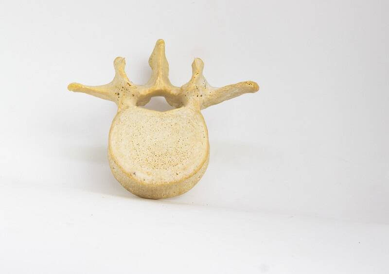
The vertebrae are the bearing support of the human body. Their vertebral foramen (opening), also called foramina vertebralis, form the spinal canal through which the spinal cord passes.
They are bones with a specific shape.
Stavec sa skladá z tela stavca, oblúku a výbežkov.
1. The vertebral body, or in Latin corpus vertebrae, is located in front. It is the main supporting part of the vertebra. Its upper and lower surfaces are flat. The vertebral discs are placed here.
The height of the vertebrae varies, with the cervical vertebrae being the narrowest. The widest ones are located in the lumbar spinal segment. Their height decreases again towards the coccyx.
2. The vertebral arch attaches at the back to the body of the vertebra. Its role is more or less protective, as it protects the spinal cord.
The arch has two parts.
One of them is called the lamina of vertebral arch, or in Latin lamina arcus vertebrae. It points towards the vertebral body. It is attached to the body of the vertebra with two pedicles, or in Latin pediculus arcus vertebrae.
The arches of the vertebrae form the vertebral foramen, or in Latin foramen vertebrae. Together they form the spinal canal - canalis vertebralis.
The pedicles have notches on their edges,i.e. at the top and bottom. These are referred to as incisura vertebralis superior (superior vertebral notch) et inferior (inferior vertebral notch). They form a structure denoting intervertebral foramen (foramina intervertebralia).
The intervertebral foramen are important due to the passage of spinal nerves exiting the spinal cord.
3. The vertebral processes protrude from the vertebral arches. Their role is important because they connect the vertebrae and enable the body to move.
Table: The vertebrae have several types of processes
Name
Latin name
Description
Spinous process
processus spinosus
projects direcktly from the back side
there is only one spinal process
can be felt on the back under the skin
muscle is attached here
important for movement
Articular processes
processus articulares
paired
upper/superior (superiores) and lower/inferior (inferiores)
located just behind the pedicle
connect the vertebra
they contain cartilage at the joint
Transverse processes
processus transversi
they protrude at the same location as the articulatar processes
paired
So what is spondylolisthesis?
Spondylolisthesis is defined as a pathological displacement of the vertebral body relative to an adjacent lower vertebra, towards the centre of the body, and thus towards the abdomen - ventrally.
+ In a later stage of the shift also in the ventrocaudal direction - pointing to the abdomen and down.
It most commonly affected area isthe lower back, more specifically the lumbar segment of the spine. The cervical segment (neck) less so.
Spondylolisthesis in the lumbar regions L5 and S1. Photo: Getty images
Vertebral displacement can occur in various forms, with the types of spondylolisthesis being important in terms of the cause, causing difficulties and also in the treatment.
A simple classification is congenital and acquired spondylolisthesis. Congenital listhesis is also divided into low-grade or high-grade dysplasia. Acquired for traumatic or post surgical or pathological and degenerative.
Various forms of classifications have been developed in the history of spondylolisthesis. Today, the Newman, Wilts, and MCnab classifications are used.
Table: Classification of spondylolisthesis
Type
Description
1. Dysplastic
about 20% of cases
in the vast majority of cases at L5 and S1 level
twice as common among women
it is often diagnosed as early as about 5 years of age
it progresses slowly, especially in the period from 10 to 15 years of age - a growth factor
it is often asymptomatic
signs and symptoms occur mainly during adolescence
2. Isthmic
about 45% of cases
familial occurrence, ie familial
unclear origin
mostly about spondylolysis - a defect of the intercostal space
the isthmus is completely interrupted or prolonged
3. Degenerative
about 20 % of cases
sometimes referred to as pseudospondylolisthesis by Junghans
comorbidity with spondylarthrosis and spondylosis
structural changes on vertebraedue tothe degenerative process
dependent on the degenerative process on disks
mostly in the higher parts of the lumbar spine
less common in sections L5 and S1
4. Traumatic
Two forms:
acute traumatic spondylolisthesis - due to spinal injuries
stress fracture - common in sports and gymnastics
for repeated specific spinal movements in hyperextension
repeated minor injuries and fatigue fracture
most common in segments L5 and S1
post-surgery
direct - after surgery, in the field of surgery, for example during disc surgery
indirect - arises in the segment above the procedure - spondylodesis (connection of vertebrae), as a result of overload and reactive hypermobility
5. Pathological
It occurs in congenital or systemic bone diseases, rheumatoid arthritis, tumor metastases
Spondylolisthesis is also classified by vertebral displacement percentage:
grade I- 25 % displacement
grade II - 25 - 50 % displacement
grade III - 50 - 75 % displacement, more than 50 % = high-grade
grade IV - above 75 %
Spondyloptosis = the body of the upper vertebra gets in front of the body of the second vertebra = above 100 %.
Spondylolysis
Spondylolysis refers to a vertebral defect in a section of its arch. The vertebral arch is damaged at the site of narrowing, which is referred to as an isthmus of the vertebral arch.
Isthmus = zúženie, úžina.
There is a congenital and an acquired form which has a higher occurrence. It can be present on one side, i.e. unilaterally, but also on both sides of the vertebra, i.e. bilaterally.
The most frequently affected part of the spine is the lumbar part, more precisely L4 and L5 less often L3.
The acquired form appears in childhood and adolescence. The cause is spinal congestion and sports activities.
It contributes to long-term repetition and sudden changes in the position of the spine during sports, namely forward bending, tilting along with rotation.
It is more common in boys.
Back pain in the affected area is one of the symptoms. However, it can be completely asymptomatic, i.e. without signs and symptoms. It is usually detected by accident during X-ray examinations.
Causes
The cause of spondylolisthesis, i.e.the shift, is not completely clear.
Various factors are suspected to play a role, for example heredity, age, race or degenerative process of the vertebrae, or even the degree of physical activity. In this context, it is further classified into specific forms.
Adolescents are reported to suffer from the condition most frequently. Before adolescence, the condition may take its course asymptomatically, i.e. without symptoms.
Progression is associated with a time of intense growth, from the age of8 - 20 years. Later on, in adulthood, the risk of vertebral shifting decreases. However, the risk rises again after about the age of 60.
It affects girls to a greater extent.
Sports, such as gymnastics, dancing and figure skating, are also reported to contribute to a higher incidence among girls. The cause is frequent flexion in the back in terms of hyperextension. The condition is also referred to as stress fracture.
This type falls under the traumatic form. The post-surgical type is also included here. It arises as a result of surgery in the spine, i.e. the intervertebral disc.
Example:
In the isthmic form, it is a defect of isthmus - in Latin pars interarticularis. This is an interruption of the vertebra in place. The cause has not been fully understood yet.
It also depends on the type. There may be a complete interruption of the isthmus or an extension (elongation) thereof. In the elongated form, the probability of shift is slightly lower.
Degenerative changes cause damage in several parts of the vertebra. Alternatively, it may be a pathological process on the bone tissue or joint surfaces, but also due to damage to the intervertebral disc.
The pathological form occurs in other diseases, such as various congenital syndromes or systemic bone diseases, rheumatoid arthritis or the oncological process on the spine.
The symptoms of spondylolisthesis are influenced by several factors. Of course, the occurrence of difficulties also depends on the specific form of olistesis.
As mentioned, in most cases, the development of pathological changes begins in childhood. However, the difficulties progress mainly during adolescence. The reason is more intensive growth.
Up to this age, spondylolistesis can be asymptomatic. Subsequently, various health problems develop as the progression progresses.
Symptoms of spondylolisthesis include:
chronic pain in the spine - lumbar and sacral areas
low back pain after physical activity
pain radiating to the limb
pain worsens standing, walking
reducing the distance walked without pain - claudication interval
posture disorders - severe kyphosis, lordosis and scoliosis
when walking or standing
deepening of lumbar lordosis
bending at the knees
limitation of movement of the affected part of the spine
back muscle stiffness
muscles weakness in the limb
tingling - paraesthesia in the limb
in severe forms of nerve root irritation, sphincter issues may arise, as in the case of cauda equina syndrome
Most frequently, relief comes after bending forward. Back pain in children is rather rare.
Problems may have a neural character, which means that there is an irritation of the nerve root. Nerve root pain is characterised by a sharp or burning character, high intensity and radiating pain to the dermatoma area.
The dermatome is part of the innervation of the relevant nerve.
There are also other neurological problems associated with the condition, including tingling sensations, i.e. paresthesia, or a loss of sensitivity or strength. The neurologist also may identify other motor, sensory and reflex disorders.
Another type of pain is pseudoradicular pain, which is characterised by pain in a specific area. As with nerve root irritation, the location of this condition is not clear and uncertain.
It can be felt on the hips, on the sides of the thighs or lower legs.
The main difference from the root syndrome is also the absence of impaired sensitivity and motor skills or weakened reflexes.
Diagnostics
Examining vertebral displacement is not difficult. The patient's medical history is the initial identificatin of the condition. A person with spondylolisthesis begins to complain of pain. In other cases, the finding is accidentally made during another examination (e.g. X-ray).
Next, the patient's clinical picture: neurological examination, the specialist examines the spine visually or by palpation. Mobility, posture, posture, gait, and reflexes are evaluated.
It is important to assess the mental condition. As chronic difficulties reduce the quality of life and significantly affect the mental state of the person.
Imaging methods are important:
X-ray
CT
MRI
perimyelography (PMG) using a contrast agent
densitometry - examination of bone tissue quality, especially in people over 70 years
scintigraphy
Imaging methods help to evaluate the overall state of the vertebral displacement. The percentage displacement and the degree of spondylolisthesis (stages 1 to 4) of the upper vertebra compared to the lower, but also the angle of displacement are evaluated.
Differential diagnosis is important. Its role is to distinguish the condition from other causes, for example inflammatory processes, disorders of the cardiovascular system, coxarthrosis, and serious oncological diseases.
Course
The course of the disease, depending on the form, is rather slow. As already described, a pathological disparity can arise in childhood for a variety of reasons.
Thus, spondylolisthesis can progress for a longer period unnoticed, i.e. it is asymptomatic. Pain is only associated as a result of higher growth rates during adolescence.
Affected persons are known to have posture disorders both when standing and walking.
At higher degrees of shift, there are also other neurological difficulties, ie the radiation of pain to impaired sensitivity or movement.
Back pain occurs between the lower edge of the ribs and the lower part of the sciatic muscles. The muscles of the spine are affected by spasms, they are stiff. Pain gets worse with movement or physical exertion.
Claudicationis defined as pain in the legs or arms that comes on with walking or using the arms. Muscle pain occurs at a higher rate of shift at about 50%.
It is associated with sensory impairment. It is understood that problems with skin sensitivity occur during the innervation of the affected nerve.
Similarly, motor properties weaken. An example is the reduction of muscle strength or tension.
Cauda equina syndrome is a serious condition.
Question: What kind of a condition is it?
It is a serious problem that arises as a result of nerve compression. This bundle of nerves is referred to as the cauda equina.
The spinal cord runs through the spinal canal along the vertebrae L1 to L2. Cauda equina, meaning "the horse's tail", is the lowest part of it.
Cauda equina syndrome is characterized by a significant impairment of motor and sensory functions. The disorder occurs at the level of the pelvic organs, pelvic floor and lower limbs.
The cause is the medial disc herniation in the area of the lumbar spine lower than the L2 vertebra. The symptoms are varied and depend on the location or extent of the compression.
An example is a sensitivity disorder in the genital area and the rectum.
An example is motor dysfunction and weakening of muscles in the lower limbs, but also a disorder in the regulation of the sphincter (opening/closing muscle) or the association of incontinence, i.e. involuntary excretion of urine and stool.
In this case, sexual dysfunction also develops.
How it is treated: Spondylolisthesis
How is spondylolisthesis, vertebral displacement, treated? Medication and surgery
Merriam-Webster medical dictionary. Retrieved 2017-09-07.
"spondylolisthesis". Farlex medical dictionary. Retrieved 2017-09-07., in turn citing:
Miller-Keane Encyclopedia and Dictionary of Medicine, Nursing, and Allied Health, Seventh Edition. Copyright date 2003
Dorland's Medical Dictionary for Health Consumers. Copyright date 2007
The American Heritage Medical Dictionary. Copyright date 2007
Mosby's Medical Dictionary, 9th edition
McGraw-Hill Concise Dictionary of Modern Medicine. Copyright date 2002
Collins Dictionary of Medicine. Copyright date 2005
Introduction to chapter 17 in: Thomas J. Errico, Baron S. Lonner, Andrew W. Moulton (2009). Surgical Management of Spinal Deformities. Elsevier Health Sciences. ISBN 9781416033721.
Walter R. Frontera, Julie K. Silver, Thomas D. Rizzo (2014). Essentials of Physical Medicine and Rehabilitation (3 ed.). Elsevier Health Sciences. ISBN 9780323222723.
Shamrock, Alan G.; Donnally III, Chester J.; Varacallo, Matthew (2019), "Lumbar Spondylolysis And Spondylolisthesis", StatPearls, StatPearls Publishing, PMID 28846329, retrieved 2019-10-28
Tenny, Steven; Gillis, Christopher C. (2019), "Spondylolisthesis", StatPearls, StatPearls Publishing, PMID 28613518, retrieved 2019-10-28
Frank Gaillard. "Olisthesis". Radiopaedia. Retrieved 2018-02-21.
Foreman P, Griessenauer CJ, Watanabe K, Conklin M, Shoja MM, Rozzelle CJ, Loukas M, Tubbs RS (2013). "L5 spondylolysis/spondylolisthesis: a comprehensive review with an anatomic focus". Child's Nervous System. 29 (2): 209–16. doi:10.1007/s00381-012-1942-2. PMID 23089935. S2CID 25145462.
"Adult Spondylolisthesis in the Low Back". American Academy of Orthopaedic Surgeons. Retrieved 9 June 2013.
Syrmou E, Tsitsopoulos PP, Marinopoulos D, Tsonidis C, Anagnostopoulos I, Tsitsopoulos PD (2010). "Spondylolysis: a review and reappraisal". Hippokratia. 14 (1): 17–21.
The secondary medical school in Nitra gave me the basis for my career in the field of health and diseases. Thanks to it, I worked for 2 years in the traumatology clinic and outpatient clinic at the Nitra Hospital. Since 2006 I was employed in the emergency medical service, where I stayed until 2017.
I completed my bachelor's degree at the University of Constantine the Philosopher in Nitra in the field of emergency health care. The bachelor's degree allowed me to continue my mission as a paramedic. In the meantime, I got a job at the emergency line 155. I have been working in pre-hospital health care until today.
I had an interest in people, health and even diseases in my childhood, which gave me the prerequisite to pursue this topic in adulthood. Studying and acquiring new information in practice provided me with a great basis for writing professional texts, in the form of articles that can be understood by ordinary people. Thus, my interest in the Health Portal has a solid foundation in years of practice and personal interest. Similarly, I am also interested in healthy eating, nutrition and overall healthy lifestyle. I fill my free time with family and sports.