- 1. Hayden J., Asbridge M., Magee K. (2018) The prevalence of low back pain in the emergency department: a descriptive study set in the Charles V. Keating Emergency and Trauma Centre, Halifax, Nova Scotia, Canada., BMC Musculoskeletal Disorders., 201819:306.
- 2. World Health Organization. Cancer Pain Relief Geneva:World Health Organization; 1986.
- 3. Chou R, Fanciullo GJ, Fine PG, Adler JA, Ballantyne JC, Davies P, et al. Clinical guidelines for the use of chronic opioid therapy in chronic noncancer pain. J Pain. 2009;10(2):113–30.
- 4. Pergolizzi J, Boger RH, Budd K, Dahan A, Erdine S, Hans G, et al. Opioids and the management of chronic severe pain in the elderly: consensus statement of an International Expert Panel with focus on the six clinically most often used World Health Organization Step III opioids (buprenorphine, fentanyl, hydromorphone, methadone, morphine, oxycodone). Pain Pract. 2008;8(4):287–313.
- 5. Ventafridda V, Tamburini M, Caraceni A, De Conno F, Naldi F. A validation study of the WHO method for cancer pain relief. Cancer. 1987;59(4):850–6.6. World Health Organization. Cancer pain relief, with a guide to opioid availability. Geneva: WHO; 1996:2nd edition.
- 7. Smith H, Bruckenthal P. Implications of opioid analgesia for medically complicated patients. Drugs Aging. 2010;27(5):417–33.
- 8. Overholser BR, Foster DR. Opioid pharmacokinetic drug-drug interactions. Am J Manag Care. 2011;17
- 9. Pratzel HG1, Alken RG, Ramm S. Efficacy and tolerance of repeated oral doses of tolperisone hydrochloride in the treatment of painful reflex muscle spasm: results of a prospective placebo-controlled double-blind trial// Pain. 1996 Oct;67(2-3):417-25.
- 10. Agarwal S, Patel T, Shah N, Patel B. Comparative study of therapeutic response to baclofen vs tolperisone in spasticity//Biomed Pharmacother. 2017 Mar;87:628-635.
- 11. Baron R., R. Freynhagen. The efficacy and safety of pregabalin in the treatment of neuropathic pain associated with chronic lumbosacral radiculopathy // Pain. — 2010. — Vol. 150, N 3. — P. 420—427.
- 12. Brevik H. Collet B., Ventetridda V. Survey of chronic pain in Europe : prevalence, impact on daily life and treatment // Eur. J. Pain. — 2006. — Vol. 10. — P. 287—333.
- 13. Chou R., Qaseem A., Snow V. [et al.]. Diagnosis and Treatment of Low Back Pain : A Joint Clinical Practice Guideline from the American College of Physicians and the American Pain Society // Annals of Internal Medicine. — 2007. — Vol. 147, N 7. — P. 478—491.
- 14. Мурашко Н.К., Середа В.Г., Пономаренко Ю.В., Довгий І.Л., Парнікоза Т.П., Попов О.В., Пацало Л.М., Терентьєва Н.В., Леснік О.Г. Вертеброгенні больові синдроми: сучасний підхід до лікування (методичні рекомендації)//Методичні рекомендації— 2013, с. 21
- 15. Muller-Schwefe GH. European survey of chronic pain patients: results for Germany. Curr Med Res Opin. 2011;27(11):2099–106. Epub 2011/09/22.
Vertebrogenic Pain Syndrome: Back Pain, Its Causes and Symptoms
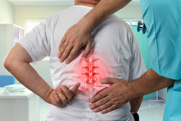
Photo source: Getty images
Vertebrogenic algic (pain) syndrome is a term that brings together a broad group of spinal disorders. The problems stem from a functional or structural disorder of the spine. They have one common symptom, namely back pain.
Most common symptoms
- Shoulder Blade Pain
- Chest pain
- Flank Pain
- Headache
- Joint Pain
- Limb pain
- Nerve pain
- Shooting pain in fingers and toes
- Pain that Radiates into the Shoulder
- Groin Pain
- Back Pain
- Spirituality
- Tinnitus
- Muscle stiffness
- Nausea
- Defence
- Tingling
- Erectile dysfunction
- Bone thinning
- Muscle weakness
- Pressure on the chest
- Head spinning
Show more symptoms ᐯ
Characteristics
Vertebrogenic pain or algic syndrome brings together a broad group of diseases that have a functional or structural basis. The common feature of back problems mainly includes pain.
Question: What is vertebrogenic pain syndrome?
Abbreviated ass VPS =
vertebro / concerning the vertebra
+ genic / source and meaning of origin
+ algic / pain
+ syndrome / a set of typical symptoms.
Back pain, pain around the neck, thoracic spine, the shin, and the sacrum or the coccyx area.
It is associated with radiating, even shooting pain to another part of the body, for example to the head, to the front of the chest, imitating heart trouble, upper and also lower limbs.
The pain may also be accompanied by other symptoms and neurological difficulties. Examples are tingling, tingling, impaired sensitivity or muscle weakness.
Back pain has plagued mankind since time immemorial, however, the present time is characterized by an upward curve.
It is reported to affect 80-90% of the population and every person will experience it at least once in his or her lifetime.
Alarmingly, back pain is one of the most common conditions limiting a person's activity and causing incapacity for work in people under 45 years of age.
It may cause disability.
It is the 5th or 6th most common cause of hospitalization.
It occurs acutely and as a chronic form, which has a considerable impact on a person's psyche.
The acute form resolves within 4 weeks in most cases and 10-40% progress to chronic problems.
According to the duration of pain, it is divided into:
- acute - up to 1 month duration
- subacute - duration 1-3 months
- chronic - lasts for more than 3 months, i.e. for years or for life
It most often affects the lumbar region of the back, followed by the cervical region, and less common are difficulties in the thoracic region.
- cervical segment 30% of cases
- thoracic segment 10 % of cases
- lumbar section 60 % of cases,
also includes the lumbosacral transition, which is the transition between the lumbar and sacral parts of the spine - polytopic form - multiple sections of the spine
The spine (backbone)
We know that the spine is made up of vertebrae. Vertebrae are small bones that join together to form a whole.
Vertebrae = there are 33 to 34 vertebrae.
The vertebrae are divided into sections of the spine:
- cervical - 7 vertebrae, vertebrae Cervicales C1 to C7
- thoracic - 12 vertebrae, vertebrae Thoracicae Th1 to Th12
- lumbar - 5 vertebrae, vertebrae Lumbales L1 to L5
- sacral - 5 or 6 vertebrae, vertebrae Sacrales S1 to S5 (S6),
they form the os sacrum - the sacrum - coccygeal - 4 or5 vertebrae, Coccygeae Co1 - Co4 (Co5)
In addition, the backbone, i.e. Lat. Columna vertebralis, also contains other components and structures.
These are the intervertebral joints, ligaments, i.e. ligaments, intervertebral discs, spinal muscles (paravertebral muscles). The vertebrae together form the spinal canal and smaller passages for nerves - neuroforamen.
Vertebral discs as shock absorbers
Intervertebral discs are important from the point of view of movement and statics. The disci intervertebrales, as they are called in Latin, serve as dampers or shock absorbers, to cushion shocks and shocks. They distribute the overload over the entire surface of the vertebrae.
The bulkiest vertebrae and discs are in the lumbar section. They are thus adapted to bear the highest loads within the entire spine. This part has a lower degree of mobility.
Vertebral discs = 23 pcs, between vertebrae from C2 - C3 to the crossing of the vertebrae L5 and S1.
They contain a ring, the anulus fibrosus, which forms a strong sheath. The other part is the nucleus, or nucleus pulposus.
The core has no vascular supply.
It is nourished mainly during movement. Movement, i.e. repeated pushes and releases, creates a flow of fluid inside the disc. During this flow, oxygen, nutrients and waste products are exchanged.
The third part of the disc, the end plate, helps in the exchange of substances. It is located on the contact surfaces of the discs and vertebrae. In addition to blood vessels, it also contains nerve endings.
Similarly, blood vessels and nerves interfere with part of the annulus of the disc. It is reported that approximately 1/3 of its outer layer.
With age, i.e. due to aging, the number of blood vessels decreases and disappears.
This results in impaired nourishment of the discs.
A significant negative factor in reduced disc nourishment is also lack of exercise, i.e. inactivity. The consequence of which is a decrease in the water content of the disc.
After birth, the nucleus has up to 90% water content.
After the age of 50, it drops to about 70%.
Fibre degradation and ring disintegration occur. A complication of this condition is dislocation of the nucleus from the disc. A herniated disc can be caused by spinal cord or spinal nerve compression.
In addition to the intervertebral discs, the vertebrae and the entire spine are connected:
- ligaments - they form the ligamentous apparatus, they fix individual vertebrae to each other (short ligaments), but also the whole spine (long ligaments)
- intervertebral joints
- special connections that are found on certain sections of the spine,
e.g. cartilaginous or immobile joints in the sacrum and coccyx - muscles, muscular system of the spine, paravertebral muscles, muscle corset
The vertebrae and intervertebral discs are shaped, the spine is curved in an S-shape, the muscular and ligamentous system is in balance, like a muscular corset.
Thanks to this and other features, it handles a functional task.
The spine supports the body, carries its weight, bears static and dynamic loads, participates in movement, balance and stabilization, and protects the spinal cord and spinal nerves.
The spine has to cope with everyday activities, plus the effects of ageing.
Examples are degenerative changes in vertebrae, joints, discs, muscle wasting and loss of ligament elasticity and decalcification, as in osteoporosis.
Spinal cord and spinal nerves
The spinal cord is about 40 to 50 centimetres long. It extends from the brain, running from the 1st cervical vertebra C1 to approximately the second cervical vertebra L2.
The Latin name is Medulla spinalis.
There is a bundle of spinal (spinal) nerves that extends from L2 in the lumbar spinal canal. It is referred to as the cauda equina, i.e. horse's tail.
The function of the spinal cord is known.
It connects the brain - the central nervous system/CNS and the rest of the human body - the periphery. It conducts nerve impulses bilaterally and also has a reflex role, thus its functions is transmission and reflex.
The spinal cord runs through the openings of the vertebrae, referred to as the foramina vertebralis, and together they form the spinal canal - the Canalis vertebralis.
The spinal nerves exit from the spinal cord. They pass through an anatomical structure - the intervertebral foramen or neuroforamen, formed by the pedicles of the vertebrae (pedicles that connect the vertebral body and the vertebral arch).
A pair of nerve roots, the fila radicularia, extends from one spinal segment.
A significant term in spinal root compression = radiculopathy.
One spinal segment is made of:
- two adjacent vertebrae
- intervertebral disc
- intervertebral openings
The spinal segments are divided into:
- 8 cervical
- 12 thoracic
- 5 lumbar
- 5 sacral
- 1 coccygeal
= 31 pairs.
Spinal nerves contain different types of fibres. These carry different information from the body to the spinal cord and brain and back to the periphery, i.e. the body, e.g. organs, muscles, bones, limbs, skin.
Innervation of a given type has a corresponding professional designation.
Muscles = myotome.
Skin = dermatome.
Internal organs = viscerotome.
Bone, ligaments and joints = sclerotome.
Viscerotome + dermatome = Head's zone. A phenomenon known in the case when, in the disease of an internal organ, the discomfort is projected to the skin area.
A well-known example:
With a heart muscle infarction, pain in the left shoulder may occur, with displacement along the side of the elbow up to the little finger.
There is also a tiny branch of nerves branching off from the nerve root that go to the intervertebral joints.
This brief introduction provides useful information to assist in understanding the causes and symptoms occurring in vertebral pain syndrome.
Causes
The cause of vertebrogenic algic syndrome is not one. It is a variety of disorders that cause pain and other symptoms.
In broad terms, these are functional or structural changes.
These relate to bones, i.e. vertebrae, joints, ligaments, muscles or nerves. And various combinations of each other. In the latter case, they are degenerative, traumatic, inflammatory and other damage to the spine.
Functional blockages of the spinal segment
Functional blockage of one or more segments. It is caused, for example, by overloading of muscles and ligaments.
The mechanism is based on the pinching of the joint sleeve between the joint plates. This condition is referred to as meniscoid entrapment.
The meniscoid is a cartilaginous articular plate. The most famous one is found in the knee - the meniscus.
Pain and reflex contraction, i.e. muscle spasm, occurs. It increases the intensity of the pain experienced.
Muscle overload can be acute, for example, in an untrained person, when lifting a load. In children, but also in adults, faulty posture, poor movements and as a result of a long-term sedentary lifestyle and muscle imbalances are common.
Summary of functional causes:
- incorrect posture
- unilateral (one-sided) spinal overload
- work in one position
- sleep on one side
- prolonged sitting, sedentary lifestyle and sedentary work
- poor exercise habits
- muscle imbalance - disbalance
- upper-crossed syndrome
- lower-crossed syndrome
- physical stress if one is unaccustomed
- heavy lifting
- back cold, blowout, air conditioning
- hypermobility
Learn more:
Facet syndrome
Sacro-iliac joint blockage
Low back pain
Degenerative diseases
In this case, the main focus is on age-related changes, and thus the effects of ageing on the body. However, in close proximity to this group there is also an association with certain risk factors.
This includes, for example, faulty posture, excessive prolonged, unilateral or incorrect spinal overload and other difficulties similar to functional blockages.
Chronic negative influence results in disease states such as:
- osteochondrosis - degenerative changes of the intervertebral discs
- spondyloarthritis - affects mostly intervertebral joints
- spondylosis - a degenerative process affecting the entire segments
- spinal stenosis - narrowing of the spinal canal
In short,...
1. Osteochondrosis is a degenerative process that results in damage to the intervertebral disc. The cause is a disorder of metabolism, blood circulation and nutrient supply.
This is manifested by the gradual drying of the disc, which loses water, and therefore elasticity and strength. Its size decreases.
This has resulted in overall instability in the segment. And from this instability stems damage not only to the discs, but to the segment as a whole, that is, it goes into spondylosis.
2. Spondyloarthritis affects the small intervertebral joints. Like osteochondrosis, this process has a negative impact on the entire segment, not just the articular surfaces.
3. Spondylosis affects the entire segment, i.e. the vertebra, disc, intervertebral joints, surrounding ligaments and muscles. Again, this is a degenerative process resulting in instability of the spine.
A significant problem in this case is the formation of osteophytes. Osteophytes are bone growths that can cause radiculopathy. In radiculopathy, it is the compression of the nerve root.
There is a similar situation. This affects the spinal canal. It is referred to as...
4. ...spinal stenosis, which can take several forms (congenital or acquired and combinations thereof). Basically, it is a reduction of the space through which the spinal cord, nerves or blood vessels pass. At a certain extent, it causes compression, i.e. squeezing of these structures.
Intervertebral disc bulging/slipping
It can also be found under the name herniated disc.
In this case, it is a disease process that results in the disc bulging out of its normal position.
It can take several forms such as bulging, protrusion, extrusion and extrusion with sequestrum.
Learn more: Spinal disc herniation.
The root of the problem lies in the irritation of the surrounding structures. As a result, various symptoms can arise.
The cause of a herniated disc may be a degenerative process in the disc or segment itself. Another type is an injury mechanism and overloading of the spine by a sudden and inadequate load, such as when lifting a load.
Radicular (root) syndrome
This disease state is caused by other diseases such as disc herniation, osteophyte formation in spondylosis or spinal stenosis.
The cause is nerve compression, i.e. a direct compression. Hence the difficulties present. In addition to pain, which spreads along the respective path of innervation, tingling, pins and needles, impaired sensitivity and muscle strength are also associated.
Root syndrome = radicular syndrome = radiculopathy.
Learn more: Radiculopathy.
Cauda Equina Syndrome
This syndrome is also caused by another problem, e.g. disc herniation or spinal stenosis, but also other conditions.
However, in this case, the place of oppression is important.
It arises below the level of the L2 vertebra, i.e. it does not cause oppression of the spinal cord, but of the cauda.
The cauda equina is a tangle of nerves that branches off from the spinal cord and passes through the spinal canal below the level of the L2 vertebra.
Learn more: Cauda Equina Syndrome.
Trauma/injury
Injury implies less serious or critical conditions. From minor injuries to severe accidents. For the spine, these are conditions such as:
- contusion, i.e. bruising of soft tissues
- distortion, i.e. stretching of muscles and tendons, e.g. in a car accident
- acceleration-deceleration injuries of the spine
- which are also known as whiplash injury
- luxation - joint dislocation
- fracture, i.e. partial or complete break
Learn more:
Spinal injury
As a result of the injury, pain is produced. A serious cause of discomfort oppression of the spinal cord and nerves. Compression can be caused by a damaged disc, but also a vertebra.
With injuries, it is also necessary to mention the pain in the coccyx.
In her case, it happens that after a fall on the buttocks, the difficulties manifest themselves with the passage of time, years. In this period, a person no longer remembers the mechanism of the injury and does not associate it with pain in the back.
Learn more: Coccygodynia - coccygeal pain, coccyx pain.
Spondylolisthesis and spondylolysis
In spondylolisthesis, it is a pathological displacement of the vertebra compared to the neighbouring vertebra. It can have several degrees and the causes are also diverse.
Learn more: Spondylolisthesis.
Spondylolysis occurs when the arch of the vertebra is interrupted, namely in its narrowed section - in the part of the isthmus. The causes are not completely clarified. There is a congenital and acquired form.
An example of an acquired is also a post-injury condition, but also as a result of overexertion in sports activity, baseball, in girls especially gymnastics.
Congenital defects
This group summarizes disease states that develop already during intrauterine development of the fetus.
They may have a different genetic basis. They occur independently, but also as a group of symptoms that are typical of various disease states and syndromes.
A congenital defect can be scoliosis, spondylolisthesis, a different number of vertebrae (lumbarization of S1 - S1 then as the last cervical vertebra, sacralization of L5 - when L5 is part of the sacrum). For example, spina bifida, i.e. incomplete closing of the spine and the membranes around the spinal cord.
Deformities of the spine
Spinal deformities mean a morbid deviation of the shape from the normal curvature of the spine. In this case, the curvature can be in the antero-posterior direction, but also in the lateral direction.
The anterior direction is represented by hyperkyphosis and hyperlordosis. Lateral deviation scoliosis.
In the kyphotic form, the disease is also reported, which arises from the involvement of the covering discs of the discs, progresses with the development of vertebral deformity. It occurs in children from 9 to 17 years of age.
Hence the name juvenile or adolescent kyphosis - Morbus Scheuermann.
Learn more:
Scoliosis
Kyphosis - hyperkyphosis
Lordosis - hyperlordosis
Osteomyelitis - spondylodiscitis
In this case, it is an inflammatory affection of the spine, more precisely of the vertebrae and disc. It can be acute, as, for example, in the dissection of a staphylococcal infection or from around the spine.
Learn more:
Spondylodiscitis
The chronic type is, for example, tuberculosis of the bones.
It is a specific infectious disease caused by the bacterium Mycobacterium tuberculosis. The bacteria enter through the respiratory system into the blood, and through the bloodstream anywhere in the body.
Including the spine.
Pain and impaired mobility are only the beginning, ending with the disintegration of the affected structure and tissue. Progression of the disease = worsening of the discomfort.
Osteoporosis
This concept is well known, namely as the thinning of bone tissue.
Diseased altered bones are subjected to numerous microtraumatizations. Minor damage leads to a change in the structure of the vertebrae. This changes their shape, deforms them.
The cause can be the post-menopausal period in women, i.e. hormonal changes, kidney, digestive or endocrine system diseases. And inactivity and sedentary lifestyle are also cited as risk factors.
Learn more: Deterioration of bone tissue.
Rheumatic diseases
This group includes damage to joints, cartilage, but also other soft parts and bones. The medical term is arthritis.
Arthritis is a disease of the joints, so it does not avoid the spine. Joint inflammation takes various forms, one of which is rheumatoid arthritis.
Learn more:
Arthritis
Rheumatoid arthritis
A specific type of the disease is chronic inflammatory disease, which mainly affects the SI joint and intervertebral joints. And this is Bechterev's disease, called ankylosing spondylitis.
Learn more:
Ankylosing spondylitis
Axial spondylarthritis
Diseases of internal organs
Just as pain from the spine radiates to other parts of the body, so it is with diseases of the internal organs. Their manifestation can be a painful back.
Examples are diseases affecting:
- Lungs
- heart
- Stomach
- Gallbladder
- Gut
- Kidneys
- Female genitals, ovaries
Tumour
A less common cause of difficulty.
The spine may be affected by oncological disease. It is either a primary or secondary tumour.
A primary tumour is, for example, an osteoma or meningioma.
A secondary tumour is in the form of metastasis or as a result of transfer of the oncological process. It happens in metastases from prostate, breast, lung tumours.
Learn more: Spinal tumours.
Psychogenic back pain
With long-term spinal pain, these difficulties also have an impact on a person's mental side. However, the path can also be reversed.
What does it mean?
Despite the fact that there are no structural changes in the spine and it is not affected by functional blockage, the person reports pain and other discomforts.
There are at least two reasons.
The person could be just pretending. In this case, the person in question is faking back pain, for a variety of reasons.
Does the person need to be determined as unfit to work? After a work injury? Or to get a disability pension?
Another form is back pain in persons with a personality that is predisposed to it.
Specifically, the hysterical personality type, when the person in question exaggerates his or her difficulties or suffers from depression.
Symptoms
The main symptom is pain. However, depending on the cause, possible concomitant symptoms are also associated.
Table: Symptoms divided by cause
| Cause | Symptoms |
| Acute blockage | Acute cervical spine blockage, i.e. Lumbago. A well-known example is pain and stiffness in the neck:
|
| Cervicocranial syndrome | CC syndrome
|
| Cervicobrachial syndrome | CB syndrome
|
| Cervicovestibular syndrome | CV syndrome, cervical vertigo
|
| Chronic neck pain | Chronic cervical segmental syndrome
Long-term pain stems first from faulty posture and muscle imbalance
|
| Cervical myelopathy |
caused by a degenerative process, with progressive ischaemia, i.e. lack of blood supply
|
| Radicular syndromes |
|
| Pain in the thoracic spine |
These are mainly difficulties resulting from intervertebral and rib joint blockage, intercostal neuralgia
|
| Chronic back pain lumbosacral region |
and therefore the lower part of the shin and sacrum, also called Low Back Pain This may also include:
|
Learn more: Buttock muscle inflammation - sciatica.
Diagnostics
History taking and clinical examination are important. The doctor asks about pain, about its location, spread, character, intensity and dependence on position and movement.
The doctor visually evaluates the spine, namely mobility, gait, posture.The condition of the skin and muscles, sensitivity and muscle strength, reflexes are ascertained by palpation (touch). It is also important to ask about the abdominal muscles and the ability to control urinary and faecal excretion.
Imaging techniques include:
- X-ray, dynamic images, different positions
- CT
- MRI
- PMG - perimyelography, contrast examination of the spine, spinal cord
- EMG - electromyography - evaluates muscles
Laboratory examination of blood and liquor.
Diagnosis in vertebrogenic algic (pain) syndrome depends on the symptoms present and the cause that provokes it.
Simple functional blockages will also be detected by a general practitioner. More serious conditions require a more comprehensive neurological examination, which is supplemented by additional ones as needed.
Doctors from several disciplines may be involved in the diagnosis. Examples are a general practitioner for adults or children, a neurologist, an orthopedist, a rheumatologist, a neurosurgeon, a radiologist, a physiotherapist. Possibly a psychologist and a psychiatrist.
Differential diagnosis is important. Internal medicine and cardiology specialists or a surgeon may also be involved.
Prevention, prevention and more prevention
As with any other disease, so with the spine doubly so. Actually, threefold.
Prevention =
- suitable bed + mattress + pillow + grid
- sleeping position and when falling asleep
- the worst position is the prone position, i.e. do NOT lie on your belly
- proper sitting position
- correct posture
- sufficient physical activity
- sport, appropriate form, such as swimming, cycling, running
- especially walking
- ergonomics at home and in the workplace
- working environment - suitable desk and chair
- limit the lifting of loads and strain on the spine
- learn the correct technique
- rational nutrition, vitamins, minerals, medicines to support joints
- overweight and obesity - weight reduction
- suitable footwear
How it is treated: Vertebrogenic Pain Syndrome
Treatment of vertebrogenic pain syndrome: medications, rehabilitation
Show moreVertebrogenic Pain Syndrome is treated by
Interesting resources

Bc. Lukáš Tóth
Healthcare worker
The secondary medical school in Nitra gave me the basis for my career in the field of health and diseases. Thanks to it, I worked for 2 years in the traumatology clinic and outpatient clinic at the Nitra Hospital. Since 2006 I was employed in the emergency medical service, where I stayed until 2017.
I completed my bachelor's degree at the University of Constantine the Philosopher in Nitra in the field of emergency health care. The bachelor's degree allowed me to continue my mission as a paramedic. In the meantime, I got a job at the emergency line 155. I have been working in pre-hospital health care until today.
I had an interest in people, health and even diseases in my childhood, which gave me the prerequisite to pursue this topic in adulthood. Studying and acquiring new information in practice provided me with a great basis for writing professional texts, in the form of articles that can be understood by ordinary people. Thus, my interest in the Health Portal has a solid foundation in years of practice and personal interest. Similarly, I am also interested in healthy eating, nutrition and overall healthy lifestyle. I fill my free time with family and sports.
View all articles by the same author


