Spondylodiscitis: The Causes and Treatment of Chronic Back Pain

Spondylodiscitis: The Causes and Treatment of Chronic Back Pain
Photo source: Getty images

Back pain can be caused by a less common condition called spondylodiscitis. The technical name refers to an infection of the spine, intervertebral discs and vertebrae.

Characteristics

Back pain is a current and common problem. It can be acute or last for a long time. The cause is usually a variety of diseases.

One less common disease is infection, otherwise osteomyelitis of the spine or spondylodiscitis.

Osteomyelitis bone infection. It is caused by various microorganisms. It destroys bone tissue.

Osteo = menas "bone". Myelitis = inflammation of the spinal cord. 

Spondylodiscitis consists of... 
Spondyl = meaning "vertebra" +
Spondylos from Greek = vertebral +
Disc - from Latin "disci intervertebralis" = intervertebral discs +
Suffix -itis = a Latin ending meaning "infection, infectious disease".

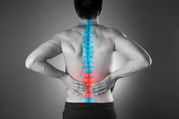
The Spine and the Vertebrae

The spine forms a supportive construction of the human body. It carries the weight of its upper part and plays an important role in movement and defense.

Vertebral column, the spine, the backbone = Lat. "columna vertebralis".

Its specific composition helps achieve the above mentioned functions. The small bones, the vertebrae along with intervertebral discs, the ligaments and the muscles form a special functional unit.

People have 33 or 34 vertebrae. Based on the spinal segment their names are:

  1. vertebrae Cervicales, cervical vertebrae = 7 vertebrae, labelled from C1 to C7
  2. vertebrae Thoracicae, thoracic vertebrae = 12 vertebrae, Th1 to Th12
  3. vertebrae Lumbales, lumbar vertebrae = 5 vertebrae, L1 to L5
  4. vertebrae Sacrales, sacral vertebrae = 5 alebo 6 vertebrae, S1 - S5 (S6)
  5. vertebrae Coccygeae, coccyx= 4 or 5 vertebrae, Co1 - Co4 (Co5)

The cervical spine is characterised by a high degree of mobility. It is attached to the skull with the first cervical vertebra.

C1, the first cervical vertebra is called the atlas.
C2 is the second cervical  vertebra called the axis. It has an odontoid process (bony protrusion) called the dens and is important for the movements of the head.

The ribs are attached to the thoracic vertebrae. These form the thorax that serves as protection of vital organs.

The lumbar vertebrae are the most resistant. They can bear large loads since they are adapted to this specific function.

The sacral vertebrae are fused into the sacrum - the "os sacrum" in Latin. The sacrum is connected to the lumbar and pelvis joints, which are referred to as SI joints, i.e. sacroiliac joints.

The coccyx, also known as the tailbone, is the final segment of the backbone. It is not just some minor part of it. It is significant.

Learn more about: 
SI joint block
Coccydynia or Tailbone pain 

It is also important to know that the vertebrae are anatomically adapted so that the spinal cord could pass through them.
The vertebral bodies and arches form the spinal canal.

Spinal cord = medulla spinalis in Latin.

The spinal nerves protrude from the spinal cord.
It is therefore a link between the brain and the rest of the body.
The function of the spinal cord is relaying nerves and coordinating reflexes.

What about intervertebral discs?

The small vertebral joints and the intervertebral discs participate in the body's movement. Thehir Latin name is "disci intervertebralis". That is where the discs get their shortened name from.

They have an important function for the body's movement. They also act as shock absorbers when walking, running, or just moving around. The individual vertebrae do not obstruct each other.

The discd come in different sizes. The largest ones are located in the lumbar region.

There are 23 of them. They stretch from the intervertebral space C2 - C3 to segments L5 and S1.

Between the surface of the vertebral body and the disc is the vertebral endplate. It plays an important role in nutrition and makes sure the vascular and the nervous system get nourished.

Question:
Why are we giving you all this information?

Answer:

A spinal infection primarily affects these parts.

An infection causes damage to the vertebrae, the intervertebral disc and the thin layer between them, i.e. the vertebral endplate.

Coming up: 
How spondylodiscitis is defined. 
The causes of inflammation. 
The symptoms accompanying the disease. 
A diagnosis as well as available treatments.

How Spondylodiscitis Is Defined

Spondylodiscitis is an inflammatory disease caused by infection, ie the entry of microorganisms into the spine.

It initially affects the intervertebral disc, the vertebral endplate and the vertebral body.

The disease causes damage to these parts. As a result, this damage causes back pain and other associated neurological disorders, depending on the extent of the affected area.

It is an uncommon disease with a reported incidence of 1: 250,000. This means 2-4% of all bone infections.

It is more common among men.
The age at which it occurs is not precisely delimited.
However, it is more common after the age of 50.

It is often confused with other diseases of the spine. Therefore, effective treatment may be delayed, which presents a risk for potential complications or even death.

The infection affects the segments of the spine to the following approximate extent:

  1. lumbar (lower back) - 45 to 50 %
  2. thoracic (torso) - 35 %
  3. cervical (neck) - 3 to 20 %
  4. the rest affects the sacral segment

Causes

What causes a spinal infection?

The disease has either an endogenous or exogenous form.
Endogenous infections originate in another inflammation in the body at a more distant site.
Exogenous infections are caused by a spinal surgery, but in another infection near the spine.

In the endogenous type, pathogenic microorganisms from another site enter the spine via a vascular pathway, i.e. via blood in arteries or veins. In this way, one or more parts of the spine can be affected.

Spread by the blood = hematogenous spread.

That includes the following infections:

  • inflammation of the digestive system
  • excretory system
  • reproductive system
  • respiratory system
  • cardiovascular system

Due to the lengthy period of diagnosis, the primary focus of infection does not appear many times.

The exogenous type arises as a complication due to surgery of the spine, vertebrae, discs or injuries. Another way is direct spread from tissue to tissue in close infection.

This type rarely occurs after a lumbar puncture or after giving an injection near the spine.

Certain accompanying conditions increase the risk of getting the disease, for example:

  • immune disorders
  • HIV
  • treatment with corticoids, cytostatics, immunosuppressants
  • drug addiction
  • diabetes
  • renal failure
  • rheumatoid arthritis
  • tuberculosis

Risk factors associated with surgery:

  • long operating time
  • extended hospitalisation
  • posterior surgical approach - a study by Levie et al.
  • revision surgery
  • higher number of people during the operation
  • increased surgical blood loss and the need for a blood transfusion
  • drainage for more than 3 days
  • obesity or malnutrition
  • comorbidities, such as diabetes
  • smoking
  • advanced age

What Microorganisms Cause Spondylodiscitis?

Infections can be caused by bacteria, fungi or, to a lesser extent, parasites.

The most common pathogen is Staphylococcus aureus, or Staphylococcus aureus. An infection with infection with an antibiotic-resistant subtype, referred to as MRSA, has also been documented.

MRSA = methicillin-resistant Staphylococcus aureus. 
A strain of bacteria resistant to certain antibiotics. 
It is also referred to as a superbacteria.
Learn more about MRSA here and here.

Staphylococci can cause more than 50% of these infections. Other bacteria include Escherichia coli (E. coli) and Enterobacter.

E. coli and Enterobacter are enterobacteria. They are found in the digestive system, i.e. in the microflora in the guts of humans and animals. They occur worldwide, commonly in the soil, water or vegetation. Some are primarily pathogenic to humans, such as Sallmonella or Shigella.

Thus, most spondylodiscitis is caused by a non-specific infection.
A specific type is tuberculosis infection, or tuberculosis of the spine.

Half of all cases of bone tuberculosis occurs in the spine.
It is formed exclusively by endogenous blood flow.

Tuberculosis is a primary lung disease.

However, the infection can spread to other parts of the body in the bloodstream. This is also referred to as the extrapulmonary form. The most common is tuberculosis of the bones and joints.

Pott disease = chronic epidural tuberculous spine infection and you can read about it here. The lower thoracic and upper lumbar vertebrae areas of the spine are most often affected.

Symptoms

The main manifestation of spinal infection is pain.
Depending on the location of the inflammation, there is pain in the lumbar area, lower back or higher, between the shoulder blades or the neck.

The pain is chronic, long-term, recurrent.

Other symptoms include reduced mobility, pain exacerbated by movement, muscle spasm, or stiff muscles around the affected spine.

It is often mistaken and is thought to be the result of another cause of vertebrogenic algic syndrome.

In some cases, there may be general symptoms of an infectious diseas, such as an increase in body temperature, fever or a septic condition. Sepsis can culminate in a shock, which is described in a separate article.

When the body temperature rises, chills, i.e. shivering, are usually present. Fatigue, weakness, nausea, and even vomiting are other general signs and symptoms that can occur in an acute infection.

An complication of infectious spinal inflammation is the formation of an abscess. It is a demarcated cavity that is filled with inflammatory fluid and pus.

Symptoms of spinal cord or nerve damage are more serious.

Neurological symptoms can take a variety of forms, from sensory issues, tingling (paraesthesia) to muscle weakness and varying degrees of paralysis. We describe them in more detail in the articles on radiculopathy, pseudoradiculopathy, spinal stenosis or the cauda equina syndrome.

Symptoms of Pott Disease

Tuberculosis of the spine was described by Pott himself, after whom Pott disease is named. In 1779, he described the so-called triad of symptoms, i.e. the 3 typical symptoms of this disease.

The triad of symptoms of Pott disease:

  1. gibbus deformity - hunch, as a result of hyperkyphosis
  2. abscess - demarcated inflammation
  3. paraplegia - lower limb paralysis

Initial symptoms may also include:

  • back pain
  • sharp pain exacerbated by movement
  • limited mobility
  • increased muscle tension
  • muscle stiffness
  • sensitivity to touch

Diagnostics

The patient's medical history is taken during the examination, as there are significant clinical manifestations. Subsequently, a standard neurological examination is performed.

Differential diagnosis is important. Its role is to accurately diagnose and differentiate from diseases such as degenerative spinal changes, but also axial spondyloarthropathy or Bekhterev's disease and Scheuermann's disease.

Scheuermann's disease = juvenile kyfosis = in children and adolescents.

In the case of spondylodiscitis, the basic examinations also include a laboratory blood test. This includes CRP, FW (blood sedimentation mainly in acute illness, in chronic may be normal), a special method is blood culture, which is supplemented by specific pathogen identification and sensitivity to antibiotics.

Subsequently, a biopsy can be performed, i.e. a needle is inserted under CT control and a sample is taken. However, this method will provide a small amount of material. More material is obtained during operational sampling.

Imaging methods, such as X-ray and scintigraphy, are important, but CT and MRI will provide the most detailed insight.

Since there is some risk for tuberculosis, it is necessary to be checked for this disease as well.

Course

The course of the disease can be acute when it manifests with initial general symptoms.

Examples are rising body temperature to fever, chills, nausea, general weakness and fatigue. These may be accompanied by a feeling of vomiting or even vomiting.

Back pain, muscle stiffness or limited mobility of the spine is at the site of inflammation.

The chronic form does not have to be portrayed by general difficulties. However, it is characterized by back pain. It may not respond to common painkillers.

It is recurrent.

During the course of the disease, neurological problems may occur which result from nerve or spinal cord irritation. Examples are radiation of pain in the limb, impaired skin sensitivity and weakening of muscles and reflexes.

If that is the case, then it is important to get immediately examined and a correct diagnosis.

Early treatment is a guarantee of success and a good prognosis.

Prognosis = predicting the likely or expected development of a disease.

How it is treated: Spondylodiscitis

How is spondylodiscitis treated? Medications and antibiotics

Show more
fshare on Facebook

Interesting resources

  • Doutchi M, Seng P, Menard A, et al. Changing trends in the epidemiology of vertebral osteomyelitis in Marseille, France. New Microbes New Infect. 2015;7:1–7.
  • Statistisches Bundesamt. Diagnosedaten der Krankenhäuser ab 2000 (Eckdaten der vollstationären Patienten und Patientinnen). Gliederungsmerkmale: Jahre, Behandlungs-/Wohnort, ICD10. Statistisches Bundesamt. 2017 
  • Sobottke R, Seifert H, Fätkenheuer G, Schmidt M, Goßmann A, Eysel P. Current diagnosis and treatment of spondylodiscitis. Dtsch Arztebl Int. 2008;105:181–187. 
  • Cheung WY, Luk KDK. Pyogenic spondylitis. Int Orthop. 2012;36:397–404. 
  • Gupta A, Kowalski TJ, Osmon DR, et al. Long-term outcome of pyogenic vertebral osteomyelitis: a cohort study of 260 patients. Open Forum Infect Dis. 2014;1 ofu107. 
  • Kehrer M, Pedersen C, Jensen TG, Hallas J, Lassen AT. Increased short- and long-term mortality among patients with infectious spondylodiscitis compared with a reference population. Spine J. 2015;15:1233–1240. 
  • Sobottke R, Rollinghoff M, Zarghooni K, et al. Spondylodiscitis in the elderly patient: clinical mid-term results and quality of life. Arch Orthop Trauma Surg. 2010;130:1083–1091. 
  • Kim CJ, Song KH, Jeon JH, et al. A comparative study of pyogenic and tuberculous spondylodiscitis. Spine. 2010;35:E1096–E1100. 
  • Jean M, Irisson JO, Gras G, et al. Diagnostic delay of pyogenic vertebral osteomyelitis and its associated factors. Scand J Rheumatol. 2017;46:64–68. 
  • Kapsalaki E, Gatselis N, Stefos A, et al. Spontaneous spondylodiscitis: presentation, risk factors, diagnosis, management, and outcome. Int J Infect Dis. 2009;13:564–569.