Pseudoradiculopathy, Pseudoradicular Syndrome: Causes of back pain
Pseudoradiculopathy and radiculopathy are painful conditions of the spine. In addition to back pain, they also combine other symptoms that spread through the body. They are similar, yet quite different, especially in terms of cause.
Most common symptoms
- Shoulder Blade Pain
- Chest pain
- Headache
- Limb pain
- Nerve pain
- Leg Pain
- Groin Pain
- Pain that Radiates into the Shoulder
- Head spinning
- Muscle stiffness
- Tingling
- Shooting pain in fingers and toes
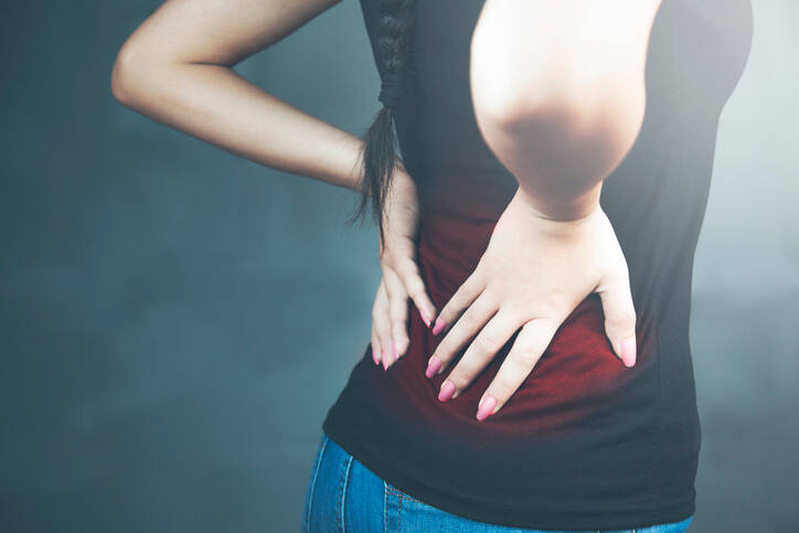
- Back Pain
- Muscle cramps
Characteristics
Back pain is a common issue these days. It is reported to affect more than 70% of the population, and up to 90% of people experience it at least once in their lives.
It is the leading cause of workplace disability in people of working age.
Overall lifestyle choices, poor posture and poor movement patterns, sedentary lifestyle and sedentary work all have a significant impact. Other examples are inappropriate office ergonomics, long-term forced posture at work, lifting loads and long-term overloading of the spine.
It often happens...
...that we feel back pain when straightening up after a forward bend, rotating, or making a twisting motion. If we are also lifting a heavy burden, that is when the problem arises.
Age does not play an important role in this case.
The problem is acute. However, back pain can persist for a long time. If it is chronic, it is already an issue that has reduced the quality of life and caused distress in the affected person.
Back pain is divided by duration:
- acute pain that lasts less than 6 weeks
- the course is not expected to be serious
- it is necessary to rule out another disease
- special treatment procedures are usally not required
- subacute pain that lasts approximately 6 to 12 weeks
- chronic pain that lasts longer than 3 months
- requires special procedures in diagnosis and treatment
- recurrent pain that subsides and returns after a certain period of time
- there is usually a break of less than 3 months during the period of worsened condition
Back pain is felt in various places, namely in the neck, chest area, lumbar or sacral region, including the coccyx. It is associated with various symptoms that worsen the overall condition of the sufferer.
Learn more: Vertebrogenic algic syndrome.
When searching for information or consulting an expert, you may come across a number of technical terms and concepts. These describe different conditions. They are incomprehensible to non-professionals and do not tell them anything useful.
It is similar to the concept of pseudoradiculopathy.
This term is used in connection with back pain and in conjunction with radiating pain into the limb.
A more precise definition of pseudoradiculopathy is given later in the article.
A clinical picture of the spine is recommended.
The spine is our body's support system and ...
The spine forms the support system of the human body. It participates in movement and ensures sufficient range of motion in all directions.
One of its functions is protection. It prevents injury to the spinal cord which runs from the brain through the spinal canal.
The spinal cord is stored in the spinal canal (canalis vertebralis) which consists of vertebrae. Together they form the spine whose Latin name is columna vertebralis.
The spine is physiologically curved. Curvatures have different names: the forward curve is referred to as lordosis. It is located in the cervical and lumbar regions.
Conversely...
The backward curve is called kyphosis. It is located in the thoracic and sacral regions of the spine.
This curvature is physiological, i.e. congenital or inborn. The second type of curve is acquired or non-congenital.
The pathological sideways curve of the spine is referred to as scoliosis.
There is a slight und unnoticeable physiological lateral or sideways curve in every human being.
The sideways curve can be briefly seen while carrying load in one hand or when standing while shifting the weight to one leg.
The spine consists of vertebrae. There are 33-34 of them.
Table: division of the spine into individual segments
| Spinal segment | Latin name | Description |
| Cervical spine | vertebrae cervicales |
|
| Thoracic spine | vertebrae thoracicae |
|
| Lumbar spine | vertebrae lumbales |
|
| Sacral spine | vertebrae sacrales |
|
| Coccyx | vertebrae coccygeae |
|
A vertebra consists of the vertebral body, arch and processes.
A. The vertebral body, corpus vertebrae, is in the front and forms the load bearing part of the vertebra. The upper and lower surfaces of most vertebrae are connected to intervertebral discs.
The height of each vertebra varies.
Cervical vertebrae are smaller in height than lumbar vertebrae. As we move towards the coccyx, their height decreases again.
B. The vertebral arch attaches to the body of the vertebra at the back. It has a protective function - it protects the spinal cord.
+ the arch has two components.
The lamina of the vertebral arch points towards the vertebral body, where it is connected by two pedicles, or pediculus arcus vertebrae in Latin.
The vertebral arches form the vertebral foramen and the vertebral canal.
The pedicles have notches above and below. They are small depressions, i.e. superior (upper) and inferior (lower). These form the intervertebral foramen (foramina intervertebralia).
Branching off the spinal cord, the spinal nerves pass through the intervertebral openings.
C. Vertebrae have processes that project from the arch. They serve to attach the vertebrae and help with the movement of the body.
Table: Types of processes
| Name | Latin name | Description |
| Spinous process | processus spinosus |
|
| Articular process | processus articulares |
|
| Transverse process | processus transversi |
|
The vertebrae are firmly but movably connected to each other. This connection is formed by cartilage, ligaments, intervertebral joints and muscles.
Table: ways of fixating the spine
| Connection type | Description |
| Ligaments |
|
| Intervertebral joints | art. intervertebrales |
| Intervertebral discs | they absorb shocks during locomotion and help the body move |
| Special connection |
for example, synchondrosis (or primary cartilaginous joint)
|
| Muscular system |
muscles of the spine along with the muscles of the abdomen, muscles of the trunk and pelvis
|
Between the vertebrae lie 23 intervertebral discs.
The discs are named after their designation in Latin, i.e. disci intervertebrales. They are not found between all the vertebrae. The first disc is located between the 2nd and 3rd cervical vertebrae (C2 and C3).
It is the last disc that lies between the last lumbar and the first sacral vertebra (L5 and S1).
The discs have the following functions:
- absorbing shock when moving, walking, running or jumping
- stabilising the spine
- maintaining balance
- balancing compressive and tensile forces
- distributing forces over the entire surface of the disc
- they are involved in all movements of the spine, such as bending or rotating the body
They adapt to the bodies of the vertebrae. They have different heights. They are higher between the cervical and lumbar vertebrae, with the highest disc being between the L5 and S1 vertebrae. In the area of lower range of motion, on the contrary, they are lower.
Noteworthy information: spinal disc herniation.
The spinal cord - a brief overview
The spinal cord is located in the spinal canals. It runs from the brain, from the first cervical vertebra C1 to the first or second lumbar vertebra L1/L2.
The spinal cord - medulla spinalis.
It is important because it functions as a reflex and signal transmission. It conducts nerve impulses, i.e. stimuli from the body to the brain and, conversely, from the brain to the body. The spinal nerves branch off the spinal cord, running to the rest of the body and limbs.
Like the brain, the spinal cord is made up of white matter and gray matter. It is enclosed in membranes or meninges. The white material is on the surface, and gray matter is inside. It has the shape of the letter H.
Neurons, i.e. nerve cells in gray matter, form the anterior, posterior spinal horns and the lateral horns. In the area of the dorsal root lies a nerve nodule called the dorsal root ganglion or spinal ganglion.
Like the vertebrae, the spinal cord is divided into individual spinal cord segments.
The spinal cord is made of 31 segments from which branch one pair of sensory nerve roots and one pair of motor nerve roots:
- 8 cervical segments
- 12 thoracic segments
- 5 lumbar segments
- 5 sacral segments
- 1 coccygeal segment
The spinal rootlets are formed by the connection of individual spinal nerves. The spinal nerves connect the spinal cord and the brain with other parts of the human body.
Their Latin name is nervi spinales.
Fibres emerging from the spinal cord are called nerve rootlets or fila radicularia, forming the dorsal roots (front) and ventral roots (back).
1. Ventral roots of spinal nerve - radices anteriores -protrude from the anterior corners of the spinal cord. These ventral roots (front) are efferent roots that areexiting the centre, the brain and the spinal cord and leading to the periphery.
2. Dorsal roots of spinal nerve - radices anteriores, protrude from the dorsal (back) corners of the spinal cord. Dorsal afferent roots - leading from the periphery - the body, back to the spinal cord and the brain.
Their functions are motor or sensory fibres: the front fibres are the motor roots, an the back fibres transmit sensory information.
Some ventral spinal roots have sympathetic fibres in a certain section, i.e. between C8 and L2. Segments S2 - S4 contain parasympathetic fibres.
The anterior and posterior roots join together in the spinal nerve, which passes through an opening in the vertebra.
This is the site causing radicular syndrome which certainly differs from pseudoradicular syndrome.
After this brief introduction, we can move on to the definition of pseudoradiculopathy.
Pseudoradicular syndrome is defined as...
Pseudoradiculopathy is a type of back pain that is associated with radiating pain to the limbs. However, the issues do not arise due to mechanical pressure against the nerve.
Thus, there is no disruption of the structure of the spinal root, i.e. the radix.
According to neurologist Dr. Karel Lewit, incomplete radicular syndrome occurs when compression is exterted on the protective layer of the root, but not the root itself.
It is manifested by pain that spreads to the periphery (such as the limbs) via its dermatome. However, the issues are not associated with symptoms as is the case wih root compression - radiculopathy (impaired mobility and sensitivity).
The dermatome is part of the innervation of the nerve.
The pain is spread unevenly along the surface and its depth. It is not always caused by either movement or a change of position. On the contrary, pain caused by radicular syndrome is very intense, sharp, burning with an easily identifiable location and radiation.
Table: basic difference between radical and pseudoradicular syndrome
| Radicular syndrome | Pseudoradicular syndrome |
| based on nerve compression | no nerve compression |
| common in disc herniation and other structural changes | most common disorder of the sacroiliac joint, lumbar-sacral joint, coxarthrosis |
| pain spreads via the dermatoma | similar spread of pain, it is boundless, does not spread below the knee |
| sensitivity disorder | without accompanying neurological difficulties, such as weakening of muscle strength or skin sensitisation |
| muscle weakness - paresis | |
| issues exacerbated by movement |
Causes
Back pain is divided into several types according to pain, as shown in the table.
Table: Types of back pain
| Type | Description |
| Non-specific pain | Unspecified
|
| Nevre root pain | Neurogenic
|
| Pain caused by organic disease |
Pain is caused by:
|
Pseudoradicular problems are reportedly caused by a functional disorder tha is based either on intervertebral joints (vertebral joint surfaces), muscles, or tendon attachments.
Problems do not arise due to compression - nerve compression, as in radiculopathy.
Each radicular syndrome is also reported to be accompanied by pseudoradiculopathy.
This is due to long-term overload and excessive overload on soft tissues and structures in the spine. Examples are conditions, such as:
- coxarthrosis (hip pain)
- SI joint blockage - sacroilic joint,
joint disease of the ilium and the sacrum - facet syndrome
- hypertonic pelvic floor syndrome
Learn more about ankylosing spondylitis and axial spondylarthritis.
Here is a summary of the most common causes of pseudoradicular problems:
- intervertebral joints - disorder at the level of small joints between vertebrae
- muscles, overload or muscle imbalance
- tendon attachments, overload
- bone - the periosteum - a membrane that covers the outer surface of all bones
- structures around the joints
- others:
- osteoporosis
- spondylosis
- coxarthrosis
- developmental and congenital disorders of the vertebrae
- vertebrogenic blockages, such as lumbago or low back pain
Symptoms
- pain, not as severe as in nerve root compression
- paresthesias, pins and needles, tingling, pricking, burning
- muschle cramps and muscle stiffness, spasms
- radiating piain - shooting towards the periphery (peripheral regions of the body and limbs)
The pain may have a dull character. The intensity does not exceed the extent as seen in nerve compression, and is not necessarily dependent on movement or change of position.
The site does not have a precise boundary. Radiating pain can be transferred via several dermatomas, i.e. along the pathway of innervation by multiple nerves.
It usually spreads in the area of the iliosacral joint, it can radiate into the groin area, gluteal muscles in the buttocks area, on the sides of the thighs.
The pain does not usually radiate below the knee.
It can be perceived as superficial or deep pain.
There are no signs of nerve compression, such as impaired sensitivity, reflexes or muscle weakness.
There is a blockage, i.e. a movement disorder, in a given segment of the spine, stiffness of the muscles - spasms, as in the case of an acute blockage, or lumbago.
Diagnostics
The purpose of diagnostics is, therefore, to figure out whether the problem at hand is a pseudoradicular issue or nerve compression, i.e. its root - the radix.
First, the diagnostic is based on the medical history, followed by a clinical examination, including a neurological examination, monitoring of posture, gait, and overall evaluation of the musculoskeletal system, reflexes and maneuvers (also called straight leg raise or Lasègue's sign, and others)
Signs of other diseases are examined, which is important for differential diagnosis.
As shown in the table, there are different causes of back pain.
Certain important imaging methods are used, such as:
- X-ray
- CT
- MRI
- EMG
A differential diagnosis includes blood tests, identification of inflammation and examination of the cerebrospinal fluid (cerebrospinal fluid) after lumbar puncture in order to confirm or rule out Lyme disease, for example.
Course
The pain may have a dull character. It usually does not reach the maximum intensity, it is less pronounced and localised inaccurately. Likewise, the spread of pain is less clear. The pain is non-specific and on a larger scale.
Radiating pain can be experienced via multiple dermatomas, i.e. along the surface as part of innervation by multiple nerves. It is also felt as deep pain.
The pain spreads from the back, across the buttock area, to the groin and the sides of the thigh. It does not shoot below the knee.
It is associated with pins and needles or other discomfort - paraesthesia, but there are no symptoms as in direct nerve compression (sensitivity disorders or muscle weakness).
Pain may sometimes be caused by a change of posture and movement. Also, one of the manifestations is limited movement in a given segment of the spine and muscle spasm or muscle stiffness.
"Simple" pseudoradiculopathy is milder, the prognosis is better and treatment is easier.
How it is treated: Pseudoradiculopathy
How is pseudoradiculopathy treated?
Show morePseudoradiculopathy is treated by
Other names
Interesting resources


