What is magnetic resonance imaging - MRI? When can it be used and when not? What is its advantage over CT scanning? And a lot of other interesting information you can read in the article.
History of MRI
The phenomenon of nuclear magnetic resonance has been described in the literature since the 1940s.
This phenomenon is used in magnetic resonance imaging. Its use emerged after 1970.
The abbreviation MRI (Magnetic Resonance Imaging) is used. Bloch and Purcell won the Nobel Prize in Physics in 1952 for this discovery.
Characteristics
Because MRI does not use ionizing radiation, it is considered safer than CT (computed tomography).
MRI is more suitable for imaging soft tissue. Bone is better evaluated with CT.
MRI is used to diagnose the tissues of the human body. It is well suited for the head, trunk, abdomen, pelvis and extremities.
A little physics
The nucleus of an atom is made up of neutrons and protons.
Each of these particles rotates on its axis in a peculiar motion called spin.
Protons are positively charged particles and each positive particle creates a magnetic field. It therefore also exhibits a magnetic moment.
This is also called the magnetic dipole moment.
A negatively charged electron will move along a closed trajectory in the electrostatic field of the nucleus. The electron on the closed trajectory will create a current crank which corresponds to the magnetic dipole moment. The resulting magnetic field acting on the electron is thus weak, but it does act.
Atomic nuclei with a positive nucleon number have no spin (do not behave magnetically). Their magnetic moments cancel out and cannot be used in magnetic resonance imaging. In contrast, atomic nuclei with an odd nucleon number retain their magnetic moment.
This includes the hydrogen atom H, which has only one proton exhibiting a relatively large magnetic moment.
Hydrogen is also the basis of magnetic resonance because of the high proportion of water in the body (60%).
3D resonance vector. Source: Getty Images
The principle of MRI
If a rotating nucleus is placed in a constant magnetic field, the magnetic moments will align with the external magnetic field. The axis of the nucleus will easily rotate about the direction where the constant field is applied.
To keep the cores in constant motion, a high frequency magnetic field is used. When the coil approaches the rotating magnetic moment, a voltage is induced in the coil. This voltage is then measured. The magnitude of the voltage measured depends on the position and type of tissue.
Protons also exhibit precessional motion (movement along the shell of an imaginary cone). The frequency of this motion is called the Larmor frequency. It depends on the strength of the magnetic field and the type of atomic nucleus.
Resonance - to measure the resonant frequency of protons (their spectrum), it is necessary to deflect the magnetization vector from its equilibrium position, thus producing a transverse vector of tissue magnetization.
The magnitude of the transverse vector is zero due to the chaotic motion of the particles. Using an electromagnetic pulse in the form of energy, we achieve this change.
Relaxation - after the end of the electromagnetic pulse, the protons return to their original energetically preferable position and their synchronous motion ceases. It can be either T1 - longitudinal or T2 - transverse relaxation.
Advantages of magnetic resonance
It is a precise imaging based on different signal intensities of soft tissues (brain, heart, cartilage, internal organs...).
It distinguishes pathologies that are not visible in other examinations.
It is a non-invasive method, so it is also suitable for pregnant women and newborns. It does not use harmful ionizing radiation like other examinations.
It provides information about blood circulation, blood vessels, etc.
Some vascular (angiographic) images can be imaged without the need for KL (contrast medium), thus reducing the burden on the patient.
Contrast agents
They are used to enhance (improve) the image. They are used to make structures visible that are not distinguishable on a normal image. Their function is to facilitate proton relaxation and thus shorten the T1 and T2 relaxation times.
This shortening leads to an improved T1-weighted image, while T2 is attenuated.
Contrast agents can be divided into paramagnetic and superparamagnetic.
Some substances contain gadolinium.
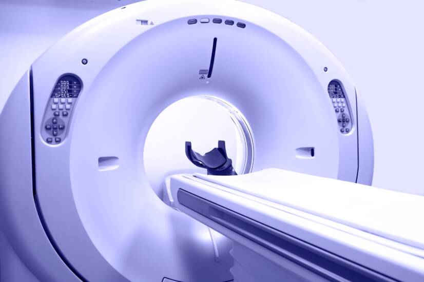
Instrumentation
The patient is placed in an MR tunnel where he is exposed to a homogeneous magnetic field. To this is added an additional magnetic field consisting of gradient coils. The examination is controlled by a computer where image data are also collected.
Special applications of magnetic resonance imaging
Diffusion MRI - Displays signal changes caused by the diffusion of water molecules in tissues. It is used in the diagnosis of aging brain anemia, Alzheimer's disease, autism, schizophrenia, or changes in the prostate.
MRA - Magnetic resonance angiography - flow of excited nuclei.
Functional MRI - The essence of the method is the change in blood flow and blood volume in the active area of the cerebral cortex (used in the evaluation of thinking, perception, i.e. the actual brain activity).
MR spectroscopy - Provides detailed information about the structure, dynamics and chemical environment of molecules.
Radiological examination. Source: Getty Images
Contraindications of MRI
Since MRI is not a radiation machine, the risks are minimal.
Children usually need to be monitored (sedated) for the duration of the scan, as they cannot stand to lie still for so long.
There is a small chance of an allergic reaction after the contrast agent has been administered.
If the patient suffers from claustrophobia or feelings of anxiety, it may be difficult for them to endure this examination.
Relative contraindications are as follows:
Metal in the body (e.g., pacemaker) is contraindicated during this examination, but titanium joint replacements, etc., are not contraindicated.
Items such as stents, replacement valves or nerve stimulators may damage the device or distort the image.
Magnetic resonance imaging examinations have been performed on pregnant women since 1980 and no adverse effects have been reported. (Usually, however, no contrast agent is used). Nevertheless, it is not clear what effect a strong magnetic field may have on a developing fetus, especially in the first trimester of pregnancy.
Some tattoos contain metal in the ink and are therefore not suitable for this examination. Piercing is prohibited.
Patients with kidney disease are not given a contrast agent during the examination (this means fewer imaging options for the radiologist).
Magnetic resonance imaging examination
Magnetic resonance imaging is a very useful machine. It helps doctors to view the inside of the body, including tissues that are not detectable by X-ray.
It is very important that the patient writes out a request form for this examination before the actual examination.
It also focuses on the presence of metals or other devices (pacemaker) in the body. This examination is safe, painless, non-invasive and reliable. However, metal in the body can lead to damage and also deteriorate the quality of the images.
Therefore, it is up to the doctor's judgement whether the test can be performed.
Staff should also be made aware of small metal fragments following an accident. Bridges and other dentures are not commonly a problem.
However, some other metals in the body could make it impossible to undergo an MRI scan. This includes pacemakers, clips to treat aneurysms and other devices that incorporate metal.
The nurse will take a history of any difficulties with regard to the MRI scan. There is the possibility of administering a contrast agent, which will be considered by the doctor.
Staff should be informed of any kidney or liver disease, previous allergy to the contrast agent or pregnancy.
Metal objects, including zippers on clothing or necklaces and earrings, cannot be worn in the scanner.
The MRI machine does not use X-rays and does not produce radiation.
However, it must be separated because of the possibility of interference from radio waves and placed in a room designed like a Faraday cage. When the machine is in use, it makes loud noises. You can alert the machine with a button at any time during the examination if you are uncomfortable.
A series of scans are performed with a short break between the scan and the next series. The subject may hear a change in sound during this time. It is normal for the sounds made by the machine to be very loud.
It is a good idea to endure the scan, although it lasts on average 30-50 minutes. It provides useful information about the patient's body and individual organs.
If you need to interrupt the examination, a button alerts the staff. One part of the machine is always open. After the examination, the images are sent to a radiologist who evaluates them.
Diagnostics
The images produced by the MRI machine are used to diagnose diseases that may be related to muscles, organs or other tissues.
If the doctor thinks that one of his or her suspicions may be involved in the diagnosis, he or she will use MRI for this purpose. The images taken are of high resolution and produce a realistic image of the body's tissues.
Sometimes an MRI scan is necessary before surgery to clarify and better understand the mechanism of the disease.
Magnetic resonance imaging is used to make diagnoses including:
Brain and spinal cord lesions in diseases such as multiple sclerosis (MS), cerebral infarction, bleeding in the brain and spinal cord, trauma, cerebral vascular aneurysms, tumors....
Organ tumors and abnormalities such as liver, pancreas, kidney, reproductive organs, spleen, bladder, heart, endocrine glands...
Heart and blood vessels, useful in the diagnosis of aneurysms (bulging blood vessels), inflammation, blockages...
Inflammatory bowel diseases such as Crohn's disease or ulcerative colitis
Liver diseases such as cirrhosis
Breast cancer
Joint and bone irregularities, tumours, abnormalities or inflammation of bones, intervertebral discs...
Functional magnetic resonance imaging can help in the diagnosis of Alzheimer's disease, schizophrenia or brain tumors - it involves visualizing the flow of a substance and its uptake in a given area altered by the disease.
MRA - MRI scan of cerebral vessels. Source: Getty Images
Claustrophobia
This is a situational phobia caused by an irrational fear of enclosed spaces. It can be triggered by triggers such as:
staying in a room without windows
being stuck in a lift
being stuck in traffic
confinement in small spaces
tunnels, caves, etc.
Other manifestations include sweating, trembling, flashes of experience before the eyes, fear, anxiety, shortness of breath, hyperventilation, increased heart rate, feeling like vomiting.
Claustrophobia can be treated with psychotherapy. Cognitive behavioural therapy or visualisation is used. Sometimes pharmacological treatment is also necessary.
Interesting facts
An MRI scan is done when there is time (as opposed to a regular CT scan, which takes a short time).
Magnetic resonance imaging sometimes fails to show the difference between the tumour tissue and the fluid (swelling). This is why further investigations are sometimes necessary (puncture etc...).
Menstruation is not a contraindication to the examination.
Before the examination, it is possible to eat, drink and take normal medication (for most devices).
After the examination, side effects such as dizziness, headache, feeling like vomiting may occur after the administration of the contrast agent. However, they are rare.
Diagnostic MRI scans can be repeated with respect to the administration of the contrast agent.
The aim of the portal and content is not to replace professional
examination. The content is for informational and non-binding purposes
only, not advisory. In case of health problems, we recommend seeking
professional help, visiting or contacting a doctor or pharmacist.