Diastasis is a functional problem of the musculoskeletal system in which the abdominal muscles are spaced and loose due to weakening of the abdominal wall. What are the causes of abdominal diastasis and treatment options?
In diastasis, the rectus abdominis muscles (musculus rectus abdomini) are spaced apart at the midline of the abdomen (linea alba).
Once the muscles are separated and released, the internal organs are covered only by the peritoneum (the sac of the abdominal organs), subcutaneous fat and skin.
The stability of the centre of the body, the mechanics of the trunk, the stability of the pelvis, and the cover of the abdominal viscera are disturbed.
Diastasis is a musculoskeletal disorder that most commonly occurs in women as a result of pregnancy and childbirth.
More than half of women who give birth experience diastasis after childbirth.
However, it can also occur in adult males or newborns.
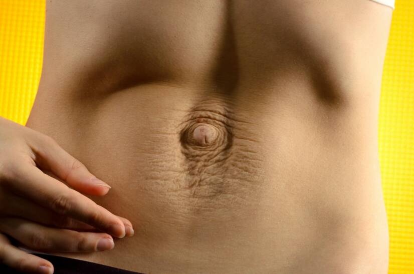
Abdominal diastasis. Source photo: Getty Images
Causes
The etiology of abdominal diastasis is attributed in some studies to congenital excessive laxity (looseness) of the ligaments and subsequent loss of strength of the abdominal ligament line (linea alba).
The most common factor in the development of diastasis is pregnancy. As a result of hormonal influences during pregnancy, the increase in uterine volume and stretching of the soft structures of the body, the straight left and right abdominal muscles are stretched apart.
In some cases, it can be the result of rapid weight gain, obesity and excessively weakened abdominal muscles.
In other cases, it may be an increase in the abdominal cavity due to another organic disease, such as abdominal ascites.
Sometimes, on the other hand, it is the result of overloading and distortion of the abdominal muscle structure due to improper technique or excessive exercise intensity.
The most common factors in the development of abdominal diastasis:
Pregnancy and childbirth
Parent over 35 years of age
Multi-parent
Hormonal influence
Weight gain
Obesity
Weakened abdominal wall musculature
Lack of compensatory physical activity
Overload and distortion of the abdominal muscles
Mechanical damage, injury, trauma
Symptoms
If the abdominal muscles are spaced apart, the body's support system is weakened at the same time.
If left untreated, diastasis can increase the risk of spinal stability and mobility problems, muscle imbalances in the spine, the risk of improper breathing patterns, and the risk of abdominal protrusion and abdominal hernias.
As the abdominal musculature is closely functionally related to the pelvic floor, diaphragm and deep spinal muscles, the deep stabilisation system of the body is disrupted.
Diastasis is often associated with back pain and instability, especially in the lumbar spine and pelvic region.
If diastasis is left untreated, gastrointestinal and digestive disorders, urinary incontinence, or menstrual cycle disorders such as painful menstruation are possible.
The most common manifestations of abdominal diastasis are:
Loose abdominal wall
Back pain
Pelvic pain
Impaired trunk mobility
Weakened pelvic floor
Digestive problems
Urinary incontinence
Menstrual cycle disorder
When the cause of diastasis is diagnosed, the patient is advised on proper movement habits for daily activities. The patient is educated on diaphragm activation and abdominal muscle awareness.
Handling heavy loads and isolated strenuous abdominal muscle exercises (sit-ups, plank) should be avoided. Slow standing up over the hip is recommended, not a swinging sit-up type movement.
Loose abdominal wall and excess skin overhang in diastasis. Photo source: Getty Images
Diagnostics
The stretch of the rectus abdominis muscle can be determined by palpation (touch). The doctor or therapist palpates the medial inner edges of the rectus abdominis muscle at the midline of the abdomen (linea alba).
If the gap between the muscles is more than 2 cm, it may be abdominal diastasis. In the higher stages of abdominal diastasis, the muscle gap may be as large as 10 cm.
The basic diagnostic procedures include measurement with a special tape measure or callipers. In the case of diastasis, this is a precise measurement of the width of the abdominal muscle spacing.
Abdominal diastasis can only be determined by aspiration (gaze examination) in individuals with very little subcutaneous fat.
As an objective diagnostic method, ultrasound, computed tomography or magnetic resonance imaging can be used to view the soft structures of the body in detail. The appropriate type of diagnostic procedure is determined by the examining physician.
Self-examination of abdominal diastasis
The home examination consists of lying on your back with your legs bent at the knees and your feet on a mat. Support your head with one hand and observe the abdominal wall with your eyes.
Press the abdominal wall above the umbilicus with the fingertips of the other hand. If the fingers go in deeper and the gap between the left and right rectus abdominis muscles is greater than 2-2.5 cm, there may be diastasis.
In this case, consultation and professional examination by a doctor is recommended.
Physiological state of the abdominal muscles and diastasis (spacing) of the left and right rectus abdominis muscles from the midline, linea alba. Photo source: Getty Images
Diastasis in pregnancy and after childbirth
During pregnancy, a large number of physiological changes occur in the female organism due to hormones. One of them is the loosening of the abdominal wall due to the stretching of the soft structures of the body.
Abdominal diastasis is experienced by a significant percentage of women who give birth. In some women, abdominal diastasis persists for a long time after childbirth.
The abdominal fascia (linea alba) may loosen or break with traction. The inner parts of the rectus abdominis muscle are spaced apart. The area of greatest spacing is usually the umbilicus.
However, the diastasis itself is not painful for the woman.
A loose abdominal wall has a great influence on the reduction of the stability of the pelvis, the spine and the weakening of the pelvic floor muscles. It is therefore recommended to undergo comprehensive rehabilitation after childbirth, the basic content of which is exercise with a physiotherapist.
For successful conservative treatment, regularity of home exercises and adherence to exercise measures are important.
With stability and strength in the abdomen, spine and pelvis, a woman can reduce the risk of incontinence and back pain at menopause.
Exercise as prevention and treatment of diastasis
The prevention of diastasis is regular activation of the abdominal wall before the actual conception of the baby.
Studies confirm that regular exercise of the abdominal wall before conception reduces the risk of diastasis. If diastasis occurs postpartum, there is also a higher probability of faster regeneration of the soft structures.
Diastolic breathing and abdominal wall strengthening
This exercise may be simple at first glance, but it is not. The starting position is lying supine on a soft mat. The legs are bent at the knees and the soles of the feet are glued to the mat.
The head is in imaginary extension of the spine and the shoulders are loosely resting on the mat.
There should be no space between the mat and the spine. Place the palms of the hands on the last ribs - on the outside of the lower abdomen. The palms of the hands are used to check the correct technique of the exercise. It is recommended to close the eyes to better perceive the breath and the movement of the hands.
Breathing into the diaphragm and abdomen, we try to spread the ribs sideways apart, creating an imaginary tire growing around the abdomen. With the palms of the hands, we check the diaphragmatic breathing sideways.
Try to keep the chest movement minimal. Inhaling, using intra-abdominal pressure, hold for a few short seconds. Exhaling, relax the abdominal wall and return to the original position. Breathing is smooth and slow.
A more difficult version of the exercise is the technique of diaphragmatic breathing and awareness of intra-abdominal pressure during inhalation and exhalation simultaneously.
The primary goal of the exercise is to keep the abdominal wall firm and activate the diaphragm throughout the continuous breathing. After a few inhalations and exhalations, the abdominal muscles are then completely relaxed.
Diaphragmatic breathing with abdominal wall activation. Inhalation: ribs move sideways away from each other and the diaphragm muscle drops down. Exhalation: ribs move back towards each other and the diaphragm muscle rises up. Photo source: Getty Images
Glute bridge with diaphragmatic breathing
The starting position is lying on the back on a soft mat. The feet are full length on the mat and the knees are bent. The hands are placed loosely on the mat next to the torso, palms down.
The head is in imaginary extension of the spine and the shoulders are naturally placed on the mat.
Inhaling into the diaphragm and lower ribs, we simultaneously lift the pelvis from the mat towards the ceiling. We activate the abdominal wall, diaphragmatic breathing. We firm the abdominal muscles and hold the top position for a few seconds (a few inhalations and exhalations).
The ideal is a simultaneous isolated contraction of the gluteal musculature (buttock muscles). Then with an exhalation we relax the musculature and slowly vertebra by vertebra we return to the basic position back on the mat.
Alternate placing the heel on the ground
The starting position is lying on the back. The head is in extension of the spine, the shoulders are lowered and placed on the mat. The arms are loosely beside the body. The palms are placed on the lower ribs on both sides.
The lower limbs are lifted upwards so that the feet and knees do not touch the mat. The lower limbs are naturally bent at the knee joints in the air. The hip joints form an imaginary right angle with the torso.
There is no space between the mat and the spine.
With a breath, we activate the diaphragmatic lateral breathing. We become aware of the intra-abdominal pressure and firm the abdominal wall. We slowly place one of the limbs with the heel on the mat and then slowly return to sleep smoothly in the air.
During the movement of the limb, we keep the abdominal wall stable, breathing smoothly and holding on with the palms of our hands. We then relax the muscles and repeat the exercise while moving the opposite lower limb.
How it is treated: Abdominal diastasis
How is abdominal diastasis treated? Will medication or exercise therapy help?
neurologiepropraxi.cz - The importance of the deep stabilization system in the context of vertebrogenic difficulties, Assoc. Paed.Dr. Pavel Kolář, Prof. MUDr. Karel Lewit, DrSc.
dspace.cvut.cz - Physiotherapy in women with diastasis of the abdominal wall.
I completed my bachelor's degree in physiotherapy at the Faculty of Biomedical Engineering of the Czech Technical University in Prague. I continued and completed my master's degree in physiotherapy at the Faculty of Health Engineering of TnUAD. I am currently pursuing my PhD rigorosis at the Slovak University of Health Sciences in Bratislava. During my studies I worked as a physiotherapist in the rehabilitation clinic of Vamed Mediterra in Prague and then as a physiotherapist at the Regional Hospital in Liberec in the Department of Neurology. During my employment I was part of the medical team in the Covid19 department. I am mostly interested in human musculoskeletal system, rehabilitation, physiotherapy in gynaecology and natural medicine. My hobbies include exercising, running, writing and managing social media.


 of the left and right rectus abdominis muscles from the midline, linea alba. Photo source: Getty Images")