- MAREŠOVÁ, Pavlína. Modern procedures in gynaecology and obstetrics. 3rd, revised and supplemented edition. Prague: ISBN 978-80-7345-709-9.
- ROZTOČIL, Aleš. Modern obstetrics. 2nd, revised and supplemented edition. Prague: Institute for Mother and Child Care.
- pubmed.ncbi.nlm.nih.gov - Diastasis of the direct abdominal muscle. Javed Akram, Steen Henrik Matzen
- pregnancybirthbaby.org.au - Separation of the abdominal muscles diastasis recti. Pregnancy childbirth
- healthline.com - Diastasis recti: What is it and how is it treated? Jane Chertoff
Postpartum diastasis: cause and symptoms? + Exercises at home
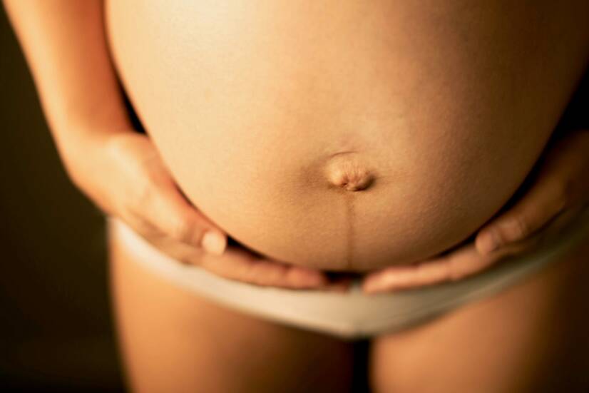
Photo source: Getty images
Most common symptoms
Show more symptoms ᐯ
Symptoms
The most common physical manifestations of postpartum diastasis:
- Loose abdominal wall
- Back and pelvicpain
- Loose abdominal overhang in the navel area
- Impaired mobility and stability of the trunk
- Weakened pelvic floor musculature
- Problems with the digestive tract
- Urinary incontinence (urine leakage)
- Disruption of the menstrual cycle
. Source: Getty Images")
Options for the prevention of diastasis
Prevention of abdominal diastasis in pregnancy and in the postpartum period consists primarily in keeping the abdominal wall stable and elastic before the baby is conceived.
An appropriate lifestyle and physical activity before delivery improve the quality and speed of postpartum recovery.
Within the period of pregnancy itself, it is possible to use the services of physiotherapy in gynaecology, where specific exercises will be recommended to the woman to correct the imbalance of the musculoskeletal system, taking into account her individual state of health.
Exercise at home - prevention and therapy of diastasis after childbirth
Ideally, the choice of exercises should be consulted with a specialist, taking into account the individual state of health. Below are suitable examples of exercises to strengthen the abdominal wall and eliminate imbalances of the pelvic muscles.
Exercise 1: Defensive breathing and abdominal wall strengthening
The following exercise may seem simple at first glance, but this is not always the case. The woman's starting position is lying on her back on a soft comfortable mat.
The legs are bent at the knee joints and the feet are resting flat on the mat. The head is straight in an imaginary extension of the spine and the arms, lowered freely from the ears, rest on the mat.
There must be no space between the mat and the spine.
Place the open palms on the last ribs from the side - on the outside of the lower abdomen. The palms serve as a check for correct exercise technique.
For a better perception of one's own breathing and the movement of the hands, it is recommended to close the eyes during the exercise. With breathing into the diaphragm and abdomen, the woman tries to spread the ribs sideways, creating an imaginary tire growing sideways around the abdomen.
The palms of the hands are used to control the diaphragmatic breathing technique. The chest moves minimally during the exercise.
Inhaling using and maintaining intra-abdominal pressure, we hold and exhale for a few seconds.
With the exhalation, we then relax the abdominal wall and return to the original position.
Do not hold the breath during the exercise.
. Source: Getty Images")
Exercise 2: Bridge with abdominal wall and pelvic floor activation
The knee joints are bent, the shoulder blades are flat on the mat. There is no space between the spine and the mat. The knee joints are level with the width of the hip joints. The arms are loosely placed close to the body, palms down.
During the exercise, the pelvis gradually rises up towards the ceiling. The shoulder blades remain supported throughout the exercise. In the upper bridge position, the pelvic floor muscles are activated and the gluteal halves contract.
In the upper position, hold and firm the abdominal wall several times using the diaphragm. With an exhalation, the practitioner lowers the mat again and releases all the activated muscles of the body.

Exercise 3: Alternating leg bends and abdominal stabilization
The starting position of the exercise is lying on the back. The head is in extension of the spine, the arms lowered from the ears loosely placed on the mat. The lower limbs are raised upwards so that the feet and knees do not touch the mat.
The lower limbs are ideally in the air, naturally bent at the knee joints to a right angle. The hip joints form an imaginary right angle with the abdomen. Again, there must be no space between the mat and the spine.
With the inhalation we activate the lateral diaphragmatic breathing, regulate the intra-abdominal pressure and strengthen the entire abdominal wall.
Alternately, we slowly place one lower limb with the heel on the mat and then smoothly return it to the second sleep again in the air.
Repeat the exercise while moving the opposite lower limb.
Pay attention to the space between the mat and the spine and to the swing speed of the exercise.
. Source: Getty Images")
Exercise 4: Plank/half plank and mid-body activation
The position consists of leaning on the forearms and knee joints (feet for the heavier version). The spine is imaginatively straight as a plank and the head in natural extension. The exerciser looks down or in front of him/her and does not tilt the head.
Pay attention to the shoulders. They are pulled away from the ears and do not fall towards the head.
During the exercise, the abdominal wall is strengthened and diaphragmatic breathing is activated. Do not hold the breath. This is a time endurance exercise.
Beware of possible relaxation of the abdominal muscles.

The most common possible mistakes in home exercises:
- Fast swinging movement
- Failure to activate diaphragmatic breathing
- Not strengthening the abdominal muscles and walls
- Loose space between the mat and the spine
- Holding the breath during exercise
- Not being aware of muscle activation
- Irregularity of exercise
- Distraction
Treatment options for postpartum abdominal diastasis
Show morePostpartum diastasis is treated by
Other names
Linea alba, diastasis of the abdominal muscles, diastasis, spacing of the abdominal muscles