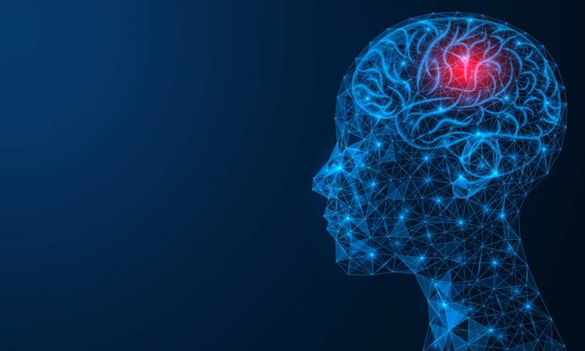
Meningitis, i.e. inflammation of the protective membranes covering the brain and spinal cord, is a neuroinfection. It affects the brain, the meninges, the spinal cord and peripheral nerves. The severity of the disease varies from asymptomatic to life-threatening.
Meningitis is a neuroinfection that can be acute, subacute or less commonly chronic. They affect the brain, the meninges, possibly the spinal cord and peripheral nerves.
The severity of the disease and clinical picture varies from asymptomatic (especially in aseptic meningitis) to life-threatening disease with a dramatic and rapidly progressive clinical picture.
Suppurative meningitis is a serious disease of the central nervous system in which diffuse inflammation is present, occasionally with the formation of a circumscribed inflammation, i.e. a brain abscess.
Acute purulent (with pus) meningitis affects the pia mater (the soft covering of the brain), the arachnoidea (the covering of the brain called the arachnoid, a bloodless space with cross-linked tissue) and the cerebrospinal fluid (the cerebrospinal fluid).
Since this is a larger group of diseases, we divide them according to several variables:
by bacterial agent,
by patient's age,
by disease origin,
by predisposing and risk factors
The most common bacteria that cause meningitis include:
Neisseria meningitidis - meningococcus
Streptococcus pneumoniae - streptococcus
Haemophillus influenzae type B
Listeria monocytogenes
Streptococcus agalactiae
Staphylococcus
Escherichia coli - E.coli
The largest group of patients are children, with a predominance of newborns and children up to 3 months of age.
If the affected child is within the 5th to 7th day after birth, meningitis is referred to as early-onset meningitis. In this case, the source of infection is still the mother and the infection comes from her urogenital tract, vagina or gastrointestinal tract.
Late-onset neonatal meningitis, from the 7th day to the 3rd month after birth, arises from the transmission of infection from the environment to the baby. Most commonly by passage from the hands of the attending staff in the hospital.
In addition to neonatal age, other risk factors include advanced age over 65 years, shunts in the liquor ducts (tubes for draining liquor), conditions following open-skull trauma, or diseases characterised by chronically weakened immunity, such as HIV and AIDS.
Causes
As it is an infectious disease, the infection is transmitted by contact with an infected person or by penetration from another focus of inflammation.
The risk of infection arises from prolonged contact with an infected person in enclosed spaces, use of a shared bathroom, smoking a shared cigarette, kissing, drinking from a single glass, increased physical exertion and exhaustion, malnutrition and genetic predisposition.
According to the cause and mechanism of occurrence, purulent meningitis is divided into two types: primary meningitis and secondary meningitis.
In primary meningitis, the bacteria enter the central nervous system through the blood, i.e. by haematogenous spread. They penetrate through the small vessels of the choroid plexus or through the vessels of the subarachnoid space.
The choroid plexus are tiny vascular structures in the brain that produce cerebrospinal fluid. The subarachnoid space is the area below the arachnoid that houses the blood vessels that nourish the brain.
The first symptoms of primary meningitis are sudden and dramatic. The most common causative agents are Neisseria meningitidis, E.coli and Streptococcus pneumoniae.
Secondary meningitis develops in another ongoing suppurative disease.
It is caused by the passage of bacteria from this primary focus, into the central nervous system. Such a primary focus and source of infection may be middle ear infection, inflammation of the sinuses, mastoiditis, or suppurative inflammation of the bones (osteomyelitis), e.g. of the skull and vertebrae.
The bacteria that cause this type of meningitis are usually Streptococcus pneumoniae (streptococcus) or Staphylococcus aureus (golden staphylococcus).
All these species of bacteria that cause meningitis contain specific components in their cell wall that trigger a cascade of inflammatory events in the body. The result is increased permeability of the blood-brain barrier, which forms a bulletproof barrier between the brain and the rest of the oraganism.
This protects the brain from infections, drugs, toxins and other substances.
Symptoms
The initial clinical signs of purulent meningitis are more or less unspecific and occur in many other diseases.
Onset or initial signs and symptoms in children and adults:
fever,
chills,
headache,
nausea,
vomiting,
stiff back of the neck
The first suspicion that it may be a neuroinfection arises only in the presence of positive meningeal signs and symptoms.
The so-called meningeal phenomena is a set of symptoms that arise when the meninges, i.e. three membranes that envelop the brain and spinal cord, are irritated by a pathological process, e.g. inflammation, or even by haemorrhage or tumour.
Nerve roots (tangles of nerves leading to the limbs) pass through the meninges, which are irritated and, when stretched, cause pain and stiffness that restricts the patient's movement.
A patient with positive meningismus cannot bend the head deeply, raise the lower limb to the chest, or stand up from lying down to sitting without the help of the arms.
Another suspicious symptom is petechiae, which are pink to purple-red spots on the skin caused by bleeding from small blood vessels.
Over the next 24 hours, the clinical picture progresses rapidly.
Epileptic seizures, varying degrees of disturbances of consciousness ranging from behavioural changes to coma, focal neurological signs e.g. limb paralysis, hyperventilation (rapid and deep breaths) and bradycardia (reduced heart rate below 50 beats per minute) may be associated.
Gradually, a combination of purulent meningitis and sepsis develops.
The blood circulation is centralized, so that most of the blood volume is concentrated in the most important vital organs, the heart and the brain, thus reducing the pressure in the periphery.
Cold and blue acral parts of the body, fingers, palms and feet are a sign that there is not enough blood circulation throughout the body. Blood is also not present in other organs, such as the kidneys, which may be failing.
decreased muscle tone (floppiness, rag-doll syndrome)
Diagnostics
When neuroinfection is initially suspected, the first diagnostic step is to perform a lumbar puncture and collect cerebrospinal fluid for laboratory analysis and testing
Before performing a lumbar puncture, an examination of the eye's background is performed. In cases of advanced cerebral edema, possible edema (swelling) of the optic nerve papilla is checked during an eye examination, which will alert that a serious complication may occur during lumbar puncture.
When the fluid is withdrawn, there is a sudden decrease in intracranial pressure and the formation of an occipital conus, i.e., the extrusion of part of the cerebellum through the inferior cranial aperture.
Such a complication is life-threatening.
Already during the collection of cerebrospinal fluid, certain changes are visible to the naked eye. In a healthy person, the liquor is similar to water, i.e. it is clear, colourless and drains quite quickly because it is thin.
In purulent meningitis, the liquor is whitish to yellowish, exceptionally it may be greenish. It oozes under high pressure, is thick and may smell.
On laboratory examination of the liquor, we find 1,000 to 10,000 times the amount of cells with hyperproteinorrhachia (increased amount of protein) and a concomitant decrease in the level of glucose in the liquor (a sign of the presence of glucose-consuming bacteria).
The liquor may be stained with a specific diagnostic dye and its smear viewed under a microscope. In such a microscopic slide we can see the causative agent of the infection.
The next examination of the liquor is a microbiological culture examination. However, this is lengthy and is used to confirm the diagnosis rather than for acute rapid diagnosis prior to treatment.
Colonies of bacteria - during microbiological examination. Photo source: Getty images.
The patient also undergoes complex examinations such as blood count, haemoculture (blood for microbiological diagnostics), biochemical blood tests to detect the level of glycaemia, hepatic enzymes or renal parameters.
X-ray of the lungs, sedimentation, biochemical and urine tests are also performed.
In patients with pneumococcal and haemophilic meningitis, head CT and ENT tests are also recommended to rule out chronic sinusitis as a cause of secondary meningitis.
If staphylococcus is the identified causative agent, an echocardiographic examination of the heart (ECHO) should be an essential investigation. This is because the source of staphylococci may be untreated endocarditis - inflammation of the inner wall of the heart and valves.
An MRI scan of the spine will show whether the primary lesion comes from inflamed vertebrae and intervertebral discs, so-called spondylodiscitis.
A dangerous symptom is oto-liquorrheaor nasal liquorrhea, i.e. liquor oozing from the ear or nose. The cause is damage to the continuity of the dura mater (hard envelope of the brain), e.g. due to trauma, or postoperatively.
A leaky dura mater leaks fluid and at the same time opens the way for the passage of bacteria to the brain area. A specific test is the determination of the so-called beta-trace protein. This will show the presence of liquor in the ear or nose, distinguishing it from other physiological fluid.
Differential diagnosis
In the differential diagnosis, other diseases with a similar clinical picture come into consideration:
The aforementioned disruption of the blood-brain membrane can allow pathogens to enter the brain where they cause inflammation. This leads to swelling of the brain - cerebral oedema. The bacteria also enter the cerebrospinal fluid (CSF), which spreads them throughout the body.
When bacteria spread through the liquor pathways, they become blocked.
The blockage of the liquor ducts prevents the outflow of the liquor, which accumulates, enlarges the brain chambers and causes the so-called hydrocephalus. Since the brain does not have the ability to expand its volume indefinitely (because it is housed in the skull), the swollen brain and enlarged ventricles begin to generate increased intracranial (intracranial) pressure.
The pressure on important brain centres produces the typical symptoms of meningitis.
Among other things, inflammatory processes in the wall of blood vessels (vasculitis and thrombophlebitis) can cause local anemia of part of the brain -brain ischemia.
All of these processes such as increased intracranial pressure, decreased blood flow through brain tissue and site of ischemia, contribute to diffuse ischemic brain involvement.
The body responds to increased intracranial pressure by systemic hypotension, i.e. reduced blood pressure in the blood vessels of other organs, thereby insufficiently circulating blood to other important organs such as the kidneys, intestines, liver and others. The patient quickly goes into septic shock, which ultimately leads to the patient's death.
Subacute course is present in shunt meningitis, tuberculous meningitis (in homeless and migrants) and cryptococcal meningitis (AIDS patients, post-transplant, immunodeficient). Pulmonary involvement is also a presenting symptom in these types of meningitis.
The mortality rate of haemophilus and meningococcal meningitis is about 5%, with pneumococcus up to 20%, with the age of the patient and associated diseases this percentage increases even more.
As it is a serious neuroinfection, even after successful management of the acute disease, permanent sequelae are common.
The principle of the occurrence of such permanent brain damage may be 2 mechanisms, namely:
inflammatory and bacterial products that are toxic to nerve cells,
cerebral edema itself.
Of the permanent neurological symptoms, the following are the most common:
facial nerve palsy
deafness
epilepsy
hydrocephalus (build-up of cerebrospinal fluid in the ventricles of the brain)
psychomotor retardation
dementia
blindness
Prevention
It is possible to protect yourself against purulent meningitis by:
1. Vaccination
Vaccination is the most effective form of prevention against infectious diseases.
It is an active immunisation (i.e. the organism itself actively produces antibodies against the causative agent), which represents long-term but also universal protection, since the vaccine is also effective against other types of meningococci, such as those that do not occur in Slovakia but pose a risk abroad.
We have several vaccines registered in Slovakia.
There are 4 vaccines registered against the invasive disease caused by Neisseria meningitidis (meningococcus).
They can be vaccinated from infancy (depending on the type of vaccine, usually from the age of 2 months) and also in adulthood or at an older age.
An effective prevention is vaccination. Photo source: Getty images.
2. Dietary measures
Listeria monocytogenes, the dangerous causative agent of purulent meningitis, is transmitted to humans from animals either by direct contact (animal husbandry) or by contaminated food.
Meat products (e.g. sausages and other cold cuts) and dairy products must be heated to at least 70 °C before consumption and then kept heated until consumption.
Listeria can grow and spread in the refrigerator.
It can survive even at low temperatures. Therefore, care must be taken when eating even chilled or defrosted foods.
It is ideal to keep these products at temperatures below 5 °C at all times.
Room temperature is not recommended for longer than 2 hours.
The most sensitive group of the population, pregnant women, the elderly and immunocompromised patients, are advised not to consume uncooked meat products and canned or unpasteurised dairy and cheese products at all.
3. Exams during pregnancy
Neonatal meningitis is caused by the passage of infection from the mother to the fetus.
An effective prophylaxis is frequent examination of the mother for the presence of group B streptococci in her organism. With a positive result, antibiotic treatment is appropriate during labour when infection occurs.
4. Protect the relatives of and the medical staff treating a person with purulent meningitis
If the causative agent of meningitis is Neisseria meningitidis, isolation of the patient from other patients and prophylactic antibiotic treatment of all loved ones and family members who have come into contact with the person is necessary.
Medical personnel who normally treat such a patient are not immediately at risk. However, if there is a risk of contact, such as splashing with infected blood or other fluid, cardiac massage or mouth-to-mouth breathing, prophylaxis is also needed for these persons.
Antibiotic treatment is administered, namely V-penicillin for one week.
van de Beek D, de Gans J, Tunkel AR, Wijdicks EF (January 2006). "Community-acquired bacterial meningitis in adults". The New England Journal of Medicine. 354 (1): 44–53.
Ginsberg L (March 2004). "Difficult and recurrent meningitis". Journal of Neurology, Neurosurgery, and Psychiatry. 75 Suppl 1 (90001): i16–21.
Ferri, Fred F. (2010). Ferri's differential diagnosis : a practical guide to the differential diagnosis of symptoms, signs, and clinical disorders (2nd ed.). Philadelphia: Elsevier/Mosby. p. Chapter M. ISBN 978-0-323-07699-9.
Centers for Disease Control Prevention (CDC) (May 2008). "Primary amebic meningoencephalitis – Arizona, Florida, and Texas, 2007". MMWR. Morbidity and Mortality Weekly Report. 57 (21): 573–27.
"Viral Meningitis". CDC. 26 November 2014.
Tunkel AR, Hartman BJ, Kaplan SL, Kaufman BA, Roos KL, Scheld WM, Whitley RJ (November 2004). "Practice guidelines for the management of bacterial meningitis". Clinical Infectious Diseases. 39 (9): 1267–84.
"Global Disease Burden 2019".
Putz, Katherine; Hayani, Karen; Zar, Fred Arthur (2013). "Meningitis". Primary Care: Clinics in Office Practice. 40 (3): 707–726.
"NHS medical conditions meningitis". NHS.
"Meningococcal meningitis Fact sheet N°141". WHO. November 2015.
Mosby's pocket dictionary of medicine, nursing & health professions (6th ed.). St. Louis: Mosby/Elsevier. 2010. p. traumatic meningitis. ISBN 978-0-323-06604-4.
van de Beek D, de Gans J, Spanjaard L, Weisfelt M, Reitsma JB, Vermeulen M (October 2004). "Clinical features and prognostic factors in adults with bacterial meningitis" (PDF). The New England Journal of Medicine. 351 (18): 1849–59.
Attia J, Hatala R, Cook DJ, Wong JG (July 1999). "The rational clinical examination. Does this adult patient have acute meningitis?". JAMA. 282 (2): 175–81.
Theilen U, Wilson L, Wilson G, Beattie JO, Qureshi S, Simpson D (June 2008). "Management of invasive meningococcal disease in children and young people: summary of SIGN guidelines". BMJ. 336 (7657): 1367–70.
Management of invasive meningococcal disease in children and young people (PDF). Edinburgh: Scottish Intercollegiate Guidelines Network (SIGN). May 2008. ISBN 978-1-905813-31-5.
Thomas KE, Hasbun R, Jekel J, Quagliarello VJ (July 2002). "The diagnostic accuracy of Kernig's sign, Brudzinski's sign, and nuchal rigidity in adults with suspected meningitis". Clinical Infectious Diseases. 35 (1): 46–52
After graduating from an eight-year high school, I knew exactly who I wanted to become in life. Studying at the Faculty of Medicine at Comenius University in Bratislava fulfilled a long-held dream and I was able to start fulfilling my mission as a doctor. After obtaining my diploma and M.D. degree, my first professional steps led to the St. Elizabeth Cancer Institute in Bratislava. At the Stereotactic Radiosurgery Clinic I worked with patients with brain tumours and eye tumours. After a year of working with oncology patients, in 2020 I moved to the Neurology Clinic of SZU St. Michael's University Hospital in Bratislava, where I am still working today. I like to spend my free time outdoors, whether cycling, hiking or just walking in the nearby forest.