Cerebral hemorrhage (cerebral hemorrhage) is divided into spontaneous and traumatic. Both conditions threaten a person's health and life. Why do they occur and how do they manifest themselves?
Brain haemorrhage is divided into two types - spontaneous and post-traumatic (traumatic).
The meaning of the division is clear from a professional and practical point of view. They differ in cause and approach to diagnosis and treatment.
Spontaneous cerebral haemorrhage
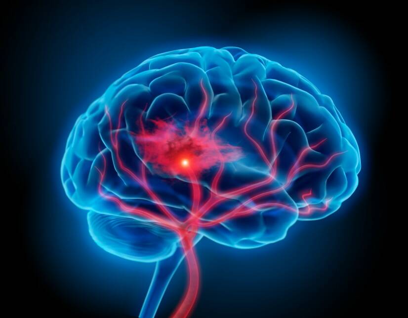
In cerebral haemorrhage, venous or arterial blood spurts into the confined space of the skull and brain.
They are divided according to the site of bleeding: intracerebral (within the brain tissue) subarachnoid (between the meninges) intraventricular (into the ventricles)
Intracerebral haemorrhage
Approximately 15% of all sudden strokes are intracerebral haemorrhages.
This is bleeding into brain tissue. The blood flows from an artery and is oxygenated.
They are uncommon but have a high mortality rate.
Subarachnoid hemorrhage
A subarachnoid hemorrhage is a bleeding emergency. The blood spurt (hematoma) is located between the meninges, specifically between the arachnoid (the arachnoid) and the pia mater (the soft membrane adjacent to the brain tissue).
It affects about 20 out of one million people per year. 5-10% of them die immediately at its onset.
Traumatic brain haemorrhage
Trauma (injury) is a sudden bodily injury by mechanical, chemical, thermal or other energy, the intensity and magnitude of which exceeds the body's resistance.
The most common causes of neurotrauma are:
road traffic accident
sports
work accidents
domestic accidents
violence
Neurotrauma is three times more common in men than in women. It occurs mainly between the ages of 15 and 25.
It causes approximately 17% of all head injury deaths.
Head and brain (craniocerebral) injuries can be:
covered
open (only the skin is broken)
penetrating (the dura mater is injured)
covertly penetrating (injury to the skull base)
Bleeding is a common secondary complication of these injuries. They are divided into intracerebral (into the brain) and extracerebral (outside the brain tissue but still within the skull).
In extracerebral haemorrhage, it is important to distinguish the location of the resulting haematoma (blood spurt). According to the localization it is divided into:
epidural
subdural
subarachnoid
intraventricular
Intracerebral haemorrhage
This is bleeding into brain tissue.
It is of arterial origin. It is caused by injury and rupture of arteries through which oxygenated blood flows under high pressure.
It occurs most commonly in the white matter of the frontal or temporal lobe.
Intracerebral hematoma is dangerous because of its rapidly increasing volume and expansive behavior. It is associated with the development of cerebral edema.
Epidural haemorrhage and subdural haemorrhage
The brain is protected by several sheaths - the meninges.
Under the diapers and under the skin are the cranial bones forming the skull.
Under this bone is the first brain envelope. Its structure is solid and hard. That's why it's called the dura mater.
The space between the skull and the dura mater is called the epidural space.
Bleeding and accumulation of blood in this space creates an epidural hematoma.
Below the dura mater is the bloodless space formed by the fascia and the liquor. It is called the arachnoidea (web).
The space between the dura and the arachnoidea is called the subdural space. Subdural hematoma is a serious complication of head injuries.
Beneath the arachnoidea is a thin and delicate membrane. It closely adheres to the brain tissue and its shape follows the brain coils, the so-called gyrification of the brain. It is called the pia mater.
Causes
What are the causes of both types of bleeding?
Spontaneous: intracerebral and subarachnoid haemorrhage
Both types of bleeding are spontaneous. They occur without any trauma, injury or blow to the head area.
They are caused by rupture of small blood vessels in the brain or rupture of a vascular aneurysm (bulge).
Cause and risk factors of intracerebral haemorrhage
High blood pressure (arterial hypertension) is one of the most common diseases of the cardiovascular system in this country and in the world. It is also a major contributing factor in the development of intracerebral haemorrhage.
Other important causes are congenital disorders of the vessel wall, such as microvascular aneurysms, amyloid angiopathy, various vascular anomalies, and others.
Cerebral hemorrhage can also occur in hematological diseases with impaired blood clotting. Coagulation may be impaired by inappropriate anticoagulant therapy. In this case, we are talking about iatrogenic damage.
Other risk factors include:
diabetes mellitus
chronic alcohol consumption
drug use
smoking
brain tumours
Vascular aneurysms as a cause of subarachnoid haemorrhage
An aneurysm is a circumscribed enlargement of a blood vessel, called an aneurysm.
The vascular wall around the aneurysm is thinner, more fragmented and is exposed to different physical and mechanical conditions. The blood does not flow straight and 'smoothly' as in a healthy blood vessel. Blood swirls form.
The combination of these two factors, together with high blood pressure or arteriosclerosis, increases the risk of aneurysm rupture with massive bleeding.
The incidence of cerebral aneurysms is approximately 1-5%. They are most common between the ages of 40 and 60 and more common in women.
The cause of subarachnoid haemorrhage is a saccular aneurysm in 75%. It is usually located in the carotid basin - in the vessels that branch from the carotid arteries in the brain.
Multiple aneurysms can occur on a single vessel.
Causes of traumatic brain haemorrhage
Injury to the skull in a head injury causes bleeding from blood vessels that are just below the skull. These are arterial or venousbleeds.
The blood accumulates above the dura mater. An epidural hematoma forms.
Epidural hematoma without the presence of skull fracture is rather rare.
About 10% of adults have epidural haemorrhage without fracture, but 40% of children have no fracture. This is due to the elasticity and pliability of the soft bones and cranial sutures in children.
Most bleeding is caused by injury to the great vessel arteria meningea media, which nourishes the dura mater.
Venous hemorrhage is caused by bleeding from the middle meningeal vein or from venous plexuses.
The cause of subdural hematoma is rupture of the bridging or cortical veins between the dura mater and the arachnoid.
They are divided into three groups according to the time from onset to the first symptoms:
acute (occurring within 3 days of injury)
subacute (manifesting between the third and twentieth day after the injury)
chronic (onset of symptoms later than twenty days after the injury)
Acute subdural hematoma accompanies more severe brain injury, which is associated with brain contusion and skull fractures.
There is a greater risk in patients who take blood thinners.
It is also more common in alcoholics and the elderly. Their brains are shrunken (atrophic) and the dilated blood vessels between the smaller brain and the meninges are more fragmented.
Rarely, a subdural hematoma may result from the rupture of a congenital aneurysm or from an arteriovenous malformation.
Chronic subdural haematoma, on the other hand, arises in mild trauma.
In up to half of the cases, the patient does not even remember the injury. It occurs in the elderly, in persons with cerebral atrophy, in users of blood thinners and in alcohol addicts.
It can occur simultaneously on both sides of the skull.
Symptoms
Symptoms of bleeding differ between spontaneous and traumatic brain haemorrhage.
What are the symptoms of spontaneous bleeding?
The symptomatology of intracerebral hemorrhage depends on the localization of the hemorrhage. The most common site of typical bleeding is the so-called basal ganglia. These are centres in the brain made up of grey matter that control motor skills.
The other typical sites are the cerebral lobes and the thalamus. Bleeding into the brain stem is very serious, accounting for 5-10% of cases.
Symptoms are similar to those of ischaemic stroke.
When such a clinical picture arises, it is not immediately clear which type of stroke is involved.
Some symptoms are more characteristic of haemorrhage than of ischaemia.
These symptoms are:
vomiting, which indicates increased intracerebral pressure
a rapidly deteriorating clinical picture due to an enlarging haematoma (blood spurt) in the brain
progressive impairment of consciousness
excruciating headache
Subarachnoid haemorrhage is manifested by a sudden agonising, explosive headache such as the patient has never experienced before.
It is accompanied by stomach upset, vomiting, confusion, brief loss of consciousness, meningeal syndrome, paralysis of the limbs and epileptic seizures.
Unbearable pain like one has never experienced before. Source of the photo: Getty Images
Post-traumatic hemorrhage and its typical symptoms
Intracerebral hemorrhage almost always manifests itself by immediate unconsciousness.
Epidural bleeding usually manifests itself two to six hours after the accident.
The first symptom is impaired consciousness.
It can take five forms:
permanent unconsciousness from the onset of injury
no change in consciousness since the injury
a brief loss of consciousness at first, then the patient regains consciousness
no disturbance of consciousness immediately after the accident, later on unconsciousness sets in
unconsciousness immediately after the injury, followed by full consciousness and then recurrent unconsciousness (only ⅓ of patients)
Later unconsciousness is not typical for epidural haematoma. It may be a manifestation of a complication such as subdural haemorrhage, cerebral contusion or cerebral oedema.
If the patient falls into a coma, asymmetrically enlarged pupils, palsy of the III cranial nerve that innervates the oculomotor muscles may occur.
Half of the patients develop paralysis of the upper or lower limbs. Paralysis of the limb on the opposite side to the hematoma and pupil enlargement. With hemorrhage in the left cerebral hemisphere, the pupil of the left eye will be enlarged and the limbs on the right side of the body will be paralyzed.
This is caused by the crossing of the nerve pathways that lead from the brain to the limbs.
Other symptoms include:
slow heart rate (bradycardia)
fluctuations in blood pressure
cardiac arrhythmias
nausea
vomiting
paleness of the face
irregular breathing
memory disorders
disorientation
The symptomatology of acute subdural hematoma is a combination of primary brain damage and pressure on the brain that creates an enlarging hematoma.
The main symptoms include unconsciousness. It may last for several minutes. Impairment of consciousness may recur after a period of full consciousness.
One of the symptoms can be unconsciousness. Source of the photo: Getty Images
An important diagnostic sign is an asymmetrically enlarged pupil on the affected side.
It appears relatively late. It is a dislocation of the temporal lobe outside its natural place, into the posterior cranial fossa. It causes pressure and traction on the nerves that exit this lobe.
Gradually, the pressure enlarges the opposite pupil. This later leads to paralysis of the limbs. If the lobe presses on the brain stem, cardiac and respiratory arrest can occur.
Other symptoms include:
speech impairment
eye movement disorders, double vision (paralysis of facial nerves, especially nerves III and VI)
headache and vomiting (symptom of high intracranial pressure)
Subacute subdural hematoma does not have such dramatic symptomatology. Manifested by slowing of thinking, somnolence, psychological problems, disinterest, depression.
Sometimes slowly worsening limb weakness or other progressive neurological problems may be present.
Chronic subdural haemorrhage presents in 90% of cases with headaches and changes in mental status, e.g:
slowed thinking
drowsiness
disinterest
confusion at night
leakage of urine
More rarely, there are general neurological problems such as weakness of the limbs, epileptic seizures, nausea and vomiting from increasing intracranial pressure.
Diagnostics
Symptoms may be indicative of a process in the skull. Despite neurological difficulties and impaired nerve function, they can lead to a preliminary diagnosis. The final word in this case is imaging.
In both cases. The difference is in the mechanism and in the assumption of the cause.
Diagnosis of spontaneous bleeding
The gold standard in the diagnosis of acute intracerebral haemorrhage is native CT of the brain (without administration of contrast medium). The haematoma appears as a bright hyperdense lesion. After 6-8 hours, a dark ring about 4 mm thick forms around the hematoma.
Within ten to twenty days, the haematoma begins to be gradually absorbed. This is associated with a gradual darkening of the hematoma on CT scans.
The CT scan of the brain is repeated several times during the patient's hospitalization. The enlargement of the hematoma, swelling and accumulation of blood in the ventricles are observed. This is called hematocephalus.
Hematocephalus is a serious complication. Blood clots can obstruct the liquor pathways and cause a build-up of liquor and further increase in intracranial pressure.
The higher the intraluminal pressure, the more dramatic the symptoms and the worse the prognosis of the patient.
Magnetic resonance imaging (MRI) of the brain can determine exactly when the bleeding occurred. This is due to the different magnetic properties of haemoglobin and the components into which it breaks down over time.
When a subarachnoid haemorrhage is suspected, a native CT or MRI of the brain should be performed immediately.
As with intracerebral hemorrhage, the hematoma appears as a hyperdense deposit between the meninges.
With a negative CT scan and persistent suspicion of the presence of bleeding into the subarachnoid space, a lumbar puncture should be performed.
The fluid collected may be visibly stained to the naked eye. Sometimes, however, it is clear.
It is subjected to a so-called spectrophotometric examination in the laboratory. The breakdown products of haemoglobin are detected. Their presence indicates that there has been a haemorrhage.
The lumbar puncture must be done with a time lag. If performed too early, the result may be a false negative.
Identifying the source of the bleeding is always part of the diagnosis. It is necessary to locate the ruptured aneurysm.
One option is to perform a CT scan of the brain with contrast medium and angiography (imaging of the blood vessels while contrast medium is being administered). The advantage is the speed of the examination. The disadvantage is the low sensitivity for imaging small vascular anomalies.
Slightly more accurate imaging of cerebral vessels is provided by cerebral angiography using digital subtraction angiography (DSA). This method reliably shows the relationships of vascular anomalies.
The disadvantages are the contrast agent load, the risk of neurological complications after the procedure and the risk associated with the invasive approach. The examination involves puncture of the femoral artery under general anaesthesia.
Differential diagnosis
It is very important to be able to correctly differentiate life-threatening acute cerebral haemorrhage from other diseases.
Psychiatric symptoms of bleeding may be mistaken for:
drug overdose
alcohol poisoning
drug use
psychiatric illness
What about a diagnosis of trauma?
Diagnosis begins in pre-hospital care when the ambulance arrives at the scene of an accident or injury.
Immediate life-saving actions include checking for consciousness, breathing and pulse. In an unconscious patient who is not breathing even after head tilt to clear the airway, cardiopulmonary resuscitation should be initiated immediately.
He's not responding, not breathing? Not breathing enough? = CPR Hands to the centre of the chest and press 5-6 cm deep, at a rate of 100 times per minute, until professional help arrives. Source of the photo: Getty Images
Examine the conscious patient and check for other injuries. After placing the patient in a stable position, the patient is transferred to the nearest hospital with continuous monitoring of consciousness, pulse and respiration. Diagnosis and treatment continues there.
The diagnostic search for the cause of unconsciousness is based on imaging examinations.
If there is no improvement in the disturbance of consciousness or if unconsciousness recurs, a CT or MRI of the brain should be performed immediately.
EPIDURAL...
On brain CT, an epidural hematoma appears characteristically as a bright, lens-shaped deposit. This lesion is localized outside the brain tissue, pressing on the skull and oppressing the corresponding cerebral hemisphere. The ventricular system of the brain is displaced.
A skull fracture is seen near the hematoma.
SUBDURAL...
CT scan in subdural hemorrhage shows a luminal hematoma in the shape of a crescent.
It is located between the skull and brain tissue. Displacement of the cerebral ventricles is present.
Chronic subdural hematoma is darker on CT than other brain tissue. This distinguishes it from acute haemorrhage.
An MRI of the brain performed in the first hours after the injury may not yet show significant changes. After a few hours, a darker lesion can be seen. Over time, it changes to light.
Course
How do spontaneous and traumatic forms of bleeding proceed? Is it possible to determine the cause from the course? Depending on the findings, of course, the treatment is chosen...
Spontaneous cerebral hemorrhage
The occurrence of intracerebral haemorrhage is usually preceded by physical exertion, psychological disturbance, agitation or fright.
Activity that raises already high blood pressure.
A clinical picture of a sudden stroke develops. It's not yet clear whether it's ischaemic or haemorrhagic.
The diagnosis is made only after an acute brain CT scan.
Clinical deterioration of neurological symptoms is usually a sign of an enlarging haematoma in the first hours. Further deterioration occurs after 24-48 hours due to the development of cerebral oedema.
With delayed diagnosis and treatment, the prognosis of the patient is poor.
The symptoms of intracerebral haemorrhage are similar to those of ischaemic stroke. The mortality rate from haemorrhage is higher.
The poor prognosis is related to cumulative cerebral oedema.
Small hematomas arising beneath the cerebral cortex have a better course. They appear at an older age. At older ages, the brain is naturally smaller. Caused by age-related brain atrophy.
In a skull with a smaller brain, there's more room for a hematoma. In this case, the enlargement of the brain does not depress important brain centers. The young brain fills the entire skull, unlike the older and smaller brain.
Subarachnoid hemorrhage from aneurysm rupture is preceded by physical exertion associated with an increase in intracranial pressure. This is the case, for example, when lifting heavy loads, having sex, with pressure on stool, with strong coughing, sneezing, agitation. It can also occur at rest, for example, in sleep.
Followed by a feeling of bursting in the depths of the head. A sudden explosive headache begins.
Patients describe the pain as sharp to stabbing, localized to the back of the head, temples or forehead.
After 12-24 hours , meningeal syndrome develops. Its symptoms are stiff neck, inability to move limbs freely, inability to sit up from lying down without the help of hands.
In this period, there may be a failure of consciousness, unconsciousness, even coma. Some patients, on the contrary, may react with restlessness, confusion, increased activity.
10% of patients have repeated epileptic seizures during bleeding.
Failure to recognize the clinical signs of subarachnoid hemorrhage can have fatal consequences for the patient. Most often, its symptoms are mistaken for cervical spine blockade, migraine attack or meningoencephalitis.
The prognosis of a patient with a subarachnoid haemorrhage is always critical. 15-30% of patients die, even if the diagnosis is made early. A poor prognosis is associated mainly with rapidly progressive loss of consciousness and severe neurological symptoms.
Residual symptoms often occur in surviving patients as a consequence of haemorrhage. These include motor disturbances, paralysis, thought disorders and psychological problems.
What is it like after an injury?
Traumatic head and brain injuries associated with intracranial bleeding fall under the care of emergency medicine.
These are sudden injuries that pose an immediate threat to the patient's life. Therefore, their course is usually rapid and acute. It requires immediate diagnosis and treatment immediately after the first symptoms.
What else is important?
You need to think about possible complications and prevention.
Complications and consequences of subarachnoid haemorrhage
- Intracerebral haemorrhage
Occurs in approximately 20-40% of patients.
Symptoms include paralysis of the limbs, sensory disturbances, speech impairment and others. Most commonly occurs in rupture of arteriovenous malformations.
- Recurrence of subarachnoid haemorrhage
Characteristically occurs already before surgery.
It is the cause of gradual deterioration of the clinical picture and eventually death of patients. The riskiest period is within two weeks of the onset of bleeding. It most often occurs within the first 24 hours.
- Cerebral ischemia
This is a very common complication. Its cause is a reflex arteriospasm of the vessels around the bleeding.
In 20% of patients, ischemia is the cause of death.
- Hydrocephalus
This is an enlargement of the ventricular system of the brain.
It appears within 72 hours of hemorrhage. It is one of the early complications. It is characterized by enlargement of the two anterior or lateral ventricles of the brain.
It is caused by congestion of the cerebral ventricles with subsequent blockage of the outflow of fluid.
With prolonged enlargement, it is chronic hydrocephalus. It is a late complication of hemorrhage and affects all 4 cerebral ventricles.
It is caused by reduced reabsorption of the liquor. Its clinical picture consists of three symptoms. These include worsening dementia, gait disturbance (so-called magnetic gait) and urinary incontinence.
- Heart rhythm disturbances
The occurrence of various arrhythmias, fibrillations or extrasystoles is characteristic of the acute phase of bleeding. Therefore, it is always necessary to perform an ECG to detect them and to further monitor the patient's cardiac action at the monitoring bed.
Prevention of intracerebral haemorrhage
The most important preventive measure is effective therapy for arterial hypertension(high blood pressure).
The following are recommended reduce salt in the diet, weight reduction, no smoking and moderation in alcohol consumption.
Prognosis after injury
Prognosis depends on the speed of medical intervention and the presence and extent of other injuries.
Epidural hematoma is a complication of head injuries. With its rapidity of development and ability to induce brain congestion (displacement of the brain lobes with their critical oppression), it is immediately life-threatening.
Without intervention, patients die within hours.
Acute subdural haemorrhage has a favourable prognosis with prompt discharge. With concomitant injury to brain tissue or delay in diagnosis and treatment, it can cause death.
Chronic subdural haematoma is usually diagnosed with a delay of more than 20 days. Therefore, it is not the cause of death immediately after the injury.
Untreated chronic subdural haematoma is the cause of various neurological complications which may be permanent.
PREVENTION is the most important thing. Examples include riding a bike safely with a helmet, not using a mobile phone when crossing the road, wearing a seatbelt when driving a car. Photo source: Getty Images
How it is treated: Bleeding in the brain
Treatment of brain haemorrhage: drugs or neurosurgery?
zona.fmed.uniba.sk - DIAGNOSTICS AND TREATMENT OF CURVIOUS BRAIN DISEASES, Juraj Modravý, Neurón plus, s.r.o., Department of Neurology, Bratislava
neurologiepropraxi.cz - SUBARACHNOIDAL BLEEDING, Viliam Porubec, M.D., Ph.D., 1st Neurological Clinic, Faculty of Medicine, University of Bratislava.
solen.sk - Diagnosis and treatment of sudden stroke, Ivan Gogolák, M.D., Ph.D., Department of Neurology, Slovak Medical University in Bratislava, Department of Neurology, FNsP Bratislava - Nemocnice Ružinov
solen.sk - REPETITORY OF URGENT MEDICINE EPETITORY OF URGENT MEDICINE Skull and brain injuries, Viliam Dobiáš Subdepartment of Emergency Medicine, Slovak University of Health Sciences, Bratislava
Petr Kaňovský et al (2020), Special Neurology, Volume 1. Traumas of the central nervous system
After graduating from an eight-year high school, I knew exactly who I wanted to become in life. Studying at the Faculty of Medicine at Comenius University in Bratislava fulfilled a long-held dream and I was able to start fulfilling my mission as a doctor. After obtaining my diploma and M.D. degree, my first professional steps led to the St. Elizabeth Cancer Institute in Bratislava. At the Stereotactic Radiosurgery Clinic I worked with patients with brain tumours and eye tumours. After a year of working with oncology patients, in 2020 I moved to the Neurology Clinic of SZU St. Michael's University Hospital in Bratislava, where I am still working today. I like to spend my free time outdoors, whether cycling, hiking or just walking in the nearby forest.