An aneurysm, also known as vascular edema, is a serious vascular disease. It affects the arteries and aortas. It is divided into right, dissecting, and false. A person can live with an unrecognized aneurysm for many years, however, its rupture immediately threatens a person's life.
An aneurysm, also known as vascular edema, is a disease of the blood vessels. It is caused by a change in the structure of the vessel wall. It most often affects the aorta and other arteries. These are mainly the arteries in the thigh or gill area and the cerebral vessels. The aorta mainly affects the abdominal part. And it is less common in the liver, kidney, or carotid artery.
Aneurysms can occur anywhere in the body. However, they mostly affect the aorta, namely its abdominal part, followed by the cerebral arteries.
The bowl of the vessel is described as an extension, in the 1.5 times normal original transversediameter (diameter). It is caused by a pathological change in the structure of the vascular wall. The exact cause of this change is not fully understood.
It is described as a multifactorial action of risk factors, which include genetic predisposition, family occurrence, male gender, or old age. This also because of atherosclerosis, inflammation of the vessel, infectious disease (syphilis), injury, tumor damaging the vessel from the outside, or congenital inferiority of the vessel wall.
A person can live with an unrecognized aneurysm for many years, or it may be detected accidentally during another examination or preventive examination. A serious complication is a rupture of a weakened concave vascular wall, which is followed by a sudden deterioration in health. Therefore, an aneurysm is also called a silent killer.
In most cases, it mainly affects older men over the age of 50. The bulging of the cerebral vessels, though happensalso in young people, and conversely, mostly women.
In men aged 65-75 years, the approximate incidence is 3-10% and a higher risk of aneurysm is for those over 80 years old.
The bulge of the vessel can have various shapes. The most common is the vesicular enlargement of the aorta . Other forms of aneurysm include, for instance, arthropod, spindle-shaped, diffuse or serpentine. Of course, size is also crucial . The size of the aneurysm itself can cause the oppression of the surrounding structures. Which is a major problem, especially with cerebral aneurysms.
In abdominal aortic aneurysm , the size of the aortic diameter is determined to be more than 3 cm . The normal width of the abdominal aorta is approximately 2 cm and the differences depend on age and gender. With an outer transverse diameter width of more than 5.5 cm , the abdominal bulge is classified as large . The small aneurysm is in the range of up to 4.5 cm .
The higher risk of rupture of the abdominal aortic aneurysm is when ranging above 5 cm.
In the case of a cerebral aneurysm, the bulge is differentiated in size into:
baby aneurysm up to 3 mm
small bulge 4 - 6 mm
middle bulge 7 - 10 mm
large bulge 11 - 25 mm
gigantic bulge over 25 mm
The distribution of the aneurysm by type is given in the table
Type
Latin name
Description
Right
verum
80% affect the aorta (heart)
where 1/3 for chest and 2/3 are abdominal
the aneurysm has all 3 vascular layers
Dissecting
dissecans
longitudinal splitting of the artery wall
intramedullary hematoma (blood clot) forms
tearing of the inner endothelium of the vessel
predominantly in the thoracic aorta
False Fake
spurium falsum
there is no dilation of the vessel wall
blood leaks through the damaged vessel into the environment
accumulates around the vessel
forms a periarterial hematoma
where it is bounded by connective tissue
especially after injury, surgery, needle injection
Arteriovenous
arteriovenous
fistula (pathological connection) between artery and vein
congenital as a joint of arteries and veins
in case of injuries
Causes
The cause of the aneurysm is not precisely elucidated. The multifactorial action of several risk factors is assumed. These include atherosclerosis, genetic predisposition, and familial occurrence. The formation is also influenced by biochemical and enzymatic factors or the hemodynamic influence at the site of the division of the aorta into smaller arteries.
Aneurysms usually occur in places where the blood vessels bend and branch, that is, where they diverge. In the area, the vessel is under the greatest hemodynamic load. In the case of cerebral edema, lower elasticity, a smaller layer of muscle and the presumption of congenital predispositions of the vascular wall also contribute.
One is not born with an aneurysm, it develops during life.
The main causes of vascular bulges and risk factors include:
atherosclerosis
inflammation of the vessel
infectious diseases such as syphilis, salmonella arteritis, TB, fungal infection
damage to the vessel from the outside, its erosion by tumor, TB, peptic ulcer
injury and injury to a vessel, for example during surgery, after needle insertion, during catheterization
congenital inferiority of the vascular wall
genetic and hereditary influences
12 times higher risk in first-degree blood offspring
connective tissue disorders
Marfan syndrome
Ehler - Danlos syndrome
biochemical and enzymatic factors
reduced amount of collagen, elastin
alpha 1 antitrypsin deficiency
higher proteolytic activity of enzymes, matrix metalloproteinases (MPPs)
hemodynamic effects
hypertension as a cause of complication, ie rupture of the edema
smoking
alcohol
age over 50 years
male
overall bad lifestyle
Symptoms
The symptoms of an aneurysm depend on several circumstances . However, the bulging of the vessel may not manifest at all . And often its first manifestation is a rupture , followed by a sudden and dramatic development of difficulties . A complication in the form of a rupture often has a difficult course ending in death or with serious permanent consequences. Therefore, the aneurysm is also called the Silent Killer .
Thoracic aortic aneurysm causes a number of problems, such as aortic valve involvement with consequent inadequate aortic function. The blood is insufficiently emptied into the aorta, ie towards the body. As a result, hypertrophy and dilatation of the left ventricle of the heart, ie enlargement of the heart, occur.
It can cause narrowing of the coronary arteries . Which causes problems such as heart ischemia , ie angina pectoris to heart muscle infarction . An aneurysm also suppresses the surrounding structures located in the chest, namely the larynx, vocal cords, trachea, bronchi or esophagus.
Interesting information: read also the articles at the Medical Center about angina pectoris , ischemic disease and heart attack .
When the abdominal aorta is affected , the highest incidence is reported in the area under the retreat of the renal arteries and in up to 1/3 of cases it continues to the lumbar arteries. A substantial amount of these abdominal bulges are asymptomatic. Subsequently, abdominal pain begins to increase and there is a risk of it breaking.
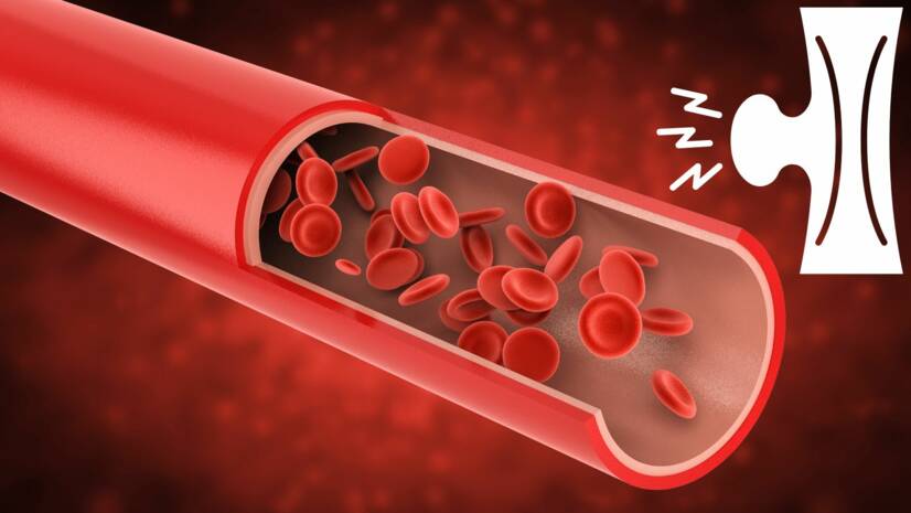
The basis of the difficulties is again the oppression of the surrounding tissues and structures, such as the intestines, kidneys or vertebrae. An aneurysm also disrupts blood flow through the vessel. This in turn predisposes the formation of thrombi - blood clots. Blood clots can cause tissue ischemia in the affected area.
Blood flow through the aneurysm site. Photo source: Getty images
A complication is also the tearing off of the clot and the transfer of this embolus, ie embolization. This is the case, for example, with an aneurysm of the popliteal artery, which is the coronary artery. Embolism in the limb causes ischemia with consequent difficulties. Non-bleeding of the limb is manifested by pain, discoloration (purple, pale, marble to gray), or a decrease in the temperature of the foot to the touch.
Swelling can also be the cause of blood loss from the body's circulation. Anemia, that is, deficiency in blood occurs. With the rupture of the aneurysm, the state of health changes suddenly. The start of intense, unbearable to severe pain, large blood loss with shock. The complication is serious, endangers the health and life of the affected person, and despite urgent surgery, 40-60% of patients die.
In the case of a cerebral aneurysm, the first manifestation of its existence is usually a rupture. This is in most cases due to an increase in blood pressure with a weakened blood vessel wall. There is a bleeding stroke that is very dangerous to human health and life and in the form of SAH / SAK (subarachnoid haemorrhage / bleeding).
Rupture is reported to occur in approximately 45% of cases of cerebral (intracranial) aneurysms. Mortality is reported in up to 44% of these bleeds. And about 10% of those affected die before medical care arrives.
In addition to hypertension, smoking, alcohol, and even pregnancy are also at risk. It is characterized by difficulties such as intense headache , meningeal symptoms, such as meningism , the opposition of the neck, inability to tilt the head and touch the chest with the chin, but also increased sensitivity to noise and light, such as photophobia . Furthermore, vomiting or impaired consciousness is associated , and thus collapse or unconsciousness.
A cerebral aneurysm causes the compression of brain tissue , as the brain is within the skull and it is inflexible. Thus, even an unbroken cerebral edema results in various non-specific difficulties . Such as:
imbalance, gait and coordination
speech disorder
vision problem, visual field loss
memory disorders
impairment of thinking, cognition and processing of information
reduction of concentration - concentration
behavioral changes
fatigue, increased sleepiness
The table lists the different symptoms of an aneurysm and its rupture according to the area of occurrence
Aneurysm site
Symptoms
Cerebral arteries
that is, the Willis circuit (vascular supply to the brain)
intense headache, described as the worst headache
meningism
stiff neck
sensitivity to light
nausea
vomiting strain
vomiting
dizziness
collapse to unconsciousness
tinnitus
visual disturbances as well as double vision
pupil dilation
speech disorders
impaired mobility
sensitivity disorder
Aorta
Thoracic aorta
the most common ascending part of the aorta possible complication:
pericardial tamponade
ischemia to myocardial infarction
chest pain
shortness of breath
impaired swallowing
speech difficulties
in dissecting thoracic aortic aneurysm :
severe to shocking chest pain
it is caused by bleeding between the layers of the vessel wall and their separation
no take of nitroglycerin and analgesics
pain radiates to the back or abdomen
shock
heart failure
as the dissection progresses towards the coronary vessels, myocardial infarction
towards the carotid arteries also an image of a stroke
the course and intensity of the difficulty depends on the site of the dissection and its progress
sudden death
about half of those affected die within 24 hours
Abdominal aorta
75% may be asymptomatic
finding accidentally at another examination
stomach ache
radiation to the back and crosses
nausea
vomiting
weight loss
anorexia
dyspeptic disorders
palpable pulsating formation in the abdomen
CAUTION is missing in obese people
and in the poor it is false positive
renal and urinary tract oppression
hydronephrosis
renal failure
image of renal colic
risk of rupture at a size over 5-6 cm, manifests itself as:
up to 40% first symptom
sudden intense pain
radiation to the back and groin
collapse
hemoperitoneum (blood in the abdomen)
shock of blood loss, rapid heartbeat and shortness of breath
peritoneal irritation
plate - like abdomen
painfully sensitive to touch
Others
risk of thrombosis to embolization
limb bleeding
pain
pulsating tactile feature
in fungal aneurysm also the risk of septic shock
fever
fatigue
increased leukocyte counts
positive finding on blood cultivation
Diagnostics
Diagnosis is based on history and clinical picture . Because a minor aneurysm may have an asymptomatic, asymptomatic course , it is detected accidentally . For example, during a preventive examination or as a by-product of another disease.
Imaging methods such as X-rays, USG (SONO), but also CT and MRI are used to diagnose the aneurysm . Angiography has its limitations in determining the overall extent and is used less frequently. Hemoculture, ie blood collection, is also added to the fungal type.
MRI of the brain. Photo source: Getty images
Some imaging forms have a limit to determine a cerebral aneurysm . However, the basis consists of CT and MRI examinations . Subsequently, CT angiography and DSA , which is digital subtraction angiography , are also selected . What is an invasive method based on CT imaging before and after injection of a contrast agent into the carotid artery. Subsequently, the cerebral circulation is drawn - a cerebral angiogram.
Course
The course is fully dependent on the type of aneurysm, and thus its location and size . Of course, on whether the bulge persists without rupture , or leads to its rupture . When the form is not cracked, it often takes place hidden and asymptomatic. Or, you experience these symptoms.
The subsequent rupture manifests itself in most cases suddenly and with intense difficulties . The same is true of the dissecting form. Early diagnosis and early treatment are therefore often life-saving . In the case of these diseases, preventive examinations and targeted aneurysm screening are of great preventive importance.
How it is treated: Aneurysm
How is an aneurysm treated? Treatment is both conservative and surgical
Baird RJ, Doran ML (August 1964). "The False Aneurysm". Canadian Medical Association Journal. 91: 281–84.
Norwood MG, Lloyd GM, Moore S, Patel N, Panditi S, Sayers RD (April 2004). "The changing face of femoral artery false aneurysms". European Journal of Vascular and Endovascular Surgery. 27 (4): 385–88.
Li JW, Wang SM, Chen XD (August 2004). "Management of femoral artery pseudoaneurysm due to addictive drug injection". Chinese Journal of Traumatology = Zhonghua Chuang Shang Za Zhi. 7 (4): 244–46.
Currie S, Mankad K, Goddard A (January 2011). "Endovascular treatment of intracranial aneurysms: review of current practice". Postgraduate Medical Journal. 87 (1023): 41–50.
Perrin, Michel (17 February 2010). "Venous aneurysms". Servier – Phlebolymphology. Retrieved 14 January 2020.
Azar D, Ohadi D, Rachev A, Eberth JF, Uline MJ, Shazly T (February 2018). "Mechanical and geometrical determinants of wall stress in abdominal aortic aneurysms: A computational study". PLOS ONE. 13 (2): e0192032. Bibcode:2018PLoSO..1392032A. doi:10.1371/journal.pone.0192032. PMC 5798825. PMID 29401512.
"Abdominal Aortic Aneurysms". The Lecturio Medical Concept Library.
Anastasiou I, Katafigiotis I, Pournaras C, Fragkiadis E, Leotsakos I, Mitropoulos D, Constantinides CA (2013). "A Cough Deteriorating Gross Hematuria: A Clinical Sign of a Forthcoming Life-Threatening Rupture of an Intraparenchymal Aneurysm of Renal Artery (Wunderlich's Syndrome)". Case Reports in Vascular Medicine. 2013: 452317.
Avinash P Mural International Journal of Medical Case Reports Vol 3 Iss. 4, pp. 1–4
Christianto B. Lumenta, ed. (2010). Neurosurgery. Heidelberg: Springer. p. 181. ISBN 978-3-540-79564-3.
Lumb, Philip (2014). Critical Care Ultrasound E-Book. p. 56. ISBN 978-0323278171.
Lindholt JS, Juul S, Fasting H, Henneberg EW (April 2005). "Screening for abdominal aortic aneurysms: single centre randomised controlled trial". BMJ. 330 (7494): 750.
Hirsch AT, Haskal ZJ, Hertzer NR, Bakal CW, Creager MA, Halperin JL, et al. (September 2006). "ACC/AHA Guidelines for the Management of Patients with Peripheral Arterial Disease" (PDF). Journal of Vascular and Interventional Radiology. 17 (9): 1383–97, quiz 1398.
Kent KC (November 2014). "Clinical practice. Abdominal aortic aneurysms". The New England Journal of Medicine. 371 (22): 2101–08.
Melissa L Kirkwood. "Iliac artery aneurysm".
Walker BR, Colledge NR, Ralston SH (2010). Davidson's principles and practice of medicine (21st ed.). Edinburgh: Churchill Livingstone/Elsevier. p. 604. ISBN 978-0-7020-3085-7.
Manasco, Hunter. "The Aphasias". Introduction to Neurogenic Communication Disorders. p. 93.
The secondary medical school in Nitra gave me the basis for my career in the field of health and diseases. Thanks to it, I worked for 2 years in the traumatology clinic and outpatient clinic at the Nitra Hospital. Since 2006 I was employed in the emergency medical service, where I stayed until 2017.
I completed my bachelor's degree at the University of Constantine the Philosopher in Nitra in the field of emergency health care. The bachelor's degree allowed me to continue my mission as a paramedic. In the meantime, I got a job at the emergency line 155. I have been working in pre-hospital health care until today.
I had an interest in people, health and even diseases in my childhood, which gave me the prerequisite to pursue this topic in adulthood. Studying and acquiring new information in practice provided me with a great basis for writing professional texts, in the form of articles that can be understood by ordinary people. Thus, my interest in the Health Portal has a solid foundation in years of practice and personal interest. Similarly, I am also interested in healthy eating, nutrition and overall healthy lifestyle. I fill my free time with family and sports.