Erysipelas is defined as an acute streptococcal disease of the skin and subcutaneous connective tissue. It is accompanied by general symptoms.
It is most common on the feet, especially on the shins (90%).
It is less common on the hands, face and neck. However, it can occur in other locations. For example, in the lumbar region in patients with artificial hip joint implants.
Erysipelas = erysipelus = rose.
The disease can affect a patient at any age. The highest incidence is between 60 and 80 years of age. It occurs in both sexes equally.
Erysipelas has a worldwide incidence. Comparing the USA and Europe, for example, the aetiology, clinical picture and treatment are similar in both cases. The incidence is higher in the summer months.
Erysipelas belongs to the group of bacterial skin infections (pyoderma). The most common causative agents of pyoderma are Staphylococcus aureus and group A β-haemolytic streptococci.
Division of gram-positive and gram-negative bacteria. Source: Getty Images
The incidence of infection depends on the following factors:
general condition of the organism - systemic diseases
local skin condition - chronic skin diseases, skin injuries
characteristics of the bacteria - virulence
influence of the external environment - temperature, humidity
Pyodermas can be divided into two groups. The first group consists of primary pyodermas, which include impetigo, furuncle, carbuncle and erysipelas.
The second group are secondary bacterial infections that further complicate pre-existing skin changes. Examples are secondary infections on eczema, ulcers, skin wounds, burns or chickenpox.
The following table gives an overview of pyoderma by causative agent
Golden staphylococcus aureus
Streptococcus pyogenes
Bullous impetigo
Non-cellular impetigo
Dermatitis exfoliativa staphylogenes
Ecthyma
Folliculitis
Erysipelas
Furunkl
Carbuncle
Hidradenitis suppurativa
Causes
The causative agent of erysipelas is Streptococcus pyogenes (beta-hemolytic group A streptococcus).
Streptococci are a diverse group of bacteria:
viridal streptococci in the oral cavity and vagina - part of the physiological flora of mucous membranes
primarily pathogenic - S. pneumoniae, S. pyogenes, S. agalactiae
conditionally pathogenic - S. mutans, S. milleri
The S. pyogenes envelope contains hyaluronic acid. This is indistinguishable from hyaluronic acid in connective tissue. The envelope is weakly immunogenic, allowing the bacteria to escape immune mechanisms.
What diseases are caused by different types of streptococci?
Streptococcus pyogenes causes angina, pharyngitis and skin infections (impetigo, pyoderma, cellulitis, phlegmon). Strains that produce erythrogenic toxin cause erysipelas.
Some strains can cause late sequelae of streptococcal infections (rheumatic fever, acute glomerulonephritis).
The causative agent of erysipelas usually enters the body at a site where the skin barrier is breached. The predilection sites on the shins and face are considered as entry points.
A certain spatial distance between the erysipelas and the site of entry of the pathogen is typical. Examples are the ragades in the interdigital spaces of the lower limbs (entry of the pathogen) and the subsequent erysipelas on the shins.
The nasopharynx is considered the source of the pathogen.
The most common factors contributing to erysipelas include minor injuries, disorders of venous and lymphatic circulation, diabetes mellitus and alcoholism. Prolonged standing or strenuous hiking for several hours may also play a role.
Predisposing factors for first attacks of erysipelas may be local, systemic or social. Social factors include poor hygiene.
In the following table, we list each of the local and overall predisposing factors
The incubation period lasts from a few hours to two days.
The disease starts suddenly with chills and fever (40 °C). Symptoms such as nausea, malaise and headache also appear.
Within a few hours, an asymmetrically localised, intensely red, flat, oedematous lesion appears on the skin. The lesion spreads rapidly into the surrounding area. The lesion is well circumscribed. It protrudes into the surrounding area in typical tongue-like projections.
Patients often experience tingling or mild pain before the redness develops. The redness spreads into the surrounding area at a rate of approximately 2 to 10 cm per day. It may reach large sizes. Regional lymph nodes may be enlarged and painful.
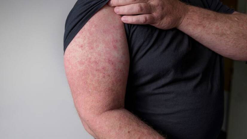
With a more severe course, vesicles, pustules, blisters or small areas of haemorrhagic necrosis may appear.
Example of erysipelas on the leg. Source: Getty Images
The most common symptoms of erysipelas include:
erythema (redness of the skin), which is flat and painful.
swelling
overheating
chills
fever
headache
lymphangitis
lymphadenitis
Tachycardia and hypotension may also occur. These symptoms signal a more severe course of the disease.
We distinguish the following types of erysipelas:
Erysipelas erythematosum - The surface of the lesion is warm and slightly painful to the touch.
Erysipelas vesiculosum et bullosum - In this type, blisters and large vesicles appear on the erythema.
Erysipelas gangraenosum - A necrotic form that occurs with weakened immunity or predisposing factors.
Erysipelas phlegmonosum - Occurs when the process migrates to the subcutaneous connective tissue. Phlegmon to abscesses may form this way.
What is a phlegmon?
A phlegmon is a violent purulent inflammatory process that takes place in the subcutaneous tissue. From there it spreads upwards (to the skin) and downwards (to the tendons and fascia). It is caused by the introduction of infection into the deeper layers after injuries, erysipelas and others.
Erysipelas on the face is very common and dangerous. It starts in the saddle of the nose and spreads in a butterfly pattern. Bilateral swelling of the eyelashes occurs.
Erysipelas can also affect the mucous membranes. Erysipelas of the larynx is very dangerous and can be fatal.
Erysipelas of the vulva is manifested by massive swelling and reddening of the labia minora, which may necrotically disintegrate. Erysipelas of the penis may also be gangrenous.
Diagnostics
Diagnosis is made on the basis of clinical signs (fever, chills, high sedimentation rate, leukocytosis). The basis of diagnosis is the history, clinical picture and laboratory examination.
Laboratory results show leukocytosis. Other inflammatory parameters such as sedimentation rate and C-reactive protein (CRP) are also elevated.
An excellent indicator of streptococcal infection is antistreptolysin O (ASLO).
What is ASLO?
Antistreptolysin O (ASLO) is a protein produced against the antigen streptolysin O. It is part of the cell wall of group A streptococci.
Elevated levels of ASLO alert us that antibodies against streptococci are circulating in our body, but also against our own tissues. The normal level in healthy adults is 200 IU/ml. In children, the level is higher (400 IU/ml).
In practice, we must assess the dynamics of the antibody response. Antibodies to streptolysin appear in the blood approximately 1 to 3 weeks after the outbreak of infection.
Maximum levels usually appear after 3 to 6 weeks. Normal levels are reached within 6 to 12 months.
Differential diagnosis
In some cases, the diagnosis of erysipelas is misdiagnosed. Most commonly, eczema, lymphedema and lipodermatosclerosis are misdiagnosed as erysipelas.
Examples of differential diagnosis are given in the following table
In the normal erythematous form, the temperature begins to drop after a week, the inflammation subsides and the process heals. Erysipelas does not leave immunity. Multiple relapses are frequent. Various erosions and fissures (ragades) are the gateway for the entry of infection.
What are the complications of erysipelas?
Complications occur especially in cases where the disease is treated late. With significant swelling, blisters form. These are filled with serous fluid. Secondary staphylococcal infection leads to their impetiginization.
Blisters are more common in elderly patients with atrophic skin. Abscess is also a local complication.
Rare complications include bacteremia and septicemia (less than 5%). Recurrence of the disease is a frequent complication (15% to 34%). The cause is persistence and untreated risk factors.
Such risk factors include:
swelling
tibial ulcer
onychomycosis
injury
minor skin injuries
overweight
Other complications include:
Myocarditis, endocarditis or pericarditis.
glomerulonephritis
rheumatic joint involvement
metastatic pneumonia
lymphoedema
local tissue devastation
phlebothrombosis
Currently, the above complications are rare. They can occur in children, the elderly and patients with risk factors. The prognosis of the disease is very good with early treatment.
How it is treated: Erysipelas - rose
Treatment of erysipelas, rose: Medications, antibiotics and topical ointments
I graduated from the University of Veterinary Medicine and Pharmacy in Košice. After graduation I stayed working at the University. Until 2019, I was leading practical exercises in the subject of Drug Technology. I was mainly engaged in the preparation of individually prepared medicines in pharmacies. During my time there, I wanted to keep learning. In 2016, I defended my rigorous thesis at the Veterinary and Pharmaceutical University in Brno. By completing the rigorous procedure I gained knowledge in the field of laws related to pharmacy and medicine. In 2017, I completed my MHA studies at St. Elizabeth's College of Health and Social Work. By studying, I gained knowledge in the field of public health. In 2021 I completed my PhD studies (specialization in parasitic diseases) at the University of Veterinary Medicine and Pharmacy in Košice. The desire to learn new things and new knowledge led me to become a co-founder of the dermatovenerology outpatient clinic. And why can I give you information from the medical and pharmaceutical field? I like to learn and get to know new things. I have focused all my studies and scientific research on skin diseases. I have also gained knowledge in the field of parasitic diseases. Writing is one of my hobbies and a kind of relaxation.