Thrombophlebitis of superficial veins: What is phlebitis and why does it occur?

Thrombophlebitis of superficial veins: What is phlebitis and why does it occur?
Photo source: Getty images

Superficial thrombophlebitis is an inflammatory disease of the superficial veins. It mainly involves the veins of the lower extremities.

Most common symptoms

Show more symptoms

Characteristics

Thrombophlebitis, more precisely superficial thrombophlebitis, is an inflammatory disease of the superficial veins. It largely affects the veins of the lower extremities.

Superficial phlebitis ranks among relatively common diseases.

Inflammation of the veins is classified as vasculitis.

Vasculitis = inflammation of blood vessels.
Vas = Latin for "blood vessel"
Phlebitis = phlebos + itis. 
Phlebos = Latin for "vein". 
... itis = the ending means "inflammation". 

Thrombo = relates to the presence of a thrombus, i.e. a blood clot. 
Thrombo-phleb-itis - inflammation of a vein with the presence of a blood clot. 

A brief overview of blood vessels

In general and abbreviated terms, blood vessels are divided into arteries and veins. 

  1. arteries are filled with oxygen-rich blood, i.e. oxygenated blood.
  2. veins, on the contrary, blood with low oxygen content, i.e. deoxygenated blood, flows in those

However, this is only lay awareness. More precisely, it is a division according to:

  1. arteries = take blood FROM the heart
  2. veins = take blood TO the heart

For example:

The pulmonary artery, the truncus pulmonalis, divides into the pulmonary arteries and in doing so conducts deoxygenated blood from the right ventricle of the heart to the lungs.

4 pulmonary veins carry oxygenated blood from the lungs to the left atrium of the heart. From there, the oxygen-enriched blood is expelled into the left ventricle of the heart and then through the aorta to the entire human body.

Artery:
From Greek "aer" = air, 
térein = contain. 

Further, arteries and veins differ slightly in their structure.

The walls of the veins do not contain as many muscle cells. At the same time, blood flows in them, which is the result of the complex interplay of the cardiovascular system. 

In the lower limbs, the situation is made more difficult by the force of gravity. The latter pulls the blood downwards. 

Small valves are therefore formed in the veins of the lower limbs, which, like in the heart, direct the flow of blood.

The return of blood from the lower limbs to the upper half of the body is aided by the deposition of the deep veins. The deep veins are located in the close course of the arteries.

So...

The muscles of the lower limbs are also helpful in blood flow by moving them. They form a so-called muscle pump, like the muscles of the heart.

Arteries + muscles = squeezing the veins and pushing the blood up towards the heart.

Therefore, regular movement is important in the circulation of blood throughout the body, not just in the lower limbs, legs.

Surely you remember...

One difference we were taught back in elementary school...

There is high blood pressure in the arteries. Therefore, when they are injured, blood spurts. And for oxygen saturation is also light.

Conversely...

In the veins, the blood pressure is low, when damaged, the blood does not spurt, but only flows out freely. And the blood is darker because it does not contain oxygen.

There are veins in which the blood pressure is lower than the atmospheric pressure around the body. Injury of large veins, such as the veins of the thigh or neck, is dangerous. There is a risk of aspiration of ambient air and air embolism

And do you know why blood from a lower limb varix, or varicose vein, can squirt?

Blood is pushed against gravity in the legs and its flow is directed by valves. And therefore, even from a small defect of a varicose vein, blood can spurt.

The superficial veins drain into the deep veins.

The deep venous system forms a kind of collecting large veins. Smaller veins and also superficial veins drain into these. These collect blood from the periphery of the human body.

In the lower limbs it is equally arranged. The superficial veins collect blood from the skin, muscles and other structures of the lower limb and then drain into the large deep veins.

The veins of the lower limbs are divided into three types:

  1. deep vein system - in depth
  2. surface vein system - on the surface
  3. system of connecting veins - connects both systems

Thrombosis is...

Thrombosis is a condition of blood clotting in the blood vessels and in the heart. 

Blood clotting is a vital act to prevent bleeding, for example, even in the case of trauma. Without haemocoagulation, as the act of blood clotting is called, we could bleed to death from even a minor injury.

Haemocoagulation is to stop bleeding = haemostasis.

However, hemocoagulation also includes fibrinolysis.

Fibrinolysis, on the other hand, is from hemocoagulation, the act of dissolving blood clots.

These two processes in the human organism take place in mutual harmony and balance.

With pathological disruption of the balance of blood clotting and dissolution of blood clots, a condition may arise:

  1. excessive blood clotting = thrombosis
  2. excessive bleeding = bleeding conditions

The condition of excessive blood clotting can have a diverse basis. The risk is mainly a partial or complete restriction of blood flow through the affected blood vessel or the detachment of a blood clot.

When part or all of the thrombus breaks away from the blood vessel wall and travels through the blood vessels, it is referred to as an embolus.

When a blood vessel in another part of the body becomes blocked, this condition is referred to as embolisation.

In thrombosis, for example, the following occurs: 
Heart muscle ischemia to heart muscle infarction.
Examples of embolisation:
Pulmonary embolism and stroke.

In addition to blood, it can also embolize another foreign object that has entered the blood vessels. Examples are fatty or tumour tissue, air or amniotic fluid, as well as a piece of severed vascular catheter.

Want to know more about thrombophlebitis?
Why does it arise?
How does it manifest?
Or briefly about her treatment?
Read on for information in the article...

Why superficial thrombophlebitis?

Phlebitis is a general term for two types of inflammation of the veins. Namely, for inflammation of the superficial or deep veins.

  1. inflammation of superficial veins = superficial thrombophlebitis (ST)
  2. deep vein inflammation = phlebothrombosis - deep vein thrombosis (DVT)

It has been reported that the inflammation of the vein can occur without associated thrombosis, i.e. without the deposition of a blood clot on the inflamed vascular wall. 

However, in the vast majority of cases, a blood clot also forms when the vein is inflamed.

The latter is the result of the response of the hemocoagulation cascade to a violation of the vascular wall. When a vein becomes inflamed, the blood vessel is disrupted.

Platelets and other components of haemocoagulation are deployed at this site. A thrombus is formed.

Therefore, inflammation of the veins is associated with thrombosis.

There are several differences between phlebothrombosis, i.e. inflammation of the deep veins, and superficial thrombophlebitis.

In deep vein thrombosis, pulmonary embolism is a common complication.

For more information, read the article Deep vein thrombosis.

In the past, it was reported that superficial venous inflammation is a benign, i.e. not serious disease.

However, this is not entirely true. It has been found...

Although it is an inflammation of a superficial vein, in a large number of cases thrombophlebitis develops into deep vein thrombosis.

Or...

Phlebothrombosis and thrombophlebitis occur together.

Therefore, even with superficial venous inflammation, there is some risk of complications. The most serious and life-threatening is embolisation to the lungs. The second condition is the development of thromboembolic disease.

It is estimated that:
ST and DVT occur simultaneously in 6-53% of cases.
ST develops into DVT in 0-33% of cases.

Both of these states have a common basis, and this is described by Virchow's triad:

  1. stasis of blood in the veins
  2. damage to the vascular wall
  3. blood coagulation disorder

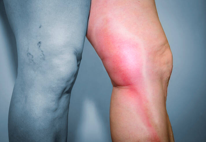
Definition of superficial thrombophlebitis

Superficial thrombophlebitis is an inflammation of the vessel wall, on which thrombus formation sets in. In this case, the thrombus is firmly attached to the vessel wall. Which reduces the risk of embolization.

It mainly affects the blood vessels of the limbs, especially the lower limbs.

Tombophlebitis is further subdivided according to which vein it affects, into:

  1. superficial thrombophlebitis affecting the varicose vein - varicocele, also known as varicose superficial thrombophlebitis
  2. superficial thrombophlebitis affecting a healthy vein

Another form is the division into primary and secondary:

  1. primary - originate and interfere in the vein itself
  2. secondary - in association with another disease

There is also a division according to whether it has a known or unknown cause:

  • unknown cause
    • idiopatická tromboflebitis saltans sive migrans
    • Mondor's disease
    • epidemic thrombophlebitis - tropical form
    • granulomatous giant cell thrombophlebitis
  • known cause
    • arises as a result of thrombosis
    • thrombophlebitis in Behcet's disease
    • thrombophlebitis in sarcoidosis
    • thrombophlebitis in oncological disease
    • thrombophlebitis in infectious disease

And at this point we are already touching on the topic of the causes of inflammation of the veins.

Causes

What is the cause of thrombophlebitis, you ask?

In some cases, it is not possible to determine the exact cause of occurrence.

Even it is difficult to determine whether the first inflammation, or vice versa, was the first thrombosis?

There is a multifactorial basis behind the damaged vessel. This was first described by the German physician Rudolf Ludwig Carl Virchow in 1856.

The Virchow's triad scores the risks of developing inflammation on three points:

  1. slowing of blood flow - stasis, or turbulence in the flow, can be caused by:
    • vascular aneurysm - aneurysm
    • varicose veins
    • heart failure
    • heart valve defect
    • limitation of limb mobility in case of injury, after surgery, also during hospitalization
    • travelling for too long
    • mechanical oppression by swelling, tumors, also in obesity
    • other neurological and internal medicine diseases
  2. imbalance of blood clotting and fibrinolysis
    • susceptibility to increased blood clotting, i.e. hypercoagulability
    • genetic factor
    • systemic diseases, inflammation of blood vessels
    • tumour - malignancy
    • infection
    • septic thrombophlebitis
    • some medicines - hormonal contraceptives
    • smoking
    • pregnancy and the postpartum period form an increased susceptibility for blood clotting
  3. damaged vein wall
    • mechanical damage to a blood vessel
    • injuries, blunt force trauma
    • surgeries
    • varicose veins
    • administration of drugs into a vein, especially central venous catheters
    • some medicines given into a vein, as in chemotherapy
    • drugs injected into a vein

Deep vein thrombosis and superficial thrombophlebitis have common risk factors involved.

Risk factors include:

  • varicose veins
  • long limb immobilization, bed immobilization
    • over 72 hours
  • long journeys - over 4 hours
  • sedentary lifestyle, sedentary occupation
  • dehydration
  • smoking
  • injury (such as injury to the head, spine, spinal cord, long bones)
    • also a direct blow to the surface of the blood vessel
  • intravenous catheters - i.v. catheters, mainly central
  • obesity
  • over 60 years of age
  • pregnancy and postpartum period
  • taking hormonal contraceptives
  • genetic predisposition and changes in coagulation factors
  • oncological diseases - malignant tumours
  • past history of venous inflammation
  • other diseases
    • ischaemic heart disease
    • ischaemic disease of the lower limbs
    • right heart failure
    • liver
    • kidneys

The cause of inflammation of the veins in many cases is mechanical damage, an example is a direct blunt blow to the surface of the vein. The situation is aggravated by the presence and trauma in the area of the varicose vein.

Another type is chemical irritation when an inappropriate or undiluted solution is injected into a vein. An example would be chemotherapeutic drugs.

Inflammation of the vein can also arise from the entry of infection - microorganisms when the vein is injured, but also when a medicine or drug is injected into the vein.

Damage to the inner lining of the blood vessel, the endothelium, triggers the blood clot formation process. Subsequent thrombosis is equally risky for the development of the overall picture of thrombophlebitis.

However, in the overwhelming majority, the varicose vein is behind the formation of thrombophlebitis. This is the cause of varicose veins in up to 90% of cases. That is a case if varicophlebitis.

The superficial inflammation of the varicose vein is preceded by a slowing of blood flow and dilation of the vessel with changes in the structure of the vessel wall. Subsequently, as well as the most common provoker of this form, it is followed by an injury to the dilated vein.

Phlebitis of a healthy vein is mainly considered in conditions after surgery, trauma, and also in oncological, hematological and systemic diseases.

And, as already mentioned, in some cases it proceeds in an idiopathic form, when the exact cause is not found.

Distribution of superficial thrombophlebitis

Form of thrombophlebitis Description
Varicophlebitis
  • thrombophlebitis varicosa
  • varicose vein inflammation
  • affects varicose veins, i.e. enlargements
  • 88% of PT sufferers have varicose veins of the lower limbs
  • occurs in 10-20% of varicose vein sufferers
  • mostly affects a single varicose node
  • from there it spreads further
  • the cause is mainly mechanical damage
    • blunt trauma or pressure on a vein
  • in immobile people
  • after surgery
  • after an injury
  • in women after childbirth and during the postnatal period
  • in oncological disease
  • possible spread to the deep venous system
Phlebitis of healthy veins   
  • non-varicose superficial thrombophlebitis
  • also known as thrombophlebitis superficialis
  • 12% of patients with PT do not have varicose veins of the lower limbs
  • as a result of:
    • injury
    • venous catheter insertion
    • infection
    • chemical damage
    • tumour
    • without known cause
    • and others, such as:
  • Thrombophlebitis venae saphenae parvaeseu magnae
  • may arise without a known cause
  • in oncological diseases
  • in blood diseases
  • after surgery 
  • after childbirth
  • after injury
  • affects a different section of the vein
  • spreads centrally, i.e. towards the large veins
  • Thrombophlebitis saltans / migrans
  • inflammation of a blood vessel in the range of 1 to 5 cm
  • travels - migrates through the body, appearing and disappearing in several places in the vein
  • jumps from one place of the organism to another
  • multiple stages of inflammation at the same time, acute and already subsiding at the same time in several parts of the body
  • after the acute inflammation disappears, a hyperpigmented band remains at the site of inflammation
  • in chronic inflammatory diseases
  • in oncological diseases
  • Mondor's disease
    • fibrotic changes of blood vessels in the upper limbs, chest and upper abdomen 
    • rare form
    • particularly in women
    • unclear cause
  • Trousseau syndrome
    • in patients with oncological disease
    • increased risk of pulmonary embolism and death
  • Thrombophlebitis venae basilicae - cephalicae
    • superficial inflammation of the veins of the upper limb
    • mainly after the injection of medication into the vein
      • application of undiluted high percentage glucose
  • Catheter thrombophlebitis
    • after venous catheter insertion
    • the risk is increased by the time the catheter is left in the vein
      • over 48 - 72 hours
    • the risk of microorganisms entering the vein increases with time
      • these are mainly bacteria such as golden staphylococcus or enterococci
    • important to rotate the puncture site, do not leave the catheter in one place for too long
    • with inflammation necessary to do culture and sensitivity
  • Thrombophlebitis purulenta
    • it is purulent thrombophlebitis
    • inflammation spreads from a purulent deposit, such as an abscess or phlegmon
    • the risk is the spread of inflammation to the deep venous system
  • Lemierre's syndrome
    • septic form of inflammation of the jugular vein - v. jugularis interna
    • for infection in the upper respiratory tract
    • risk of spread to the lungs and liver
    • caused by the bacterium Fusobacterium necrophorum

Learn more in the following articles: 
Chronic venous insufficiency
Chronic venous disease

Symptoms

The manifestation of superficial thrombophlebitis is a group of difficulties, these are the same in the case of inflammation of a healthy vein or in the case of varicose vein involvement.

In the case of varicella inflammation, the local involvement tends to be more pronounced.

Characteristic symptoms of thrombophlebitis are:

  • redness during inflammation of the vein
  • pain and tenderness at the site of inflammation 
  • the vein is rigid to the touch and movable against the substrate
  • swelling at the site in case of mild course
  • transiently present hyperpigmentation area - brown dark pigmented streak
  • stiffening persists for several weeks to months, after the thrombus has dissolved in the vein
  • increased skin temperature during inflammation

Swelling of a larger extent and of the whole limb is not present in superficial thrombophlebitis.

Outside of the general manifestations, there may be associated difficulties that mark a specific type of inflammation, as shown in the table. An example is the migratory form, in which the inflammation and hardening of the vein moves or jumps to different sites.

Diagnostics

Diagnosis of thrombophlebitis itself is not difficult. Clinical examination is sufficient. Of course, medical history is also important.

Superficial inflammation often runs along with deep venous thrombosis.

Therefore, it is important to do a duplex sonographic examination. It is performed on both lower limbs. It should be done as soon as possible after the diagnosis of superficial thrombophlebitis.

This examination is all the more significant the more the larger veins are affected. Such as the vena saphena magna and vena saphena parva.

Vena saphena magna = the great saphenous vein. 
Vena saphena parva = the small saphenous vein. 
Veins running from the shin to the femoral veins. 

Inflammation can extend further than the inflammatory skin changes seen. At this time, the risk of deep vein involvement is also increased.

We look for the cause, such as also Bürger's or Behcet's disease.

Particular caution should be taken with recurrent inflammation.
We look for risk factors, genetic predisposition and congenital thrombophilia and associated diseases

Course

Superficial thrombophlebitis typically takes place by painful hardening of the vein. It is also sensitive to touch.

In the course of an inflamed vein, the skin is reddened, with increased temperature and swollen.

Redness + pain + swelling = inflammation of the vein wall and surrounding area.

The inflammation lasts for several days and then subsides. Inflammation of a small extent with a mild course may not manifest itself with significant discomfort and the vein is sensitive mainly to touch.

The acute stage and local symptoms subside within 7 to 14 days.

Redness of the skin also subsides. However, the skin may be temporarily darker in that area. This is hyperpigmentation - a brown area along the vein.

Painless hardening of the vein is present for several weeks to months. And, therefore, for a period of time until the blood clot dissolves

In thrombophlebitis, the blood clot is firmly adhered to the vessel wall. This reduces the risk of thrombus detachment and embolization.

However, even in the case of this form of inflammation of the vein there is a risky complication. This is the spread of inflammation from the superficial vein to the deep venous system.

Consequently, in the present case, the higher the deep vein thrombosis occurred, the higher the risk of embolization.

As already mentioned, there are specific forms of phlebitis, which also have a specific course.

An increase in body temperature is not symptomatic of superficial thrombophlebitis.
Then it is necessary to think about another disease.

Increases in body temperature and general manifestations may be present with thrombophlebitis of greater extent and closer to the larger veins. Pain on movement at the site of the affected limb is also associated.

How it is treated: Thrombophlebitis of superficial veins

Treatment of thrombophlebitis of superficial veins: Medication, compression and physical therapy

Show more
fshare on Facebook

Interesting resources

  • "Thrombophlebitis: MedlinePlus Medical Encyclopedia". medlineplus.gov. Retrieved 23 September 2016.
  • Torpy JM, Burke AE, Glass RM (July 2006). "JAMA patient page. Thrombophlebitis". JAMA296 (4): 468. doi:10.1001/jama.296.4.468. PMID 16868304.
  • Jinna, Sruthi; Khoury, John (2020). "Migratory Thrombophlebitis". StatPearls. StatPearls Publishing. PMID 31613482.
  • "Thrombophlebitis Clinical Presentation: History, Physical Examination, Causes". emedicine.medscape.com. Retrieved 23 October 2016.
  • Raval, P. (1 January 2014). "Thrombophlebitis☆". Reference Module in Biomedical Sciences. Elsevier. doi:10.1016/B978-0-12-801238-3.05368-X. ISBN 9780128012383. Retrieved 2 January 2021.
  • Philbrick, John T.; Shumate, Rebecca; Siadaty, Mir S.; Becker, Daniel M. (23 October 2016). "Air Travel and Venous Thromboembolism: A Systematic Review". Journal of General Internal Medicine22 (1): 107–114. doi:10.1007/s11606-006-0016-0. ISSN 0884-8734. PMC 1824715. PMID 17351849.
  • "Superficial Thrombophlebitis: Background, Pathophysiology, Etiology". eMedicine. Medscape. 12 July 2016. Retrieved 23 October 2016.
  • Varki, Ajit (15 September 2007). "Trousseau's syndrome: multiple definitions and multiple mechanisms". Blood110 (6): 1723–1729. doi:10.1182/blood-2006-10-053736. ISSN 0006-4971. PMC 1976377. PMID 17496204.
  • Tamparo, Carol D. (2016). Diseases of the Human Body. F.A. Davis. p. 292. ISBN 9780803657915. Retrieved 23 October 2016.
  • Saultz A, Mathews EL, Saultz JW, Judkins D. Clinical inquiries. Does hypercoagulopathy testing benefit patients with DVT?. J Fam Pract. 2010 May. 59(5):291-4.
  • Theodoro D, Blaivas M, Duggal S, Snyder G, Lucas M. Real-time B-mode ultrasound in the ED saves time in the diagnosis of deep vein thrombosis (DVT). Am J Emerg Med. 2004 May. 22(3):197-200.
  • Blaivas M, Lambert MJ, Harwood RA, Wood JP, Konicki J. Lower-extremity Doppler for deep venous thrombosis--can emergency physicians be accurate and fast?. Acad Emerg Med. 2000 Feb. 7(2):120-6.
  • Nunn KP, Thompson PK. Towards evidence based emergency medicine: best BETs from the Manchester Royal Infirmary. Using the ultrasound compression test for deep vein thrombosis will not precipitate a thromboembolic event. Emerg Med J. 2007 Jul. 24(7):494-5.
  • Crisp JG, Lovato LM, Jang TB. Compression ultrasonography of the lower extremity with portable vascular ultrasonography can accurately detect deep venous thrombosis in the emergency department. Ann Emerg Med. 2010 Dec. 56(6):601-10.
  • Pomero F, Dentali F, Borretta V, Bonzini M, Melchio R, Douketis JD. Accuracy of emergency physician-performed ultrasonography in the diagnosis of deep-vein thrombosis: a systematic review and meta-analysis. Thromb Haemost. 2013 Jan. 109(1):137-45.
  • Blanco P, Volpicelli G. Common pitfalls in point-of-care ultrasound: a practical guide for emergency and critical care physicians. Crit Ultrasound J. 2016 Dec. 8 (1):15.
  • Mulcare MR, Lee RW, Pologe JI, Clark S, Borda T, Sohn Y, et al. Interrater reliability of emergency physician-performed ultrasonography for diagnosing femoral, popliteal, and great saphenous vein thromboses compared to the criterion standard study by radiology. J Clin Ultrasound. 2016 Jul 8. 44 (6):360-7.