Pityriasis versicolor is otherwise also called tinea versicolor. It is defined as a superficial, non-inflammatory or almost non-inflammatory disease. It belongs to the superficial mycoses. These arise due to infection with parasitic fungi.
It accounts for approximately 20% of all mycotic infections.
Superficial cutaneous mycoses are divided according to their aetiology into:
The main causative agent of the disease is the yeast Malassezia furfur.
In 1846, E. Eichstedt was the first to describe the finding of mycotic elements on deposits of the skin disease pityriasis versicolor. The name Microsporon furfur was not used for these microorganisms until 1853.
The name Malassezia furfur was first used by the Frenchman Henri Baillon in 1889.
The disease mainly affects post-pubertal adolescents or young adults.
It is thought to be caused by increased sebum production in these ages. The yeast thus has an excellent breeding ground for its existence.
The disease occurs mainly during the summer months. Factors that cause the disease to occur during the summer months include higher temperatures, sweating, wearing unbreathable clothing and shoes, and sports activities.
The incidence of the disease is highest in subtropical and tropical climates. In tropical countries, the incidence of the disease is up to 50%.
In northern regions such as Sweden, only 1% prevalence is recorded.
It is mainly caused by insufficient evaporation through the skin due to synthetic clothing and poor hygiene.
It affects both sexes equally. It is a low-infectious disease. A person can become infected either by direct contact with the skin of a sick person or by transmission through clothing.
Causes
The causative agent of the disease is the lipophilic yeast Malassezia furfur. This yeast is a common component of the skin surface of healthy skin. It feeds on sebum.
In people who suffer from increased sweating or work in environments with elevated temperatures, the yeast can multiply and cause disease.
This is what Malassezia furfur looks like. Source: Getty Images
Did you know that...
Most species of malassezia are part of the normal microflora of human and animal skin. On average, we can identify 2-3 different species of malassezia from the surface of one area of human skin.
Predisposing factors include:
excessive sweating (hyperhidrosis)
occlusive clothing
seborrhoea
immune disorders
The influence of heredity has also been considered. Sunscreens and oils may have some influence on the development of the disease. The lipids they contain serve as a substrate for the multiplication of the yeast Malassezia furfur.
Lipophilicity is a characteristic feature of all Malassezia furfur species. Almost all Malassezia species are dependent on external lipid intake. Their cells are unable to produce saturated fatty acids.
These fatty acids are essential for the life of the yeast. They are necessary for the formation of the cell wall. A rich source of fatty substances is sebum and human skin. Skin is a suitable natural environment for the growth of this yeast.
Malassezia yeasts produce several metabolites that play an important role in their interaction with the host organism.
For example, blockage of a key enzyme (tyrosinase) of melanin production leads to reduced melanin production in the host melanocytes and the formation of hypopigmented patches on the skin.
Did you know that...
In general, regardless of the type of lipophilic yeast, approximately 80% to 100% of healthy adults are colonized with malassezia.
Pityriasis versicolor can be a manifestation of intense sweating in vegetative dystonia, hyperthyroidism, tuberculosis, and in HIV-induced infection.
Symptoms
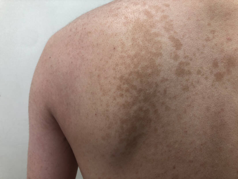
The appearance of the symptoms of the disease can vary. Hence the name versicolor = variegated. Light brown spots appear on unpigmented skin. White spots appear on pigmented skin.
The disease manifests itself as sharply demarcated spots. At first, they are the size of a lens. Later, coffee-white to dirty yellow or brown spots appear. They have a map-like structure.
The typical manifestation of the disease is that the lesions peel off. This symptom is an important diagnostic sign. It distinguishes other non-peeling pigmentary disorders (vitiligo).
Symptoms of the disease on the back and shoulders. Source: Getty Images
In the winter months, the lesions are darker (brownish). In the summer months, especially after sun exposure, they are lighter than the surrounding skin or white.
The predilection area is the mid-chest and back. These areas contain a large number of sebaceous glands. From these areas the manifestations spread to the sides of the trunk.
Sometimes the symptoms of the disease may appear in the navel area, on the inner sides of the thighs and on the shoulders.
Diagnostics
Diagnosis of the disease is based on the typical clinical picture, course and scaling. For the diagnosis, the evidence of fungi in the native preparation of scraped scales is decisive.
How do the lesions peel?
In pityriasis versicolor, the lesions peel off gently. This fine scaling is particularly pronounced when the scales are scraped off with a wooden spatula. This is a relatively simple diagnostic manifestation that distinguishes other pigmentary diseases that are not scaly.
Identification of the causative agent is an important aid in differential diagnosis and in making an accurate etiologic diagnosis. It is also the basis for determining effective treatment.
Hair, scales, blister covers and nail fragments are examined microscopically. Culture examination is a necessary complement to microscopic examination. Histopathological examination of tissue is used especially in deep mycoses.
The course of the disease is chronic. Symptoms initially appear mainly on the flanks, trunk, chest or around the waist.
Tiny spots form at the follicle outlets, approximately 1 cm in size, which may merge into large areas.
The colour of the deposits varies from person to person. Even in the same person, the deposits may not be the same colour. The most characteristic colour is pale yellow. Sometimes it may have a reddish tinge.
Comparison of normal skin and skin with signs of disease. Source: Getty Images
How it is treated: Pityriasis versicolor
Treatment of pityriasis versicolor: Drugs, antifungals and topical treatment
prolekare.cz - Knowledge about the life and activity of lipophilic yeasts of the genusMalassezia from dermatologists' outpatient clinicsI. Cycle of morphological states of lipophilic yeasts on the skin of pityriasis versicolor, B. Ernest; N. Boháčová
researchgate.net - Pharmacist's intervention in prevention and treatment of selected skin diseases, PharmDr. Lucia Masaryková, Assoc. MUDr. RNDr. Magdaléna Fulmeková, CSc. and PharmDr. Ľubica Lehocká, PhD.
ncbi.nlm.nih.gov - Tinea Versicolor, Mehdi Karray; William P. McKinney
I graduated from the University of Veterinary Medicine and Pharmacy in Košice. After graduation I stayed working at the University. Until 2019, I was leading practical exercises in the subject of Drug Technology. I was mainly engaged in the preparation of individually prepared medicines in pharmacies. During my time there, I wanted to keep learning. In 2016, I defended my rigorous thesis at the Veterinary and Pharmaceutical University in Brno. By completing the rigorous procedure I gained knowledge in the field of laws related to pharmacy and medicine. In 2017, I completed my MHA studies at St. Elizabeth's College of Health and Social Work. By studying, I gained knowledge in the field of public health. In 2021 I completed my PhD studies (specialization in parasitic diseases) at the University of Veterinary Medicine and Pharmacy in Košice. The desire to learn new things and new knowledge led me to become a co-founder of the dermatovenerology outpatient clinic. And why can I give you information from the medical and pharmaceutical field? I like to learn and get to know new things. I have focused all my studies and scientific research on skin diseases. I have also gained knowledge in the field of parasitic diseases. Writing is one of my hobbies and a kind of relaxation.