- Preventive dentistry: Limeback Hardy, collective
- Dentistry for non-dentists: Mazánek Jiří, kolektiv
- cspzl.dent.cz - Leukoplakia of oral mucosa: Riznič M., Konečná A., Ďurovič E.
- clevelandclinic.org - Leukoplakia
- nhs.uk - Leukoplakia
- healthline.com - What are the symptoms of leukoplakia?
- mayoclinic.org - Leukoplakia
Leukoplakia: white spots in the mouth? What are the risks?
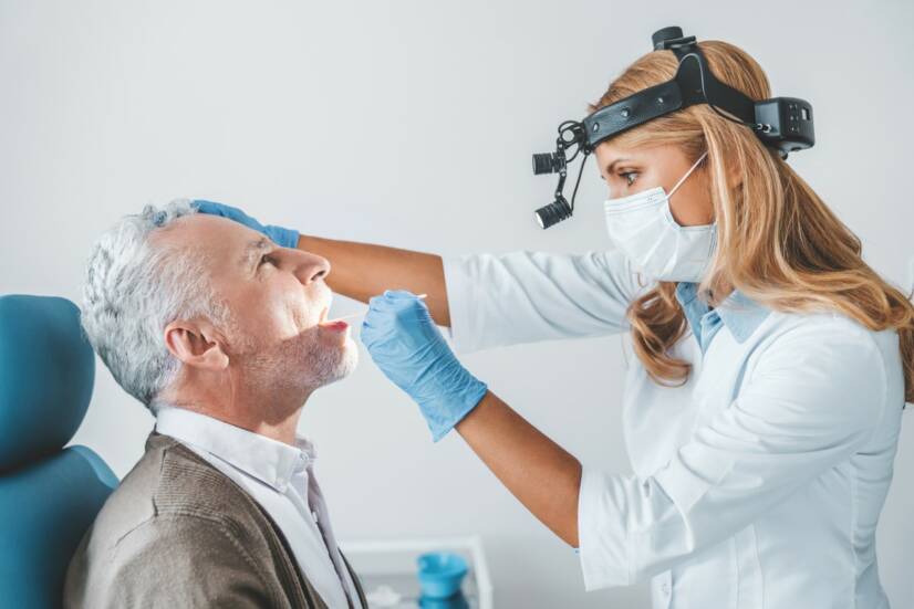
Photo source: Getty images
Leukoplakia is a skin disease. It manifests itself on the mucous membrane in the form of white plaques, which most often occur on the mucous membrane of the oral cavity. Sometimes these spots occur elsewhere on the mucous membrane.
Characteristics
Leukoplakia occurs on the mucous membranes of the oral cavity as a white area that cannot be wiped off and is five millimetres in diameter.
The mild form of leukoplakia is usually harmless and often resolves itself within a few weeks.
The highest incidence of leukoplakia is in Asian countries.
Most types are non-cancerous but require proper diagnosis and monitoring.
Some types of white and red spots may indicate potential cancer.
They occur in middle and older age, more commonly in men. Only 1% are in younger patients under 30 years of age.
Leukoplakia can develop into squamous cell carcinoma, which belongs to the group of skin tumours. It develops into cancer in about 3 to 17% of people over a 15-year period.
The likelihood of developing cancer depends on a number of factors such as the size, shape and form of the abnormal cells forming the leukoplakia.
Leukoplakia is divided into two main types
The homogeneous type is in most cases a white thin patch of uniform colour. Its surface may be smooth, wrinkled or cornified.
The non-homogeneous type is a white or white-red patch. Its shape is irregular. It may be flat but also convex, raised and with projections.
The inhomogeneous form is more likely to become cancerous.
Causes
In most cases, the cause of the occurrence is not precisely known.
Hyperkeratosis is the excessive production of keratin, thickening and excessive keratinisation of the mucous membrane.
Leukoplakia is classified according to the cause of its occurrence
- leukoplakia of unknown origin
- due to smoking, chewing tobacco
- as a result of thermal, chemical, mechanical and other irritation, prolonged use of alcohol, friction of the mucosal surface or tongue against broken, sharp teeth, ill-fitting dentures
- as a result of inflammation caused by Candida albicans
- as a result of anaemia and in immunocompromised persons
Prevention.
- Proper and thorough oral hygiene, care of teeth and gums
- Regular visits to the dentist for preventive check-ups
- When wearing dentures, ensure that the dentures fit well
- A balanced diet with plenty of fresh fruit and vegetables
- Limit smoking, alcohol and chewing tobacco
Read:
Symptoms
Leukoplakia occurs in the oral cavity on the gums, inside the cheeks, at the base of the mouth, under the tongue, on the tongue. It can also occur on other mucous membranes of the body, for example, in the vagina.
It is most often manifested by white patches caused by a defect in the cornification of the mucous membrane, which are thickened and cannot be wiped or scraped off.
In most cases, they are not painful, do not cause any complications and go unnoticed for a long time.
What does leukoplakia look like?
- The spots on the mucous membrane are white, grey or grey-blue in colour and cannot be wiped off.
- Their shape is irregular, flat. Some have a raised surface.
- They are hardened or thickened in places.
- The hairy appearance is typical of hairy leukoplakia.
- Sometimes they are with raised red spots that may develop into cancerous changes.
According to the symptoms, disorders of cornification of the mucous membrane are recognized on:
- Preleukoplakia
- Leukoplakia homogeneous (leukoplakia simplex)
- Leukoplakia mottled (inhomogeneous)
- Erythroplakia (erosive, nodular or verrucous leukoplakia)
Preleukoplakia
Preleukoplakia is manifested by patches that are not completely white. They may also be grey to grey in colour with indistinct margins blending with the colour of the mucosa.
Many people do not even know about this problem because it does not show any symptoms.
This type belongs to the mild reactions of the oral mucosa.
If the cause is detected and treated, this problem will disappear.
Homogenous leukoplakia
Manifested by a horny plaque that is palpable and visible. It may occur without whitish discoloration, but more often a whitish discoloration of the mucosa is associated.
Sometimes the plaque is convex above the level of the mucosa or there are multiple plaques of small size with irregular margins.
The convex type has a white or greyish-yellow colour.
If the plaque is larger, it is more likely to be thickened. This will cause later development of fissures.
In the case of frequent fissures, growths may appear on the nipples. These are called leukoplakia verrucosa.
Verrucous leukoplakia
Verrucous leukoplakia (leukoplakia verrucosa) is a rare but aggressive form of leukoplakia with a high recurrence rate.
Its occurrence is associated with Epstein-Barr virus (EBV).
The foci of white plaques are raised, hard and rough. They may contain deep grooves from which bleeding occurs.
Hairy leukoplakia
Oral hairy leukoplakia is also associated with Epstein-Barr virus (EBV).
It can develop in people with weakened immune systems and in people with HIV/AIDS.
It is manifested by white hairy patches. It is most common on the tongue and can occur in other areas of the oral cavity. This type does not become cancerous.
Speckled leukoplakia
When areas with mucosal thickening are affected, mucosal defects or ulcers of varying depth are formed.
These leukoplakia tend to be both yellow and yellow-brown.
They occur more frequently in smokers on the lower lip or tongue in combination with peeling of the lip.
Erythroplakia
A circumscribed patch of red colour.
This type of leukoplakia has a velvety smooth surface.
However, it can also appear as irregular patches with a red, granular surface on which white or yellow dots are visible.
This type is also called granular erythroplakia.
How does a person with leukoplakia feel?
Subjective discomfort is individual and depends mainly on the form of leukoplakia.
Symptoms are influenced by the size and location of the leukoplakia in the oral cavity.
Preleukoplakia does not cause any complications or symptoms. Sometimes painful peeling from the affected mucosa may occur, especially in smokers.
Homogenous leukoplakia may manifest itself by the sensation of a foreign body on the mucous membrane of the cheeks. If it occurs on the tongue, taste perception may be disturbed.
Speckled leukoplakia and erythroplakia are manifested by burning of the mucous membranes, often preceded by a short sharp pain. A pinching and burning sensation may be induced by the consumption of acidic foods.
In leukoplakia verucosa (verrucous form), small warty formations form on the mucous membrane. They may cause an increasing sensation of foreign body. When the tongue is affected, there is a loss of taste sensation.
Diagnostics
When you are examined by a dentist or otolaryngologist, your doctor may notice changes in your oral cavity. You may not even know about them because some types of leukoplakia do not cause any complications.
During the examination, a medical history is taken to find out what may have caused the leukoplakia. For example, it could have been an ill-fitting denture, a sharp tooth that irritates the mucous membranes, fungal diseases in the oral cavity or repeated cheek biting.
The presence of yeast, EBV, other viruses and microorganisms in the body is detected.
If the cause is not found and the white spots do not disappear by the 4th week, a biopsy, a tissue sample, is taken and sent for analysis. Taking a tissue sample for histological examination is the most accurate method to accurately determine the status of leukoplakia.
When a neoplasm is found, immediate diagnosis and follow-up is necessary to prevent it from developing into cancer.
Mucosal swab cytology, which looks at keratin-producing cells, has proven to be a useful investigative method.
Therapeutic histology is also recommended. This involves surgically removing the altered mucosal section.
Course
Leukoplakia does not cause permanent damage to the tissues in the oral cavity, but it does increase the risk of oral cancer.
It can take several weeks or even months to develop.
Milder forms disappear spontaneously after a few weeks.
What is the course of leukoplakia?
- In the initial stages, it is a disorder of cornification that adheres to the surface of the mucosa.
- Its duration of cornification is long and the person undergoes repeated follow-up examinations.
- Gradually, its extent, size, enlargement and density may change, possibly accompanied by color changes and discomfort.
Where they may appear:
- On the gums
- On the inside of the cheeks
- Under the tongue
- On the sides of the tongue
- On the lips
How it is treated: Leukoplakia
Treatment of leukoplakia: conservative, medication and surgery
Show moreLeukoplakia is treated by
Interesting resources
Related

Bc. Ingrid Šajgalová
Healthcare worker
I graduated from the Secondary Medical School in Nitra, which gave me the basis for a career in healthcare. After school I worked for three years in the surgical department and then in the department of Anaesthesia and Intensive Care Medicine. In addition to my employment, I completed my bachelor's degree at the Faculty of Health Care in Banská Bystrica in Nursing and completed my specialisation studies in Anaesthesiology and Intensive Care Medicine. Since my childhood I was determined to become a health professional and help people with their health problems. Continuous education and study of new professional topics related to health care, which is constantly evolving, and gaining practical experience, helps me to write professional articles for this portal, which is available to everyone. My hobbies are multifaceted, I am also involved in healthy nutrition, overall healthy lifestyle. I spend my free time on education, creative work, handicrafts in cooperation with my daughter, thanks to which, we do not know boredom.
View all articles by the same author


