- wikiskripta - First week of embryo development
- wikiskripta - Chorionic gonadotropin / Human chorionic gonadotropin (hCG)
- thebump.com - 3 Weeks Pregnant
- mayoclinic.org - 1st trimester pregnancy: what to expect
Week 3 of pregnancy: how does a new life emerge?
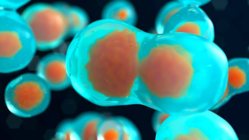
Photo source: Getty images
The union of egg and sperm has created a single cell that begins to divide incredibly quickly.
Only now does the real pregnancy begin.
Article content
In the third week of pregnancy, the real miracle of new life is happening.
For the next seven days, your newly fertilized egg will be dividing and travelling at the same time.
For the next seven days, your newly fertilized egg will be dividing and travelling at the same time.
The period of egg division is particularly sensitive to harmful agents, so it is still important to consider every activity with pregnancy in mind.
Avoid infections, alcohol, drugs or chemicals.
How is new life created?
Day 1: Fertilization
Fertilization of the egg by sperm occurs in the wider part of the fallopian tube.
This union results in the formation of a single cell, which we call a zygote.
The zygote contains different genes, the hereditary information from both parents, i.e. 23 chromosomes from the mother and 23 chromosomes from the father.
This creates a new unique being with a chromosome number of 46 XX (female) or XY (male).For more information about the egg, sperm or zygote, read this article:
Week 2 of pregnancy: When does ovulation occur?
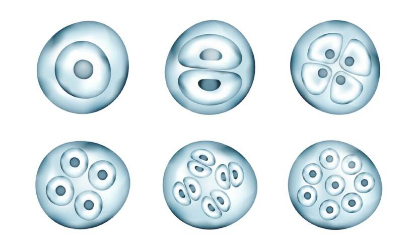
Day 2: Cell division
Approximately 20-30 hours after joining, the zygote begins to divide.
A single cell divides into two equal parts with the same number of chromosomes through a complex process.
This division is also called the furrowing or striation of the zygote.
The zygote first divides into two blastomeres. These further divide into four, from four to eight, from eight to sixteen, and so on.
The egg begins to divide into a number of cells that simultaneously shrink and fuse.
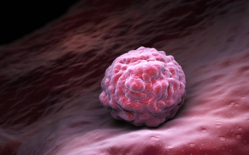
Day 3: Morula
When the embryo is made up of about 12 to 15 cells, it resembles a mulberry tree in appearance and is called a morula.
It carries complete genetic information about eye color, hair, sex, etc.
The morula moves down the fallopian tube and keeps dividing and dividing.
All this time (from fertilization to the morula stage) the fertilized egg travels from the fallopian tube to the uterus.
We say the egg migrates.
It is helped by the inside of the fallopian tube, which is thickened and contains what is called the ciliated epithelium.
It is the cilia that make it easier for the fertilized egg to travel towards the uterus.
However, there are also situations in which the egg gets stuck in the fallopian tube.
In this case, an ectopic pregnancy, a so-called tubal pregnancy, can occur.
Read more:
Ectopic pregnancy
How does an ectopic pregnancy manifest itself?
Days 4 and 5: Blastocyst
The egg enters the uterus.
The uterine lining is swollen under the influence of hormones, well blooded and ready for the egg to nest.
The embryo develops a cavity containing fluid.
With the help of this fluid, the morula divides into two parts:
- One part, called the trophoblast, forms on the outside of the egg. Eventually, it forms the placenta, which is the organ that nourishes the fetus throughout pregnancy.
- The second part is called the inner cell mass, which is the precursor of the embryo.
An egg divided in this way is technically called a blastocyst.
All the cells of a blastocyst are the same at this stage.
Meanwhile, the egg continues to develop and improve.
The blastocyst floats freely in the uterine cavity for two days.
It is rapidly increasing in volume and improving.
Days 6 and 7: Nesting
Usually on day 6 after fertilization, the blastocyst "attaches" to the uterine lining.
Most often with its embryonic pole.
The trophoblast begins to proliferate and differentiate into two layers:
- inner layer: cytotrophoblast - composed of single cells
- outer mass: syncytiotrophoblast - the mass where the boundaries between cells are lost
The cells of the trophoblast release an enzyme that dissolves the surface cells of the uterine lining. Nesting begins.
On the seventh day, one week after fertilization, the egg finalizes its nesting in the uterus.
The blastocyst burrows deeper, implants itself, dissolves the endothelium of the uterine blood vessels and comes into contact with the maternal blood.
The syncytiotrophoblast begins to produce the hormone hCG (human chorionic gonadotropin).
This keeps the corpus luteum active and enters the mother's blood.
Rising levels of hCG mean that:
- the egg has nested after a difficult and arduous journey
- the pregnancy is fine and everything is going according to plan
It takes approximately 13 days for the egg to firmly nest in the uterus.
This process can cause very light bleeding lasting 1 to 2 days on days 11-12 after fertilisation.
Read also the article:
How does fertilization occur and how long does it take for the egg to nest?
The hormone hCG
Human chorionic gonadotropin (hCG) is a glycoprotein hormone produced in pregnancy by the developing embryo shortly after conception and later by part of the placenta. Its function is to prevent the disintegration of the corpus luteum in the ovary and to maintain progesterone production.
Interesting facts about hCG:
- it can be produced in the body not only during pregnancy, but also by the presence of a certain type of tumor
- is an important cancer marker
- it is not known whether it co-causes cancer or whether its presence is the result of tumour formation
- in gynaecology and obstetrics, hCG is therefore referred to as a non-specific marker of pregnancy
The level of hCG can be detected from blood or urine.
Testing from urine is most often done on the basis of a home pregnancy test:
- reported borderline sensitivity ranges from 20 to 100 mlU/ml, depending on the test manufacturer
- in early pregnancy, results are more accurate when testing from the first morning urine, in which hCG levels are highest
- if the urine is too dilute, the hCG concentration may not match the blood concentration and the test may be a false negative
The blood test requires 2-4 ml of blood to be drawn.
Determining the amount of hCG in the blood is important for:
- monitoring the development of germ cell tumours
- determining adequate medical care after miscarriage
- diagnosis and subsequent treatment of ectopic pregnancy - if the hCG level reaches 1500 IU/ml and the vaginal ultrasound does not reveal an embryo, an ectopic pregnancy is very likely
The hCG level is also tested as part of the biochemical genetic screening in the second trimester of pregnancy.
We are talking about the so-called triple test. Its aim is to exclude certain chromosomal defects and birth defects.
The production of hCG increases rapidly in early pregnancy.
Until approximately the 9th week of pregnancy, the hCG level should double every 2 to 3 days.
The maximum increase is observed between the 80th and 90th day of pregnancy.
After that, its production decreases (in the 4th month). After the 25th week, the hCG level remains stable until delivery.
Its excretion in the urine ends approximately seven days after delivery.
Table of usual hCG levels in blood serum
| Levels of hCG in pregnancy | Level of hCG in mIU/ml | |
| Number of weeks since last cycle | Minimum value | Maximum value |
| 3 weeks | 5 | 50 |
| 4 weeks | 5 | 426 |
| 5 weeks | 18 | 7 340 |
| 6 weeks | 1 080 | 56 500 |
| 7-8 weeks | 7 650 | 229 000 |
| 9-12 weeks | 25 700 | 288 000 |
| 13-16 weeks | 13 300 | 254 000 |
| 17-24 weeks | 4 060 | 165 400 |
| 24-40 weeks | 3 640 | 117 000 |
A value lower than the normal hCG limit may signal an ectopic pregnancy, a dead fetus or an impending miscarriage.
On the other hand, elevated hCG values signal multiple pregnancies. They also allow early diagnosis of trisomy 21 - Down syndrome (along with the determination of other specific pregnancy hormones).
You might be interested in:
Abortion: what are the types and stages of abortion?
What happens to you?
In the first few days after fertilization, it is challenging to distinguish the initial signs of pregnancy from other problems or premenstrual syndrome.
You may experience:
- sensitive and engorged breasts
- Fatigue
- Irritability
- bloating
- increased basal temperature
- headache or dizziness
- abdominal pain
- nausea
We refer to all of these symptoms as uncertain signs of pregnancy.
We will discuss them in more detail in our article on the 4th week of pregnancy.
Read also:
Gallery
Interesting resources
Related

Mgr. Lucia Hlinková
Healthcare worker
She graduated from the secondary medical school in Nitra and midwifery at JLF UK in Martin. She has a specialization in Instrumentation in the operating room.
She worked as a surgical nurse for six years at the Gynaecology and Obstetrics Clinic in Martin and for two years at the Chest Surgery Clinic in Bratislava. For a shorter period of time she also practiced in the field of laser and aesthetic medicine. She is currently on maternity leave.
View all articles by the same authorThe aim of the portal and content is not to replace professional
examination. The content is for informational and non-binding purposes
only, not advisory. In case of health problems, we recommend seeking
professional help, visiting or contacting a doctor or pharmacist.


