Impetigo is characterised as a superficial infection of the skin caused by streptococci or staphylococci. The name of the disease comes from the Latin word impetere = to attack. It occurs most often in children. However, it can also affect adults.
The danger of the disease stems from the rapid spread of the infection. Within two days it can spread throughout the patient's body.
The incidence of the disease varies according to the season. The best conditions for the spread of the infection are in the summer months. The bacteria that cause impetigo multiply better in a warm and humid environment.
In the past, we used to see the highest incidence in the summer. Nowadays, people like to travel for warmth in the cooler months, so the disease is increasingly common all year round.
Patients with more severe atopic eczema are colonised with golden staphylococcus. Approximately 20 to 40% of the population are asymptomatic carriers of this bacterium (mostly in the nose and perineum) and are a potential source of infection.
Impetigo is classified as a bacterial skin infection (pyoderma). Staphylococci and streptococci are considered to be the most common causative agents of pyoderma. They are able to enter the bloodstream and lymphatic tract.
They also cause widespread coagulopathies or vasculopathies.
Pyoderma can be divided into two groups.
The first group consists of primary pyodermas, which include staphylogenic and streptogenic impetigo, furuncle, carbuncle and erysipelas.
The second group is secondary bacterial infections that further complicate pre-existing skin changes. Examples are secondary infections on eczema, ulcers, skin wounds, burns or chickenpox.
Primary bacterial skin infections are divided into:
superficial - affecting the epidermis and dermis (impetigo contagiosa staphylogenes, impetigo contagiosa streptogenes)
deep - spreads to subcutaneous tissue and muscle (erysipelas, perianal streptococcal dermatitis)
nail-bound - paronychia coccogenes
Table 1: Superficial skin infections can be divided into two subgroups, namely complicated and uncomplicated
Complicated superficial skin infections
Uncomplicated superficial skin infections
Staphylococcal Scaled Skin Syndrome (SSSS)
Furunkul
Infected pressure defects
Furuncle
Ulcer in chronic venous insufficiency
Ecthyma
Infection of the diabetic foot
Folliculitis
Lymphangitis
Abscess
Erysipelas
Impetigo
Hidradentis suppurativa
How do pyodermas arise?
The occurrence of pyodermias generally depends on the presence of a microbe with a certain pathogenic capacity and on the defences of the skin and the whole organism. Among the pathogenic microbes, staphylococci and streptococci are the main ones that cause pyodermias.
The skin's defences against microbes are complex, consisting mainly of the barrier function of the stratum corneum, which is very resistant to infection.
A very important mechanism of skin defence against infection is the production of keratinocyte antimicrobial peptides, which are able to kill a wide range of Gram-positive and Gram-negative bacteria, fungi and viruses.
The skin is colonised by a physiological microflora that produces antibacterial substances. It follows that bacteria, viruses and fungi can only penetrate the skin if the barrier function of the skin is compromised.
Causes
Impetigo can be caused by Streptococcus pyogenes (2% of cases) and Staphylococcus aureus (70% of cases). In the remaining 28% of cases, mixed infection with both pathogens occurs.
Both germs are localised in the nasopharyngeal region in many healthy people (carriers). Transmission to the skin occurs mainly by scratching with the nails.
Human-to-human transmission of impetigo occurs mainly in people who live in close contact (contact infection).
The course of the disease is influenced by various factors:
skin condition - chronic skin diseases (atopic dermatitis), mechanical damage to the skin
general state of the organism - corticosteroid treatment, malnutrition, systemic diseases
external environment - high temperature, humidity
properties of bacteria - virulence, synergism with other bacteria
Factors that contribute to the development of infection include:
Non-bullous impetigo accounts for more than 70% of all forms of impetigo. In the past, staphylococci were considered the causative agent of the disease. Later, the preference for a streptococcal origin of the disease became more common.
It is a very contagious disease.
It often causes epidemics among children.
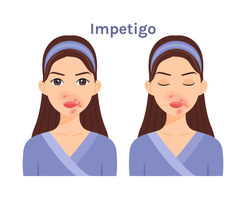
Symptoms of non-bullous impetigo arise on injured skin (pinches, burns, abrasions). It mainly affects uncovered areas of the body such as the face (around the nose and mouth), the capillitium (hairy part of the head), the neck and the hands.
At first, taut hemispherical vesicles form on the skin, surrounded by a narrow inflammatory rim. The contents of the vesicles are clear. These skin manifestations are rarely visible. The thin cover of the vesicle breaks down within a few hours. Exudate oozes from the eroded base. This gradually dries into thicker and more extensive honey-yellow crusts.
Symptoms of the disease spread rapidly to the surrounding area, where fresh vesicles and new erosions with honey-yellow crusts form again. The confluence of adjacent lesions produces arcuate to oval-shaped, sharply demarcated deposits.
Sometimes regional nodules enlarge. Complications of non-bullous impetigo can also be the so-called corners of the mouth. Rhinitis promotes the spread of the causative agent. Impetigo therefore often begins under the nose.
Nebulous impetigo in a 5-year-old boy. Source of the photo: Getty Images
If the disease is left untreated, it can persist for a long time. Numerous lesions can cause general symptoms. General symptoms include fever, nausea and lack of appetite.
Post-infectious glomerulonephritis is a dreaded complication, occurring in approximately 4% of patients. Kidney disease is caused by specific streptococci.
Bullous impetigo occurs mainly in children but can also occur in adults. The cause of the disease is always those strains of Staphylococus aureus that produce epidermolytic toxins.
The typical symptom of bullous impetigo is the formation of flat, clear blisters 4-9 mm in size. Later, the contents of the blisters turn grey-white.
The blisters form in the deeper layers of the epidermis. Their cover is therefore thicker and they persist longer. After the blisters have eroded, eroded areas are formed, lined by the epidermal remnant of the cover. Over time, a thin grey-yellow scab forms on the moist erosions and after its fall-off, a transient pigmentation remains.
Sometimes the centre of the blister cover adheres and fuses to the base of the blister. But at the periphery the process spreads to the surrounding area. In this way, adjacent blisters may fuse to form spreading formations. The disease heals without scarring.
Diagnostics
The diagnosis of impetigo is based on:
a thorough medical history
physical examination (clinical picture, extent of changes)
laboratory findings
complementary X-ray imaging
What are the diagnostic principles for each type of impetigo?
Non-bullous impetigo
On the basis of the typical clinical picture, the disease is usually easy to identify, especially in children with nasal secretions and honey-yellow scabs. Microbiological, clinical-chemical and serological diagnosis is also important.
Bullous impetigo
Clinical diagnosis is usually easy. Large blisters with purulent opacities and eroding areas are characteristic. An important step in the diagnosis is to identify the original atopic eczema.
What is the differential diagnosis?
The differential diagnosis includes blistering diseases of various origins. The most common infectious skin diseases that resemble impetigo include varicella, herpes zoster and herpes simplex.
For non-infectious causes, confusion may occur in grade II burns, a hypersensitivity reaction to insect bites.
Non-bullous impetigo
In patients suffering from this type of impetigo, haemolysing streptococci and only sometimes staphylococci are detected. The culture of streptococci depends on the time elapsed between the collection of the material and the start of the culture.
Herpes simplex, especially on the transitional parts of the skin and mucous membranes, is polycyclically circumscribed. However, it may be impetigo. The facial manifestations of secondary syphilis are symmetrical and have a basal infiltrate.
Bullous impetigo
In pemphigus syphiliticus syphiliticus neonatorum in the newborn, the blisters are localized on the palms and soles. In epidermolysis bullosa hereditaria, the mechanical mode of origin is in the foreground.
In bullous impetigo, blisters are almost never observed. Clinical manifestations include honey-yellow scabs.
Examples of the differential diagnosis are described in the following table:
small blisters on inflammatory base, crusting after drying; erosions on mucous membranes
Eczema numulare
round, sharply demarcated lesions with wetting and crusting
Course
Impetigo deposits most often appear on the face, neck and extremities. But they can appear anywhere on the body.
First, tiny macules (spots) appear. These quickly turn into blisters with a narrow inflammatory rim. The cover of the blisters cracks easily and their yellow contents dry out. This is how the typical honey-yellow or reddish-brown crusts form.
An example of what impetigo can look like. Source: Getty Images
How long does impetigo last?
In the case of an uncomplicated course, the disease lasts 7-10 days.
Sometimes spots or hyperpigmentation may appear as a result of the inflammation. These usually disappear within a few weeks to months.
Impetigo is very contagious.
Children suffering from impetigo are not allowed to go to nursery, school or after-school clubs for the duration of treatment.
Using disinfectant soaps and washing powders, using your own towel and ironed laundry will reduce the risk of infecting other family members.
How is impetigo transmitted?
Transmission of the disease occurs through three routes:
direct skin contact between a sick and healthy child.
through bacteria-contaminated objects (toys, towels, cutlery, etc.)
by autoinfection
How it is treated: Impetigo
Treatment of impetigo: Topical ointment, systemic drugs and antibiotics
solen.cz - Impetigo - diagnosis and treatment, MUDr. Táňa Pappová, PhD., MUDr. Andrea Kozárová, PhD., Department of Dermatovenerology, Comenius Medical Faculty in Martin, Comenius University in Bratislava and University Hospital Martin
solen.cz - Impetigo vulgaris, MUDr. Stanislava Polášková Children's skin outpatient clinic, Dermatovenerology Clinic of the University Hospital of Veterinary and Pharmaceutical Sciences, Prague
prolekare.cz - Infekce kůže a měkkých tkání, Autoři: R. Gürlich 1; V. Adámková 2; J. Ulrych 3; H. Brodská 4; V. Janík 5; J. Lindner 6; E. Havel 7, Působiště autorů: Chirurgická klinika FNKV a 3. LF Univerzity Karlovy přednosta: prof. MUDr. R. Gürlich, CSc. 1; Klinická mikrobiologie a ATB centrum VFN Praha a 1. LF Univerzity Karlovy primář: MUDr. V. Adámková 2; I. chirurgická klinika VFN a 1. LF Univerzity Karlovy přednosta: prof. MUDr. Z. Krška, CSc. 3; Ústav lékařské biochemie a laboratorní diagnostiky přednosta: prof. MUDr. T. Zima, DrSc. 4; Radiodiagnostická klinika FNKV a 3. LF Univerzity Karlovy zastupující přednosta: MUDr. L. Večeřová 5; II. chirurgická klinika kardiovaskulární chirurgie VFN v Praze a 1. LF Univerzity Karlovy přednosta: prof. MUDr. J. Lindner, CSc. 6; Chirurgická klinika FNHK a LF HK přednosta: MUDr. M. Leško, Ph. D 7
prolekare.cz - Enzyme treatment of skin and soft tissue infections, Authors.
praktickelekarenstvi.cz - The most common bacterial skin infections in children, Štěpánka Čapková, Department of Dermatology for Children, University Hospital in Motol, Prague
I graduated from the University of Veterinary Medicine and Pharmacy in Košice. After graduation I stayed working at the University. Until 2019, I was leading practical exercises in the subject of Drug Technology. I was mainly engaged in the preparation of individually prepared medicines in pharmacies. During my time there, I wanted to keep learning. In 2016, I defended my rigorous thesis at the Veterinary and Pharmaceutical University in Brno. By completing the rigorous procedure I gained knowledge in the field of laws related to pharmacy and medicine. In 2017, I completed my MHA studies at St. Elizabeth's College of Health and Social Work. By studying, I gained knowledge in the field of public health. In 2021 I completed my PhD studies (specialization in parasitic diseases) at the University of Veterinary Medicine and Pharmacy in Košice. The desire to learn new things and new knowledge led me to become a co-founder of the dermatovenerology outpatient clinic. And why can I give you information from the medical and pharmaceutical field? I like to learn and get to know new things. I have focused all my studies and scientific research on skin diseases. I have also gained knowledge in the field of parasitic diseases. Writing is one of my hobbies and a kind of relaxation.