A hemangioma is a benign tumor formed from blood vessels. In childhood, there are congenital hemangiomas and infantile hemangiomas. Congenital hemangiomas are visible immediately after birth, infantile hemangiomas appear later, most often during the first year of life. In adulthood, hemangiomas are most commonly found on the chest, neck, face, and also on internal organs. According to the type of blood vessel from which they arise, they are divided into capillary and cavernous hemangiomas. When multiple hemangiomas occur, the disease is called hemangiomatosis.
A haemangioma is a benign tumour that arises from blood vessels. It can form wherever blood vessels are located.
They are often found on the skin and mucous membranes. Because they are superficial, they are also very quickly seen and diagnosed.
However, haemangiomas can also occur in internal organs such as the liver, kidneys, brain, spleen and lungs. They can be intramuscular, i.e. intramuscular. As bone also has blood vessels that nourish it, haemangiomas can also form in bone.
There are many forms of hemangiomas. They are either solitary or multiple. If there are multiple hemangiomas in the body, it is a disease called hemangiomatosis.
Hemangiomas by type of blood vessel
Capillary and cavernous hemangiomas occur in adulthood. If multiple hemangiomas occur, it is the aforementioned hemangiomatosis.
Capillary haemangiomas arise from the thinnest blood vessels - capillaries. These blood vessels have a very narrow lumen and nourish the thinnest parts of the human body, i.e. the skin and mucous membranes. Most capillary haemangiomas occur on the head and neck.
Due to the abundant blood supply, they are deep red in colour and only a few millimetres in size.
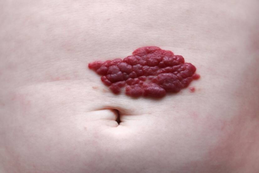
Cavernous haemangiomas arise from blood vessels that have a wide lumen. The name is derived from the word 'cavern', meaning cavity. They are usually found on the skin of the chest and are red or blue in colour.
Haemangiomas by site of origin
Intramuscular hemangioma is located in muscle tissue and can arise at any age. However, it is most commonly found in young adults.
Capillary hemangiomas occur in muscle tissue, more rarely cavernous hamangiomas. Any muscle in the body can be affected.
Because they are located inside the muscle, they are not visible. Some may cause swelling in their surroundings, which increases when the muscle is active, and the muscle may then become painful.
Bone hemangiomas are found inside the bones, most commonly in the skull or spine. The typical age for their occurrence is 50 to 70 years. Both capillary and cavernous types of lesions can occur in the bones. They cause no discomfort and are usually an incidental finding.
Hemangioma of internal organs is rarer than the above. It can develop at any age and most commonly in the liver and intestines. It does not require treatment and causes no inconvenience.
Haemangiomas in childhood
Haemangiomas can arise at any age, but haemangiomas of childhood are characterised by so-called congenital and infantile haemangiomas. These are the most common benign tumours of childhood.
Haemangiomas arising immediately after birth are congenital haemangiomas, while haemangiomas arising later in childhood are infantile haemangiomas. They are known as 'strawberry moles' because of their typical appearance.
Infantile hemangiomas are characterized by a later onset. They grow rapidly, enlarge and then spontaneously disappear.
Infantile hemangiomas affect approximately 4 to 5% of newborns. They are more common in Caucasian (European) infants compared to other races.
Female infants are up to 5 times more likely to be affected than male infants. They are also more common in preterm infants, infants born at low birth weight, infants with postpartum hypoxia, and infants of older mothers.
The vast majority of cases are sporadic, but there is some familial inheritance and occurrence in generations of families. Although we know that infantile hemangiomas can be inherited, no specific gene responsible for the transmission of this genetic mutation has yet been discovered.
They are often found on the head and in the hairy part. Source: Getty Images
Causes
The cause of hemangiomas is still unknown.
In childhood haemangiomas, it is thought that hypoxic stress increases the concentration of growth factors. This leads to endothelial cell proliferation and blood vessel growth.
In adults, there are no known risk factors for the growth of sporadic haemangiomas. In hereditary haemangiomas, there is a genetic predisposition to the development of these blood lesions.
Symptoms
Hemangioma can be present at birth, but is slightly more common during the first few months of a child's life.
The first signs of a haemangioma look like a small flat red mole anywhere on the body. The most common place for them to appear is on the face, scalp, chest or back.
A child usually has only one "mole"; adults may have more scattered over the body. Children born of twins or triplets may also have multiple hemangiomas.
During the first year of a child's life, a hemangioma grows quite rapidly. It may take on different shapes, for example, as a lobulated, spongy or rubbery bump that may rise above the level of the skin.
After this phase of rapid growth and enlargement, the hemangioma comes to a stop and stabilises in size, the so-called plateau phase. After this period, the hemangioma slowly begins to disappear.
Most red moles disappear by the fifth to tenth year of life. After the hemangioma disappears, the skin may be slightly discoloured or slightly raised where the hemangioma used to be.
Apart from the typical appearance and cosmetic problem, hemangiomas do not cause any other problems, even if they are located elsewhere on the body or internal organs.
Complications
The occurrence of hemangiomas is associated with some complications that depend on the age of the patient, the size and location of the hemangioma.
The most common complications include:
The most common complication is ulceration, i.e. the formation of an ulcer in the hemangioma. This is the most common complication of hemangiomas and affects 1 in 10 patients.
Amblyopia (blunt vision) is caused by hemangiomas located on the eyelid
Astigmatism, which is an irregular curvature of the eye's cornea that causes blurred or double and blurred vision
Other ocular complications include myopia, retrobulbar involvement and lacrimal duct obstruction
Airway obstruction
Difficulty with breastfeeding in lip hemangiomas
Cosmetic disfigurement of the face, e.g. in the case of the tip of the nose (Cyrano's nose), ears and mouth
Hemangioma on a child's thigh. Source: Getty Images
Diagnostics
Most paediatric haemangiomas are diagnosed on the basis of their typical appearance. When differential diagnosis and trying to distinguish haemangiomas from other tumours, we always start with the least invasive examination methods possible.
Ultrasound (USG) with a Doppler signal (used specifically in the examination of blood vessels) will confirm increased blood flow in the lesion, which is a typical feature of hemangiomas.
USG can distinguish between capillary and cavernous hemangiomas, but cannot determine the extent, depth and relationship to surrounding anatomical structures.
Magnetic resonance imaging (MRI) or computed tomography (CT) is more appropriate to determine the depth to which the hemangioma extends.
If multiple hemangiomas are found on the skin, it is advisable to complement the USG or MRI examination of the abdominal organs to verify whether hemangiomas are also present in this location.
Laboratory tests from the blood include coagulation parameters and blood counts, especially for further consideration of appropriate treatment.
In case of doubt about the correct diagnosis, a skin biopsy can be performed and the sample subsequently submitted for histopathological assessment.
Course
Infantile (so-called infantile) hemangiomas have a characteristic course, which has three phases:
1. Early proliferative or growth phase.
During this first phase, which lasts approximately 3 months, very rapid growth occurs. In the next five to eight months, growth slows down but continues. Deep infantile hemangiomas take longer to grow than superficial ones. Their growth is observed for 9 to 12 months.
2. Plateau phase
This phase is characterized by a resting period when the hemangioma does not continue to grow and does not increase in diameter. This period lasts approximately six months to a year.
3. Involution phase
After a period of quiescence, a phase in which the haemangioma shrinks, i.e. involutes or regresses. The lesions in this phase are soft, can be squeezed and change colour from bright red to purple to grey-blue.
Involution occurs during the first year and lasts for several years.
The healthy skin that replaces the haemangioma after the involution phase may be completely normal, but it may also have defects or scars.
Common skin defects include deposits of fatty tissue, telangiectasia (dilated capillaries that are visible as red threads), thin and patchy skin.
How it is treated: Hemangioma
Treatment: how to treat hemangioma in children and adults?
After graduating from an eight-year high school, I knew exactly who I wanted to become in life. Studying at the Faculty of Medicine at Comenius University in Bratislava fulfilled a long-held dream and I was able to start fulfilling my mission as a doctor. After obtaining my diploma and M.D. degree, my first professional steps led to the St. Elizabeth Cancer Institute in Bratislava. At the Stereotactic Radiosurgery Clinic I worked with patients with brain tumours and eye tumours. After a year of working with oncology patients, in 2020 I moved to the Neurology Clinic of SZU St. Michael's University Hospital in Bratislava, where I am still working today. I like to spend my free time outdoors, whether cycling, hiking or just walking in the nearby forest.