Inflammation of the breast is manifested by a painful and uncomfortable sensation in the breast. It most commonly affects breastfeeding women, but can also occur in non-breastfeeding women and men of any age. It is often caused by an infection that develops in the breast tissue.
Mastitis refers to any inflammation of the breast accompanied by an uncomfortable and painful manifestation in the breast, occurring mainly in the period of breastfeeding.
In men, it occurs very rarely and may be related to other changes in the breast.
Inflammation affecting the mammary gland is called mastitis, and inflammation of the nipple is called thelitis.
Anatomical structure of the breast
The breast and mammary gland go through various changes during life depending on age, menstrual cycle, pregnancy, breastfeeding and hormonal changes.
Breast skin is delicate, on top of which is the areola, which is more pigmented in colour and contains sweat ducts and sebaceous glands (Montgomery glands).
In the centre of the areola (in Latin: areola mammae) is the nipple (in Latin: papilla mammae), into which the milk ducts (in Latin: ductus lactiferi) enter.
The mammary gland is composed of 15-20 lobules, which are embedded in adipose tissue. The lobules extend into the nipple.
The mammary glands respond to all hormonal fluctuations during the menstrual cycle, pregnancy, breastfeeding and the age of the woman.
The male mammary gland is made up of a small amount of connective tissue, in which there are tiny glands without outlets.
Inflammatory breast changes affect women at any age, which decreases with increasing age, but inflamed breast tissue can also occur in men.
Puerperal mastitis
Puerperal mastitis is the most common form of inflammation of the mammary gland in the postpartum period, which is associated with lactation, in which the nipple is the entrance of infection.
The incidence of mastitis occurs in 1-5% of breastfeeding mothers.
Bilateral mastitis during breastfeeding occurs in 25% of cases.
In most cases, there is inflammation in only one breast, which is confined to only one segment of the mammary gland.
The cause is mainly due to poor breastfeeding technique associated with breast engorgement, but this reason is not enough to cause infection.
It is usually the result of a blocked milk duct, in which the release of milk has not occurred. Milk accumulating, behind the blocked duct, can get into the surrounding tissue and cause its inflammation.
Infection in breast inflammation may or may not be present.
Non-puerperal mastitis
Inflammation that is not related to lactation with an interval of several years after delivery is referred to as non-puerperal mastitis.
It occurs more often in women between 20-40 years of age.
Idiopathic granulomatous (lobular) mastitis is a rare form. Many times it appears several years or even decades after birth.
It is characterized by excessive formation of granulation tissue, the cause of which is hyperprolactinemia (increased blood levels of the hormone prolactin). It is manifested by the growth of a painful mass in the breast, which is accompanied by reddening of the skin.
Plasmocytic mastitis appears several years after childbirth. It is a nonpurulent interstitial inflammation with the presence of plasma cells accumulated in the inflammatory bed. The breast is red, painful and the inflammation is accompanied by thick secretion from the nipple.
Diabetic mastopathy in women with type 1 diabetes is a rare and severe form of breast inflammation that causes swelling, pain and redness.
It can affect one or both breasts at the same time. It is more common in pre-menopausal diabetic women, but can also occur exceptionally in men with type 1 diabetes.
Tuberculous mastitis is a rare disease in countries with a high prevalence of pulmonary and extrapulmonary tuberculosis.
It occurs in less than 1% of breast diseases. It is manifested by the appearance of a unilateral hard lump and the lymph nodes are often enlarged.
Other types of breast inflammation
Inflammation of Montgomery glands is an inflammation of the skin glands around the areola. It is manifested by painful redness of the glands and swelling. It usually resolves on its own without treatment in a few days.
A breast atheroma is a skin cyst that can occur anywhere on the skin with a larger number of sebaceous glands. It is mainly a sebaceous gland cyst filled with a thick, whitish-yellow mass with an unpleasant odor.
Mondor's disease is a form of thrombophlebitis with painful hardening of the skin at the site of the affected vein.
It most often affects the superficial vein on the breast. In most cases, this symptom resolves spontaneously and local anti-inflammatories (drugs to reduce inflammation with a pain-relieving effect) can be applied to the pain.
Causes
The most common causative agents of breast inflammation are streptococci, staphylococci, and in some cases enterococci and other bacteria.
Causes of inflammation
Blocked milk duct – if the breast is insufficiently emptied, one of the milk ducts can become blocked and its blockage can cause infection.
Nipple fissures – small tears on the nipples.
Poor nipple care before and after breastfeeding.
Milk retention – retention of milk in the breasts due to insufficient pumping and emptying of the breast
The entry of bacteria into the breast most often passes from the baby's mouth during breastfeeding.
Bacteria can enter the breast through a small tear in the skin or through the opening of the milk duct. The milk remaining in the breast after insufficient emptying provides very good conditions for bacterial growth.
Other causes
Damage to the nipple, for example, by piercing, eczema or skin disease
Breast implant
Risk factors
Previous bout of mastitis during breastfeeding
Sore breasts with tears
Wearing a tight bra during breastfeeding, which puts more pressure on the breasts and this can restrict the flow of milk in the breasts
Inadequate breast hygiene before and after breastfeeding
Smoking
Weakened immunity
Breast inflammation prevention
Complete emptying of the breasts during the breastfeeding period
Breastfeeding should be done by the baby sucking the milk completely from the breast and then attaching it to the other breast
The correct technique of sucking the baby to the breast
In case of a cracked or damaged nipple, use a silicone nipple protector
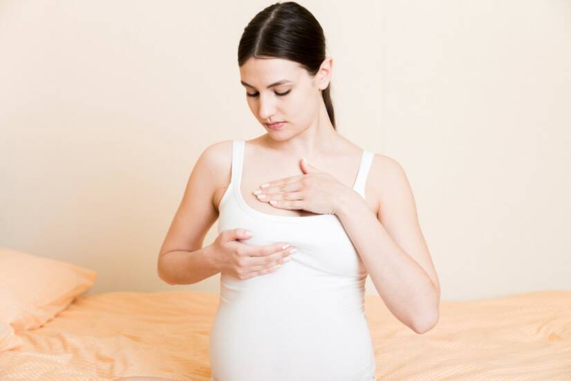
Proper breastfeeding technique reduces the risk of puerperal mastitis. Photo source: Getty Images.
The disease is diagnosed on the basis of visible symptoms, breast tenderness and also on the basis of a patient’s medical history.
If it is not mastitis during the period of breastfeeding, it is necessary to determine not only the possible causes, but also the risk factors and impending consequences that may arise from inflammation.
Correct diagnosis is very important because mastitis could be confused with, for example, inflammatory breast cancer.
To exclude other physiological changes in the breast, an ultrasound is also performed to show the site of inflammation in the mammary gland and the extent of inflammation, so that some other diseases with similar symptoms can be ruled out.
Ultrasound - Ultrasonography also helps to locate an abscess located in the breast.
Ak nedojčíte môže sa vykonať mamografia aj na vylúčenie rakoviny prsníka. If the person is not breastfeeding, a mammogram may also be done to rule out breast cancer.
Course
The course of inflammation is usually very abrupt and sudden.
Locally, a painful lump or swelling with reddening of the skin develops, followed by a temperature rise of about 40° with chills, headache, loss of appetite.
In puerperal mastitis, the interval of onset of inflammation from delivery is a few days up to 5 months, in most cases appearing between the 2nd - 3rd week after delivery.
Acute mastitis – caused by injury to the nipple during breastfeeding, most often provoked by golden staphylococcus. Milk retention also contributes to the infection, which helps to spread the infection.
There is swelling of the breast and enlargement of the lymph nodes. The breasts are red, painful, and the general condition is manifested by increased temperature to flu-like fever. There is palpably firm, painful tissue in the breast.
Chronic recurrent mastitis refers to recurrent inflammation of the breast during breastfeeding with subsequent abscess formation. The cause is usually the administration of inappropriate antibiotics, or an obstruction in milk emptying, such as a cyst, fibrosis, fibroadenoma.
When the abscess empties spontaneously into the milk, it is necessary to stop breastfeeding and express the milk.
Complications in mastitis
Recurrence of mastitis – in untreated or neglected cases with a large abscess, several abscess foci may develop.
Antibiotic treatment and drainage of the largest abscess deposit, which overlies small deposits, can cause recurrence (relapse).
Therefore, after 10-20 days of treatment, repeated breast sonography is performed to exclude the abscess deposit left behind.
Chronic mammary fistula, periareolar fistula – an abscessed inflammation of the mammary gland near the nipple can cause decomposition or damage to the milk duct.
The infection thus affects the milk ducts and spreads to distant sites, posing a complicated therapeutic problem due to the persistence of focal infection.
Another cause of a chronic mammary fistula (drainage duct from a suppurative lesion) may be infection of the scar after surgery. A fistula with oozing inflammatory contents is visible.
With treatment, the condition calms down, the ligaments multiply due to healing of inflammation, which can cause recurrence of inflammation. This condition can last for years. Therefore, removal of the fistula from the inflamed area is necessary, followed by drainage and administration of antibiotics.
How it is treated: Mammary Gland Inflammation
Ako sa lieči mastitída? Lieky, antibiotiká, masti i chirurgia
Berens PD (December 2015). "Breast Pain: Engorgement, Nipple Pain, and Mastitis". Clinical Obstetrics and Gynecology. 58 (4): 902–14. d
Spencer JP (September 2008). "Management of mastitis in breastfeeding women". American Family Physician. 78 (6): 727–31
Ferri, Fred F. (2009). Ferri's Clinical Advisor 2010 E-Book: 5 Books in 1. Elsevier Health Sciences. p. 593. ISBN 9780323076852.
Buttaro TM, Trybulski J, Bailey PP, Sandberg-Cook J (2007). Primary Care: A Collaborative Practice. Elsevier Health Sciences. p. PT1608. ISBN 978-0323078412.
The Worldwatch Institute (2015). State of the World 2006: Special Focus: China and India. Island Press. p. 36. ISBN 9781610916332.
Ratcliffe, Stephen D. (2008). Family Medicine Obstetrics. Elsevier Health Sciences. p. 634. ISBN 978-0323043069.
Michie C, Lockie F, Lynn W (September 2003). "The challenge of mastitis". Archives of Disease in Childhood. 88 (9): 818–21.
Dixon, J Michael; Pariser, Kenneth M. "Nonlactational mastitis in adults". UpToDate. Retrieved 2019-08-02.
Zhang, Ying; Sun, Xiaoying; Li, Kexin; Wang, Xiaomin; Cai, Lijun; Li, Xin; Zhou, Min (2018-05-02). ""The Therapy of Elimination First" for Early Acute Mastitis: A Systematic Review and Meta-Analysis". Evidence-Based Complementary and Alternative Medicine. 2018: e8059256.
Cusack L, Brennan M (December 2011). "Lactational mastitis and breast abscess - diagnosis and management in general practice". Australian Family Physician. 40 (12): 976–9.
Kvist LJ, Larsson BW, Hall-Lord ML, Steen A, Schalén C (April 2008). "The role of bacteria in lactational mastitis and some considerations of the use of antibiotic treatment". International Breastfeeding Journal. 3
I graduated from the Secondary Medical School in Nitra, which gave me the basis for a career in healthcare. After school I worked for three years in the surgical department and then in the department of Anaesthesia and Intensive Care Medicine. In addition to my employment, I completed my bachelor's degree at the Faculty of Health Care in Banská Bystrica in Nursing and completed my specialisation studies in Anaesthesiology and Intensive Care Medicine. Since my childhood I was determined to become a health professional and help people with their health problems. Continuous education and study of new professional topics related to health care, which is constantly evolving, and gaining practical experience, helps me to write professional articles for this portal, which is available to everyone. My hobbies are multifaceted, I am also involved in healthy nutrition, overall healthy lifestyle. I spend my free time on education, creative work, handicrafts in cooperation with my daughter, thanks to which, we do not know boredom.