Gangrene is a condition in which a certain part of human tissue dies. It is often a manifestation of secondary changes such as desiccation or infection.
Gangrene is a dangerous, even life-threatening condition. Its essence is the death of the affected tissues, in which there is not a sufficient supply of nutrients and oxygen. Infections with bacteria living without the need for air (anaerobic) are common.
What is gangrene?
Gangrene is necrosis modified by secondary changes. Most commonly desiccation and infection.
There are 3 types of gangrene:
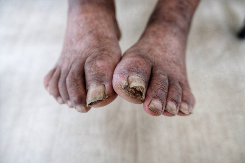
Dry gangrene (gangraena sicca) - It occurs most often on the extremities when a blood vessel is occluded. It is caused by insufficient oxygen supply. The colour of the concretions gradually changes from purple to black to dark brown. The skin loses water, elasticity and resembles parchment paper.
Moist gangrene (sphacelus) - Necrosis modified by putrefactive bacteria. Necrotic tissues have a moist appearance, smell and break down. The colour is green, caused by bacteria acting on haemoglobin. When bacteria overgrow, a serious, life-threatening condition - sepsis - can occur.
Gas gangrene (gangraena emphysematosa) - Necrosis modified by the gas-forming bacteria clostridia. It occurs after an injury when the clostridial infection gets deep into the wound (e.g. soil is introduced into the wound). Tissue is swollen, may suppurate. You can feel bubbles of gas that seem to burst when touched.
Once a certain type of bacteria gets into the surrounding tissues and bloodstream, any gangrene poses a fatal danger.
Risks of gangrene
The factor that most often increases the risk of gangrene is the narrowing of blood vessels - atherosclerosis. Gradual narrowing can lead to a limited oxygen supply and later to complete tissue death.
Diabetes is among the important factors. It is well known that diabetes puts patients at risk of the aforementioned bacterial infections.
The risk is also increased by:
Inflammatory diseases of the blood vessels
Reduced immunity (chemotherapy, HIV, drug addiction)
Burns
Frostbite
Smoking
Obesity
Diabetic gangrene
The term diabetic gangrene encompasses a wide range of acral lesions on the diabetic leg, from minimal signs to gangrene of the entire leg. It is one of the most serious complications of diabetes.
In diabetics, foot involvement is 20 to 50 times more common than in healthy individuals.
Factors causing diabetic gangrene
For patients being treated for diabetes, many factors determine the outcome. Some of the most common include:
Neuropathy - damage to the nervous system (motor, sensory, visceral)
Circulatory disturbances - inadequate blood supply and subsequent lack of vascularization of tissues
Hyperglycaemia - fluctuations in blood sugar levels causing earlier onset of later complications
Restricted joint mobility - together with sensorimotor neuropathies causing reduced muscle tone, stiffness, numbness, thus creating more pressure when standing or walking
Diabetic gangrene is a serious consequence of diabetic foot syndrome. It often leads to amputation of the affected limb. It can also develop from a minor injury or often an underestimated blister.
Prevention of these disorders is regular foot care and visits to a diabetologist.
Diagnosis and treatment of diabetic gangrene
The diagnosis is made by the doctor on the basis of the medical history. Patients often neglect mild manifestations, the so-called warning signs, and come to the outpatient clinic at an advanced stage of the disease.
History - Based on the interview, the doctor obtains the necessary information about the patient's current and previous condition.
Physical Examination - By sight and palpation, the doctor uses vision to determine the extent and stage of the disease. By palpation, he or she determines the tenderness and swelling in the area.
Laboratory examination - A blood sample is taken and sent for determination of inflammatory parameters (CRP, leukocyte count, FW).
Imaging - Determines the extent, degree and depth of involvement.
X-ray (X-ray examination) - Used to determine the extent on solid structures (bone).
USG (ultrasonography) - Used to detect abnormalities in the bloodstream.
CT (computed tomography), MRI (magnetic resonance imaging) - Used especially in high grade disease to determine the involvement of deeper structures.
Swab - Using special sterile brushes, a swab is taken from the affected area and sent for bacteriological examination (to detect the presence of anaerobic, aerobic or gas bacteria).
Initial stage of gangrene of the lower limb. Source: Getty Images.
Treatment of diabetic gangrene
The treatment of gangrene tends to be long-term and difficult. From the possibility of cure to radical surgery. The most important and first step is to compensate for diabetes.
Medical treatment - Administration of antibiotics through the blood (into a vein).
Local surgical treatment - Depends on the local findings. Surgical treatment is performed under local anaesthesia if necessary. In the case of incipient gangrene, diabetic foot or tibial ulcer, dead skin and subcutaneous tissue is removed. In some cases, bone is also removed. Wound lavage with antibiotic solutions is also performed.
Total surgical treatment - If the finding is advanced and previous procedures cannot be used or are ineffective, the affected limb must be amputated.
Interesting fact: For anaerobic infections, a stay in a hyperbaric chamber is recommended. The patient is confined in a chamber where a high concentration of oxygen is maintained. This is not conducive to the spread and multiplication of this type of bacteria.
Fournier's gangrene
Fournier's gangrene (FG) is a life-threatening gangrene of the external genitalia, perineum and perianal area. Currently, this definition includes affections of the same nature in women. However, it is more common in the male population.
The disease was first described by Jean Alfréd Fournier in 1883.
FG is a typical necrotizing fasciitis caused by various bacteria (staphylococci, streptococci, coliforms, pseudomonads, bacteroides, fusobacteria, pseudomonads) from the skin, urethra, vagina or rectum.
Causes of FG
An important prerequisite for the development of FG is impaired (mainly cellular) immunity and reduced immunity of the affected individual. Local disease or trauma in the genital area favours the development of bacterial infection.
Risk factors influencing the development of FG
Table of risk factors causing FG
Immunodeficiency states
Urological causes
Surgical causes
Dermatological causes
Less common causes
Diabetes mellitus
Scrotal abscess
Periproctal abscess
Occurs mainly in developing countries
Hernioplasty
Chemotherapy
Surgical interventions
Perforated appendicitis
Vasectomy
HIV
Prostate biopsy
Diverticulitis
Neonatal circumcision
Corticoid therapy
Urethral stricture
Rectal cancer
External injuries (scratches, insect bites)
Chronic alcoholism
Extravasation of urine during endoscopic instrumentation in the lower urinary tract
Anal fissures and fistulas
Obliterative endarteritis pudenda externa and interna
Diagnosis and treatment of Fournier's gangrene
The diagnosis of FG is usually made on the basis of clinical picture and history. The main symptoms are most often pain at the site of involvement, swelling and in later stages fever.
History - assessment of possible risk factors for FG
Physical examination - palpation, palpation of affected areas
Crevo - In ileotic conditions where there is choroidal obstruction. Rarely occurring gangrene of the colon in the autoimmune disease lupus erythomatosus
She graduated from the Secondary School of Nursing in Nitra and completed her Master's degree in nursing at the University of Medical Sciences in Nitra. After school, she worked as an operating room nurse in the Central Operating Rooms at the Nitra University Hospital since 2012.
The aim of the portal and content is not to replace professional
examination. The content is for informational and non-binding purposes
only, not advisory. In case of health problems, we recommend seeking
professional help, visiting or contacting a doctor or pharmacist.