- Functional anatomy: Ivan Dylevsky
- Nursing in Surgery II: Slezáková Lenka a kolektiv
- Pediatric Neurosurgery: Krahulík David, Brichtová Eva, collective
- AD MANUS Orthopedici - Treatment of positional deformities from the perspective of an orthopaedic technician: Andrej PLŽ
- AD MANUS Orthopedici - Plagiocephaly: MUDr. Hana Sameková
- fyzioklinik.sk - Correct positioning of the child
- Ortopedickymagazin.sk - Ing. Plž Andrej, biomedical engineer. Plž Andrej
- kidshealth.org - Flat heel syndrome (positional plagiocephaly)
- childrenrenshospital.org - Plagiocephaly
- nhs.uk - Plagiocephaly and brachycephaly (flat head syndrome)
- ncbi.nlm.nih.gov - Brachycephaly
- verywellfamily.com - What to know about dolichocephaly
- verywellfamily.com - What is positional plagiocephaly?
Plagiocephaly in children. What positional head deformities are we familiar with?
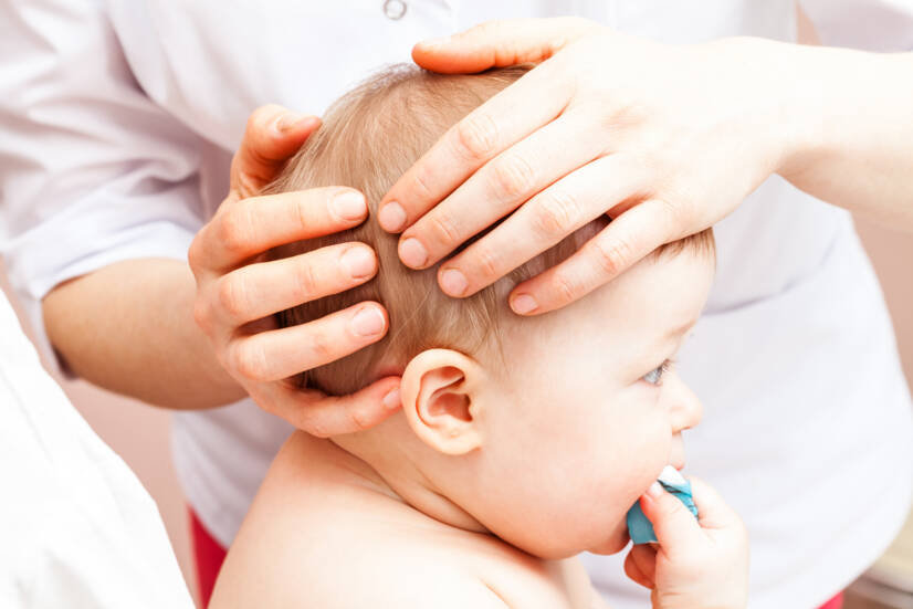
A child's head is still pliable and in many cases head shape deformities will straighten themselves out. However, in some cases this is not the case and head shape deformities arise that can affect the child's psyche at an older age.
Article content
Plagiocephaly is a broader term for head deformities caused by flattening.
Why it occurs and how it can be prevented in children can be found in this article.
Head deformities often occur in newborns and infants due to external influences such as:
- Intrauterine - While still developing in the intrauterine space, when pressure was exerted on the head before birth, for example, with a reduced amount of amniotic fluid surrounding the baby.
- Peripartum - During labour, in complications where obstetric forceps or vacuum extractors were used to speed up the expulsion phase of labour.
- Postpartum - After birth trauma, poor positioning, especially in babies with force reflux and in premature babies.
The newborn's head often looks slightly deformed after birth and in the first weeks of life. This is quite normal due to pressure during intrauterine development and after birth through the birth canal.
Deformities or asymmetries of the skull are reported in 1 in 300 babies born.
In some babies the deformity is increased, especially in premature babies, in babies with reflux and in babies who do not tolerate certain positions at all.
The head can be shaped nicely by correct positioning.
When sleeping on the back, tension in the neck muscles can prevent the head from turning sideways, putting one side of the head at risk of flattening.
Skull
A child's skull is different from an adult's skull, not only in size but also in shape.
A child's skull has a larger brain part compared to the facial part.
The skull not only protects the brain, but in infants, when the skull bones separate, it allows for volumetric growth and brain development.
In the first 6 months, the skull grows rapidly and doubles in size. By the age of 2 years, it increases up to threefold. From the age of 7 years, its growth slows down and by the age of 16-18 years, the development of the cranial vault stops.
The bones of the skull develop through ossification - ossification from the base by connective or cartilaginous tissue. Ossification begins in the fetus during intrauterine development.
The bones of a newborn's skull are still very soft, thin and flexible, joined by sutures at the head that are wide. The bones of a premature baby's skull are even softer than those of a premature baby.
The cranial sutures are the ligamentous joints of the bones of the skull. The sutures, if they do not ossify, increase the flexibility of the skull and allow movement of the vault.
They allow the bones to move against each other. This plays an important role in the passage through the birth canal to which the skull adapts.
In newborns and infants, fontanelles - bands of connective tissue - are found between the bones of the skull where the sutures join.
Two important fontanelles, called the lesser and greater fontanelle, are palpable and are important indicators of intracranial pressure.
The large fontanelle is also called the frontal fontanelle. It is located between the forehead and the two parietal bones at the top of the head. It is diamond-shaped and disappears by the end of the second year.
The small fontanelle is located between the temporal bones and the occipital bone. It is triangular in shape and disappears by the 3rd month of life.
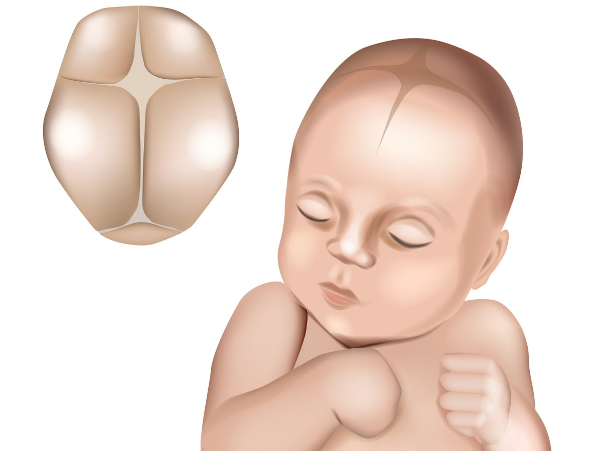
There are two other fontanelles on the skull that cannot be palpated, the sphenoid fontanelle, located in the temporal fossa, and the mastoid fontanelle at the back of the skull behind the ear.
Why does the head deform?
The cause of deformities is craniosynostosis or positioning.
The skull of a newborn in the first few months of life has the ability to form and is therefore susceptible to deformities due to the external environment.
A flattened head often develops in babies who spend a lot of time in the same position, most often during sleep, due to the consequent tightening of the neck muscles. Weak or tight neck muscles prevent the baby's head from rotating, putting pressure on one spot.
A deformed head from positioning does not affect the child's brain development and growth. The child has no pain or other symptoms. He or she develops normally like any other child. It is only a cosmetic problem that can cause a decrease in self-esteem and self-worth in adulthood.
The paediatrician checks the child at regular check-ups and notes the shape of the child's head.
If the pediatrician finds uneven head growth, he or she will send the child to an orthopedist. The orthopedist will not only look at the symmetry of the lower extremities, but also observe the shape, size and formation of the skull.
Diagnosis will reveal craniosynostosis (premature fusion of the bones of the skull) or hydrocephalus (excessive accumulation of cerebrospinal fluid inside the skull), which are among the serious diagnoses that affect a child's brain and psychomotor development.
Deformities are more common in boys than in girls and are usually due to the child's higher birth weight.
In children aged 1-4 months, deformities can be corrected by correct positioning.
At later ages, correction of the head shape requires a specialist in orthopaedics, orthopaedic prosthetics, neurosurgeon, surgeon and rehabilitation.
Forms of skull deformities
The severity of the deformities is determined by degrees of severity, by cranial measurements and by calculations with the creation of 3D photographs using a special scan.
Plagiocephaly
This is an irregular flattening of the head. It is often associated with deformation of the pinnae and is sometimes also associated with a displacement of the front of the forehead.
This deformity is often associated with a disability of the cervical spine that does not allow full movement. The child therefore holds the head in one position for long periods of time.
Flattening of the head occurs when the child's skull is still soft and there is repeated pressure on the head to one side. Most often it develops because of regular sleeping in one position.
This type of head shape is characterized by flattening on one side. The other side, on the other hand, is protruded.
In plagiocephaly, the unevenness of the head shape is visible.
The head does not have the correct shape, the ears may be unevenly aligned, one ear of the child is visibly displaced.
On the side of the posterior flattening, the forehead and ear are displaced forward. The eye and face may also be displaced forward, causing unevenness in the face.
When viewed from above, the head looks like a parallelogram.

Brachycephaly
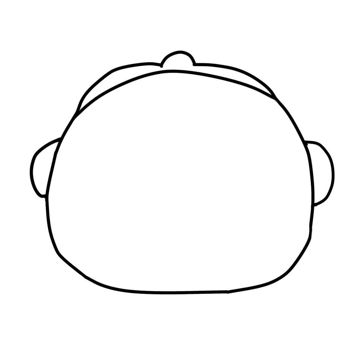
The entire temporal part of the head is symmetrically compressed.
The head has a broad shape.
Compression of the head may be asymmetrical, e.g. compression of the lateral part of the head.
This deformation of the skull causes the head to have a smaller ratio of length to width. There is a shortening of the skull in the anteroposterior dimension.
Symmetrical brachycephaly is a deformity that results in poor proportionality of the skull.
The flattening is posterior to the middle of the skull.
The head is broad with prominence of both temporal bones.
The head is taller and flatter laterally. The forehead is large and sometimes displaced forward.
It is often found in children who lie only on their backs and do not turn their heads.
Asymmetric brachycephaly is a combination of plagiocephaly and brachycephaly.
It is usually associated with immobility of the cervical spine.
The back of the head is irregularly flattened. One side is flatter than the other.
The head may be higher on one side and unusually broad. The ear, forehead, eye and part of the face on the side of the greater flattening may be slightly displaced forward.
The most common head deformity in children is the asymmetrical brachycephaly type.
Symmetrical brachycephaly is the second most common deformity.
Dolichocephaly
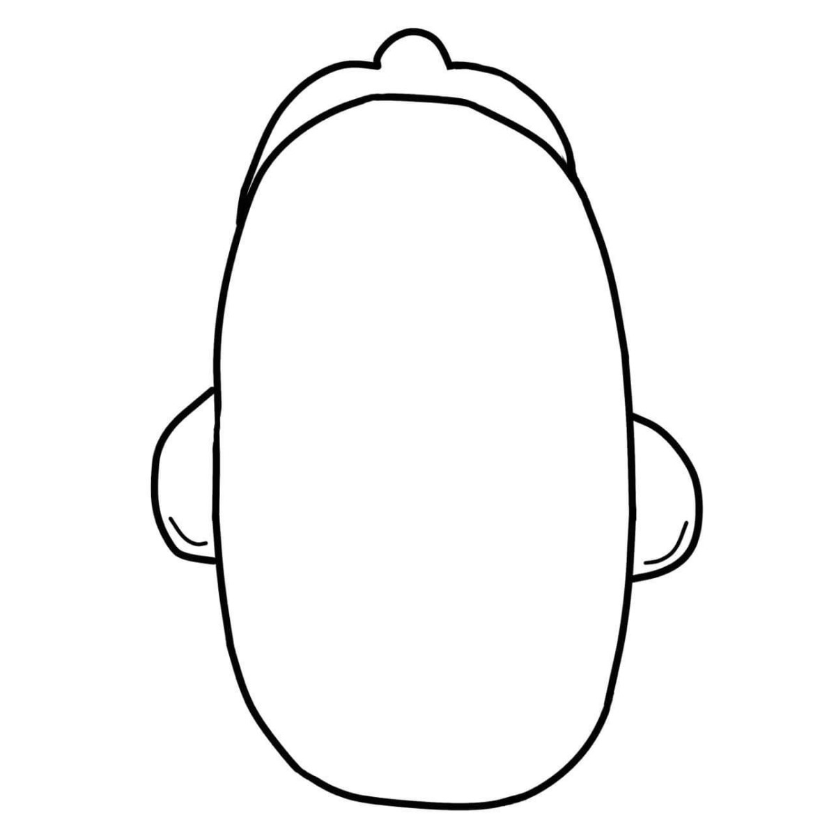
The shape of the head is narrow and long. There is compression of the cephalothorax at the sides and the skull elongates towards the front.
This deformity often occurs in premature babies who are positioned only on their side in the incubator.
Turicephaly
Is a vertically elongated skull shape, often the result of vacuum extraction at birth.
The head has a convex temple and the head is triangular in shape with a rounded top. After birth by extraction, the head flattens itself within a few days.
.jpg)
Craniosynostosis
Flattening can also be caused by too early fusion of the cranial bones.
Craniosynostosis is a birth defect in which the skull bones fuse prematurely, before the brain has fully developed. The brain continues to develop, but stops growing due to the skull not stretching. This results in an abnormal head shape.
As a result of premature fusion of one suture, various deformities can occur. Such deformities can affect brain development, increase intracranial pressure and damage the cerebral cortex and optic nerve. This can lead to blindness.
Craniosynostosis can be of two types:
The primary is premature fusion of the cranial bones.
Secondary is part of a syndrome (Apert, Cruz...).
How do I know if my child has a flattened head?
Head flattening occurs most often in babies in the 2nd-3rd month.
Flat spots begin to form on the head, either on the sides or on the back of the head. In the area with regular pressure, there is no hair on the scalp at the pressure point and a bald spot appears.
The deformity is best seen when viewed from the top of the head.
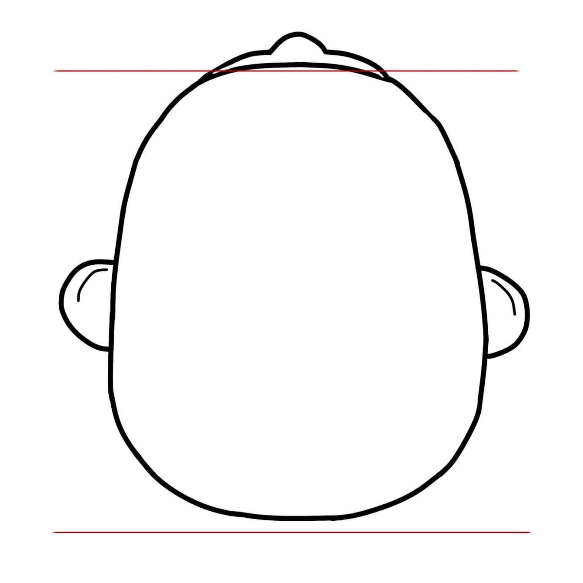
Some deformities may also cause deformation of part of the face. One ear may appear to be more forward than the other. The forehead may be more protruded or, in a lateral deformity, the forehead may be more bulging on one side.
How to prevent skull deformities?
Parents should be instructed on positioning and occupying the child during the day.
Therefore, it is important for parents to monitor the growth and shape of the head even at home. Proper positioning is important when the child is sleeping, at feeding time or during the day when the child is awake.
The newborn and premature baby cannot hold the head and it tilts. It is still weak and its posture is uneven.
However, a healthy baby must be able to turn his head to either side.
If it only turns to one side, this is called head predilection. This should not take more than 6 weeks. Especially in premature babies, for whom a corrected age is calculated (if the baby was born two months earlier, these months should be subtracted for a certain period of time and the date of birth should be used).
For premature babies, the position of the baby is very important, as the baby is very weak and cannot turn its head.
Positioning and physiotherapy exercises are recommended to prevent head deformities.
In most cases, flat head naturally improves with growth. As the child grows and gains strength, he is able to position himself, turn his head and roll over onto his tummy. This strengthens the neck and back muscles.
Lying on the tummy
Since 1992, it has not been recommended to position the baby on the tummy due to the finding that this position increases the incidence of sudden infant death syndrome. This has also increased the number of skull deformities, as many parents do not position the baby and the baby is still only laid on its back.
The question is whether to lay the baby on the tummy. Many physiotherapists recommend the tummy position, which reduces the risk of the head creeping.
However, place the baby on his tummy when he is awake. Then there is no danger. Start by doing the procedure several times a day for one or two minutes. You can gradually increase the time.
The basic principle is not to put the baby on his tummy when he is tired. Then he will be weepy or inactive. This leads to muscle tension, irritability or a bad mood.
A baby with reflux should be placed on his tummy several times during the day to strengthen his abdominal muscles, improve his digestion and counteract colic.
While laying on the tummy, the child exercises, lifts the head, strengthens the neck muscles and activates the upright position.
When lying on the tummy, the baby should rest on his forearms, the buttocks should be pressed against the mat in line with the head. The hands should be clenched into fists and relaxed. This position forms the first support, which is important for the proper development of the spine.
Changing the baby's position in the cot
Change your baby's position in the cot regularly.
Most parents, especially right-handed parents, carry their babies in their left arms and lay them on their left side in the crib. The baby is usually looking out into the room, so he or she only has his or her head laid on one side at all times.
Turn him in the crib or put a toy in his field of vision that you switch each time. This will force him to turn his head from side to side.
Carrying in your arms
Many parents do not prefer to carry their baby in their arms.
However, this too is an option to relieve head flattening. Regularly carry and lift your baby, reducing the pressure of the head support.
Carrying your baby in a scarf is healthy and beneficial. This position is also good for shaping the head and trains the postural muscles needed to support the body, which tend to shorten.
Positioning during sleep
Change the position of your baby's head during sleep by supporting it with a small towel or nappy. This puts pressure with the head against the mat, even on the side of the head where the deformity is forming.
There are also head pads on the market that hold the head in one position and less pressure is put on the back of the head.
Placing the child on the pad
Avoid placing your child on hard pads for too long, such as car seats that have hard plastic covered with only a thin layer of padding.
Place the baby on a soft cushion with padding on the sides so that he or she does not roll over. Make a sort of nest with the sides of the whole area padded. There are also various positioning nests for babies that are soft and do not press on the baby's skull.
Remodelling brace
If the deformity in an older child is longer than 4 months, a KRO (cranial remodelling orthosis) is required, which has a success rate of up to 95%.
Treatment with KRO should be consulted with a neurologist or neurosurgeon. In some cases, this method is contraindicated, for example in hydrocephalus.
The KRO orthosis is a special helmet that is hard on the outside and lined with a soft lining on the inside. The helmet does not compress the child. It does not cause any pressure on the skull, it only compresses it slightly and the part where the deformity is located is the gap between the skull and the helmet.
It is a challenging treatment. It is used for moderate to severe deformities of the child's head.
KRO is aimed at properly guiding the growth of the skull.
The most suitable period is 4-7 months of age, which reduces the treatment to 4-5 months. The older the child is, the longer it takes to properly rebuild the child's skull, due to the closing of the slits and the growth of the skull. Treatment with a brace is possible up to the age of 18 months.
Interesting resources
Related
