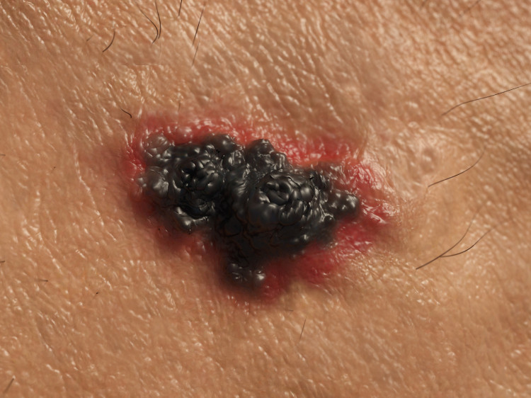
Why are doctors warning? Cases of malignant melanoma of the skin are on the rise!

Malignant melanoma of the skin is one of the most malignant cancers ever. Its insidiousness lies in the aggressiveness of the disease and its ability to rapidly metastasize. Patient survival and overall prognosis are directly dependent on early diagnosis and early initiation of treatment. Insidious forms of this disease and late diagnosis can mean very early death of the patient even within half a year.
Article content
The number of melanoma cases has been increasing enormously in recent years, especially among young people, not because of a lack of public awareness, but because of a disregard for preventive measures and indifference to their health.
What is malignant melanoma and why is it something to worry about?
Malignant melanoma of the skin is one of the most aggressive cancers ever. It is a malignant disease affecting the skin, but rarely occurs on the mucous membranes or the eye. It is a malignant cancer characterised by its high aggressiveness and its ability to metastasise in a relatively short period of time.
Of course, the course of the disease and treatment options depend on the specific type of melanoma and the stage at which it is found.
In addition to malignant melanoma, there are two other less aggressive types of skin tumour. Basal cell carcinoma is characterised by slow growth and only local tissue damage compared to melanoma. It looks like a small nodule that can develop into an ulcer. It is not painful and the likelihood of metastasis and death is minimal.
The second is squamous cell carcinoma.
This is a hard nodule that scales on the surface and can also form an ulcer. Untreated, it grows and can also metastasize.
Incidence of the disease
Melanoma affects women more than men and is increasing at a rate of about 5% each year. Men have a worse overall course and prognosis. Its incidence has tripled in recent years.
It is a myth that melanoma appears after the age of 45. New cases of melanoma in young people are constantly increasing. The only positive thing is that the rising incidence is predominantly of the less aggressive type of melanoma.
Based on the many years of observations, statistics and research currently available, doctors are increasingly warning the general public. They are drawing attention not only to the increased incidence of the disease, but also to the factors that trigger it.
The course of the disease and possible complications
Melanoma metastasises very quickly. Metastases are most often located in the skin, subcutaneous tissue, regional lymph nodes, lungs, brain and liver. Metastases in other parts of the body or organs are possible but rare.
These include bone or organs of the digestive system (stomach, duodenum, pancreas, intestine).
Risk factors for melanoma
- Genetic predisposition - is only proven in first-degree relatives and accounts for about 10% of the causes of melanoma
- UV radiation - is the most common cause of malignant melanoma
- Other factors - include smoking, alcohol, HPV virus, immune deficiency (HIV), immunosuppressive therapy, previous skin lesions (non-healing wounds), scars (post-surgical)
How do you know if it may be malignant melanoma?
Malignant melanoma occurs anywhere on the skin, sporadically on mucous membranes or the eye. About one-third of cases arise in the site of an existing mole. Two-thirds arise on intact skin (no previous mole). It looks like a normal mole at first, but changes size or colour as the disease progresses.
It is important (especially in the case of a patient at risk) to monitor pre-existing moles on the skin and changes in them or the appearance of a completely new mole-like formation. There are mainly 5 basic variables that can indicate malignancy.
It is its shape, growth rate, size and coloration of the formation and wetting or bleeding.
1. Shape - The specific sign is asymmetry, i.e. the irregularity of the shape of the lesion. Typical moles are usually spherical or elliptical in shape, which cannot be said of melanoma.
It is an atypical irregular lesion of various shapes. It resembles a stain on clothing. The edges are saw-toothed, which is one of the most characteristic features of melanoma.
2. Growth - Like most cancers, melanoma is characterized by rapid growth. If you had a mole on your body and suddenly it started to grow quite rapidly, you need to beware.
Also, a newly formed growing lesion is a bad sign. Basically, anything new, atypical and rapidly growing on your body should be watched for. In such a case, you need to see a doctor as soon as possible and start with tests as part of a differential diagnosis.
The growth into the surrounding area is visible to the naked eye and elevations are also observable. However, what is not visible and can only be detected by special examination methods is the depth of the tumour.
3. Size - Most moles are smaller in size.
Of course, there are also larger moles, but they have been on the body for many years without changes in growth, shape or colour. Such moles do not pose a risk and are not an indication to seek medical attention.
However, if the mole exceeds 6 mm and continues to enlarge, malignant cutaneous melanoma should be considered.
Melanomas are usually not smaller.
4. Color - Most often it is a brown skin lesion.
It may have several shades of color.
Gradually as the disease progresses, it is dark brown to black in color. The surrounding area is often grayish, as if smoky.
5. Others - The tumour is very often itchy, not painful and the surrounding area is often inflamed (pinkish red in colour). It may swell spontaneously but more often bleeds.
In appearance it resembles a non-healing inflamed ulcer with black pigmentation.
Let's take a look at all the summer problems together:
Our health in summer - sun, heat, injuries and illnesses
Diagnosis
Diagnosis of malignant melanoma is not difficult. Yet there are many cases where patients seek medical attention only in the later stages of the disease.
Melanoma initially resembles a normal mole in appearance, which typically changes as the disease progresses. Changes are observed in its size in width (spreading into the surrounding area), thickness (examined by skin sonogram), colouration, jagged edges, wetting, bleeding or ulceration.
Therefore, people should be cautious. If they find a mole on their body that resembles melanoma in its specific features, they should see a doctor as soon as possible.
Early diagnosis can save lives.
Clinical visual examination of the patient
The visual diagnosis of malignant melanoma uses certain criteria to help doctors or even ordinary people determine whether a particular skin lesion has features typical of melanoma.
The criteria are easy to remember. The first six letters of the alphabet (A, B, C, D, E, F), which are also the initial letter of the symptom in English, serve as a mnemonic aid.

ABCDEF malignant melanoma criteria
- Asymmetry (asymmetry of the lesion) - The tumor lesion has an irregular shape
- Border notching (irregular toothed border) - The melanoma is irregularly bordered, characterized by the presence of various protrusions. Its edges resemble the teeth of a chainsaw or a gear wheel.
- Color variegation (irregular coloration) - The lesion is usually dark brown in color. As the disease progresses, it either darkens to black or fades (the fading of melanoma does not indicate a retreat of the disease). Several shades can be observed simultaneously on one lesion.
- Diameter - A malignant lesion is characterized by its rapid growth. It reaches 6 mm or more in diameter.
- Elevation - The tumour rises slightly above the surface of the skin and is palpable. It may flake off and cause itching or bleeding.
- Funny looking lesion - A malignant melanoma differs significantly from its surroundings, giving the impression of a very different lesion.
Anamnestic data
Anamnestic data can also lead us to a definitive diagnosis, indicating the possibility and increased risk of skin cancer in a particular individual. An important fact in the family history is the occurrence of malignant melanoma in a first-degree relative.
About 10% of cases are likely to have a genetic basis.
However, the most common risk factor for melanoma is UV radiation.
Therefore, people who are or have been exposed to direct sunlight or artificial UV radiation in tanning beds for a long time are at risk of developing this cancer.
Tanning beds have a high contribution to the development of melanoma in young people. Indifference and ignoring the facts leads to an increase in the disease.
Just as UV radiation damages the skin cells, other factors (mechanical, physical, chemical) can also weaken them. Damaged and more sensitive skin is more susceptible to various skin lesions, including cancer.
This may include previous burns, burns, scars, etc. However, it does not mean that a tumour will immediately grow in the post-operative scar. Many other factors are involved simultaneously, which together create a high risk of melanoma.
Dermatoscopic examination
Is a non-invasive diagnostic, examination method using a special device called a dermatoscope that helps to better detect the malignancy of a mole/disease.
In simple terms, a dermatoscope is actually a magnifying glass that allows the doctor to see the lesion in more detail.
Ten to twenty times magnified and directly illuminated, it allows him to see what was previously invisible to the naked eye.

With digital dermatoscopy, the doctor is able to magnify the area to be examined as in conventional dermatoscopy and at the same time take pictures of it. The individual images are stored in a computer where the doctor can later view them or compare them with more recent images during a follow-up examination. This makes it easier for him to monitor the progression and regression of the condition (development of the condition - deterioration, improvement).
Any suspicion of malignant melanoma should be examined with a dermatoscope before the biopsy (taking the sample) itself. Often the dermatoscopic examination will rule out skin cancer and the biopsy itself is not necessary.
This reduces the number of unnecessary invasive examination methods and direct interventions on the body. The patient can then leave the outpatient clinic without an unpleasant scar.
Sonographic skin examination
A sonographic skin examination is a non-invasive diagnostic method. Using a special probe applied to the area to be examined, the doctor obtains an image on a computer monitor that shows the current state of the skin and subcutaneous tissue.
This allows the thickness of the melanoma, its vascularity and the sentinel node to be assessed. Sonography is most often used before the actual surgical procedure (removal of the tumour). This is to assess the extent of the surrounding tissue that needs to be excised.
Histological examination
Histology is the only invasive method of examination. It is also the key to making a 100% definitive diagnosis. It is invasive because a tissue sample is taken from the suspected lesion and then observed under a microscope at the microscopic (cellular) level. The taking of the sample is technically called a biopsy.
For moles, excision, which is the cutting out of a piece of tissue, is most commonly performed. Staining techniques are used during the examination to aid in the final diagnosis.
The tissue is then sent to the pathology department where it is further examined by an experienced pathologist who assesses under a microscope the condition of the melanocytes (skin cells), the presence and condition of the cell nuclei, the number of mitoses (cell divisions ⇒ cell growth), the general condition of the tissue, pigment deposition and other parameters.
Typical features for malignancy are atypical melanocytes, excessive and rapid number of mitoses (uncontrolled growth), irregular deposition of pigment granules, presence of tumor-infiltrating white blood cells (lymphocytes), and others.
Histological examination gives us more precise information about the tissue under examination. On the basis of this examination, a definitive diagnosis is made, namely the demonstration of malignancy or the exclusion of cancer.
In the event of a positive finding, the most appropriate treatment for the type and stage of melanoma is initiated. The treatment is proposed by the oncologist in consultation with the pathologist and is initiated only with the written consent of the patient, after sufficient information.
Treatment of malignant melanoma
The treatment of malignant melanoma depends on the assessment of the tumour tissue by the oncologist and pathologist. Surgical removal of the tumour is most often preferred. If necessary, chemotherapy with the most appropriate cytostatic for the specific type of melanoma is initiated.
Biological therapy is also chosen.
The therapy is proposed by the doctor. The patient must be given a precise explanation of his/her condition, the expected course of the disease, the possible complications related to the underlying diagnosis, as well as the complications and side effects of the treatment itself, to which he/she must agree.
Surgical extirpation of the tumour bed
Surgical extirpation of the tumour bed (surgical removal of the tumour) is still the first-line treatment for malignant melanoma of the skin.
The lesion, but also the surrounding and adjacent subcutaneous tissue, is removed to prevent recurrence of the disease if there are tumour cells in the vicinity.
The size of the surgery depends on the size and thickness of the tumour. Also, the risk of recurrence at the site of the previous extirpation is higher for thicker tumours on average.
The thickness of the tumour can be determined by sonographic examination of the skin prior to removal. Lymphoscintigraphy is used to determine the location of the so-called sentinel metastasis and its subsequent removal (located in close proximity to the tumour bed).
This minimises the risk of subsequent disease flare-ups and metastasis.
Chemotherapy
Chemotherapy is the treatment of cancer using chemicals - drugs also called cytostatics. Today, a wide range of cytostatic treatments are available. Their primary goal is to kill (poison) cancer cells. The downside is that although the biological difference between cancer and healthy cells is large, the metabolic difference is small.
Therefore, although chemotherapy kills mostly cancer cells, it does so at the cost of attacking and damaging healthy ones (to a lesser extent).
Cytostatics mainly "confuse" the cells of the human body that are most similar to tumour cells at certain points. These are mainly healthy human cells with a natural ability to grow rapidly (tumour cells also grow rapidly, hence the mistake).
These are, for example, the cells of the hair follicles, bone marrow and digestive tract. The side effects of chemotherapy, which occur in almost all patients, are based on this fact.
Side effects of chemotherapy:
- general weakness
- malaise, fatigue
- dizziness
- collapses
- excessive sleepiness
- reduced physical performance
- frequent infections, reduced immunity, fevers
- changes in mucous membranes (mouth, gums)
- food aversion
- loss of body weight
- nausea
- nausea, feeling like vomiting
- vomiting, heartburn
- stomach pain
- diarrhoea/constipation
- renal impairment
- excessive hair loss
For this reason, chemotherapy is better tolerated by young and healthy individuals and thus has a higher chance of cure. Due to age and associated diseases, when an older and more vulnerable person is treated with cytostatics, the chances of cure are minimised.
Older people with many other secondary diagnoses find it harder to tolerate unwanted damage to healthy tissues. The course of treatment is more complex and it is more difficult to recover.
Radiotherapy
Radiotherapy is often used in the treatment of cancer. It is a therapy that destroys cancer cells using ionising radiation. It works by irradiating the cancer lesion with radioactive radiation, which kills the cancer cells.
There are two types of irradiation, namely local (the radiation acts locally on the tumour) or total irradiation of the patient. In the total type of radiotherapy, the whole body is exposed to radiation. It is preferred for invasive, diffuse and imprecisely circumscribed tumours or for advanced stages of cancer with metastasis.
Disease prognosis
The prognosis of the disease as such is generally unfavourable. Malignant melanoma is one of the most aggressive tumours that rapidly forms metastases. The condition develops rapidly. If left untreated, it can end in the death of the patient.
The most important prognostic factor is the stage of the disease and the presence of metastases. There are five stages of malignant melanoma. The stages are further divided into subgroups (0, IA, IB, IIA, IIB, IIC, IIIA, IIIB, IIIC and IV)
However, if caught early, it is curable. Treatment and survival therefore depend primarily on the progress of the cancer.
Early stages without metastases can be surgically removed, while the patient remains in the oncologist's dispensary. He or she should attend regular check-ups and examinations, which will detect the lesion early in case of recurrence. This will prevent further disease progression. Advanced stages of the disease with metastasis formation are a bad sign.
Treatment is difficult and the condition in many cases ends fatally (death).
Related


