Human bones have a density that is reflected in their strength. A painless densitometry test detects bone density values that change over the course of a lifetime.
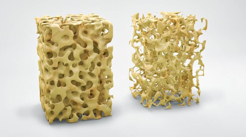
Bone mineral density increases with age until adulthood. The highest density persists until menopause, when it begins to decrease and osteopenia to osteoporosis occurs.
Densitometry is an investigative method that measures the density of bones and their minerals.
Reduced bone density is classified as a metabolic bone disorder or metabolic osteopathy. It develops slowly and lasts for months to years. The most common metabolic bone disorders are osteoporosis and osteomalacia.
Bone and its structure
Bone has a supportive function, a protective function, a hematopoietic function, a storage function for calcium and also a storage function for ions essential for life such as calcium, phosphorus, magnesium, sodium.
The surface of the bone is covered with a fibrous membrane called periosteum. It is richly vascularised and the nerves pass through it. It is of great importance for the nutrition of the bone. It covers the whole bone except the joints.
Beneath the periosteum is cohesive bone tissue, which forms the hardest part of the bone.
Bone tissue is the foundation of bone.
The underlying bone tissue is mineralised and consists of compounds of calcium, phosphorus, magnesium and sodium.
The minerals of the bone are bound to collagen fibres. The fibres are either in the form of beams to give maximum strength or in layers forming plates.
In the long bones of the body, in their central part, there is a cavity of bone marrow. This, together with the spongy beams, is filled with bone marrow.
Bone marrow is the factory for the production of blood. It makes red blood cells, white blood cells, platelets and monocytes.
Reduced bone density
Decreased bone density occurs for a variety of reasons.
Osteoporosis
Osteoporosis, popularly known as bone thinning, is one of the disorders of bone metabolism. It is a loss of bone tissue in which calcium is lost from the bones. This leads to fractures, especially in the elderly, as the bones become thinner and less strong.
To treat it, there are medications that are administered according to the condition and extent of osteoporosis.
Osteoporosis is classified as a silent disease that is asymptomatic in most people. However, some people notice that something is wrong because their spine hurts and they lose height.
The symptoms of osteoporosis are:
Back pain
loss of height
curvature of the spine
What causes osteoporosis
Reduced movement, sitting
Low calcium consumption
Bad habits such as smoking, alcohol, black coffee, sweetened coloured drinks
Hereditary factors
Gender: women are 3-4 times more likely to develop osteoporosis than men
Chronic diseases such as insulin-treated diabetes, inflammatory rheumatic diseases, kidney and digestive diseases
Certain medications: corticosteroids, heparin, antiepileptic drugs
Classification of osteoporosis
Primary osteoporosis is the most common consequence of aging and is divided into:
Type 1 (post-menopausal) occurs in women aged 55-65 due to a lack of the hormone estrogen and in men due to a decrease in testosterone, which leads to bone loss. The loss of bone mass disrupts the structure of the bones, the bones become thin, brittle and easily broken.
Type 2 (senile osteoporosis) is caused by a decrease in bone formation. It occurs in people over 70 years of age due to a decrease in parathyroid hormone and calcium absorption through the intestine, as well as a decreased concentration of vitamin D
Secondary osteoporosis occurs at the same time as another disease or treatment:
cancers such as lymphoma, leukaemia, myeloma, mastocytosis
as a result of taking medication for other diseases such as thyroid hormones, glucocorticoids, antiepileptics, heparin, cytostatics, etc.
Local osteoporosis is localized to only some parts of the bone or to several bones.
Juvenile osteoporosis occurs in children aged 8-14 years, sometimes in younger children during a period of rapid growth.
Read also.
Osteopenia
Osteopenia is a condition between osteoporosis and healthy bones. It is the beginning of osteoporosis.
Osteopenia means that your bones are weaker than normal healthy bones.
Osteopenia often begins to develop around age 50.
With diet, appropriate exercise, medications, and proper nutrition, your bones can be maintained and protected from osteoporosis.
Osteomalacia
Osteomalacia is popularly known as bone softening. It is characterized by insufficient mineralization of bone tissue.
They are caused by calcium and phosphorus deficiencies due to inadequate food intake or excessive losses, such as in kidney disease.
Another cause may be vitamin D deficiency, where the bone is unable to mineralize sufficiently, even though the body has enough minerals.
Examination of bone density
A bone density test is indicated by a specialist doctor who is an orthopaedic surgeon, rheumatologist, endocrinologist and gynaecologist.
Why is a bone density test performed?
The test reveals reduced bone density before a bone fracture occurs.
The test determines the risk of developing a bone fracture.
It confirms that you have a diagnosis of osteoporosis and determines its severity.
Monitors the effectiveness of osteoporosis treatment.
Bone density is measured using X-rays after passing through bone tissue. Compared to a conventional X-ray machine, this is a negligible exposure to radiation.
The bone density test estimates how strong and dense your bones are.
X-rays measure the amount of minerals and calcium in your bones. The more you have, the less likely you are to fracture. This means your bones are strong and solid enough.
Density is measured on the bones that are most likely to fracture. The forearm area, the upper end of the femur, the tibia, the tibial vertebrae.
Bone density results are expressed in g/cm2 and on the initial T and Z, which are standard deviations.
The examination and its values are applicable to all age groups, both adults and children.
The distribution of the result is classified into 3 grades:
Normal healthy bone
Osteopenia
Osteoporosis
To monitor the bone density status of untreated and medically treated patients, their condition and, if necessary, improvement after medication is repeatedly monitored.
Densitometry is only an ancillary examination. A thorough history taking and clinical and laboratory examination are necessary to establish the diagnosis.
The examination takes only a few minutes.
Bone density measurements are performed to assess bone loss, predict fracture risk, monitor and record the rate of bone loss, evaluate the effectiveness or failure of treatment and also to improve the patient's attitude towards his/her condition.
Diagnosis of osteoporosis
How is osteoporosis diagnosed?
Osteoporosis is diagnosed in three ways:
Bone mineral density using a DXA (densitometer) test if the early value is less than -2.5. The norm is 0.
Fracture - If you are over 50 years old or have had a fracture in your spine, wrist, hip, shoulder, ribs, or pelvis, your bones are weaker and require further testing.
FRAX is a fracture risk calculator. You enter your bone density value measured by DXA scan, your age, gender, height, weight and 7 other questions (e.g. previous fractures, smoking, medication, rheumatoid arthritis, alcohol...) into the calculator. The calculator will calculate the probability of different types of fractures in the next 10 years.
Instruments for measuring bone density
The instruments differ in the principle of measurement, measurement of different areas of the skeleton, radiation load and accuracy of measurement.
Therefore, not every method is suitable for a specific diagnosis, for example for the detection of osteoporosis or the measurement of the dynamics of bone changes.
DXA densitometric examination
The standard in bone densitometry is dual energy X-ray absorptiometry - DXA.
It is mainly used to diagnose osteoporosis. According to the WHO (World Health Organization), it is the most accurate and fastest method.
It uses X-ray sources that provide accurate bone resolution based on the absorption of X-rays by bone tissue.
In this examination, the mineral content of the bone per cm2 (g/cm2) is assessed and compared with that of a healthy population. Deviations from the average value are assessed. This number is referred to as the T-score.
The Z-score calculation represents the deviation between the same age group. The higher the deviation, the less mineralized or osteoporotic the bone is.
There are 2 types of DXA instruments, namely central and peripheral.
Central DXA is an examination that focuses on the bones of the spine and hip.
The peripheral DXA test measures bone density in the heels, fingers and wrists.
A woman examined with a densitometric device. Source: Getty Images
Peripheral densitometry
Forearm SXA, DXA measurements.
The SXA measurement was one of the first instruments to measure bone mass. Classically, measurements were taken on the peripheral bones, especially the forearm. Only one beam of radiation was used and the forearm had to be placed in a tub of water.
DXA is safe and has no contraindication in which the examination cannot be performed. However, the examination is not recommended in pregnancy, especially in the first trimester. Also, if a contrast agent is used before the examination or in the short period before it, at least 7 days.
Peripheral DXA is measured using two radiographs.
Lateral DXA is used to assess deformities in the vertebral region. The advantage is the simultaneous measurement of BMD (bone density) and the detection of fractures.
Some types of DXA also allow you to assess bone health, not just by measuring density. It also measures:
Vertebral fracture assessment (VFA) is a lateral view of the spine that can reveal fractures or crushed bones in the spine that you may not even be aware of. This is good for making a more accurate diagnosis and starting follow-up treatment.
The trabecular bone score (TBS) is the internal structure of the bone in the spine at a microscopic level. The higher the number, the better.
Full Length Femoral Imaging (FFI) is a technique for obtaining an image of the entire femur, not just the area around the hip that is examined in a standard DXA. It can show thickening of the bone that could lead to an atypical fracture.
Hip structural analysis (HSA) looks at the size, shape and configuration of the hip to determine the likelihood of a fracture.
Whole-body densitometry
Whole-body DXA provides accurate information on the proportion of bone mass, non-fat body tissue, and also the amount of body fat.
The examination scans the whole body. For better analysis, the body image can be divided into smaller parts such as the left, right, upper limb, trunk and lower limb.
The results are evaluated by measuring three types of tissues, namely adipose tissue, muscle and soft tissue, and bone tissue. They are measured in grams or percentages.
How is the test performed?
The examination is painless, quick and does not put any strain on the patient.
During the examination, the patient lies on the bed for a few minutes (approximately 20 minutes).
Two X-rays are taken of the bone, either of the femoral neck, lumbar vertebrae or wrist.
One beam images the soft tissue content and the other the absorption of the beams, i.e. the hard structure of the bone.
The more minerals there are in the bone, the fewer rays pass through the bone to the detector. Based on this, the computer evaluates the difference between the X-rays sent and received.
The values are compared with those of healthy individuals of the same age and sex to produce a Z-score.
The T-score reflects the severity of osteoporosis.
Preparation for densitometric examination:
No special preparation is required
It is not recommended to take calcium supplements at least 24 hours before the examination
Do not undress during the examination, just lie down. The only condition is not to wear buttons or zippers around the bone to be examined
Follow-up examinations while taking medication are recommended every 1 or 2 years. If you do not have osteoporosis, it is recommended every 2 years, especially for women in or after menopause.
Other examination methods
Quantitative ultrasound (QUS) is measured based on the propagation of the ultrasound wave in the bone tissue. The ultrasound machine measures the speed of sound propagation in the bone (SOS) and the attenuation of the sound wave (BUA). The machine evaluates this combination of parameters obtained.
Ultrasound densitometry allows the examination of peripheral parts of the bone (pelvic bones, forearms and finger joints). This device is based on the measurement of ultrasound waves that pass through the bone part under examination. The amount of bone minerals is examined, which tells about the quality of the bone.
The ultrasound measuring system is:
dry - the probe directly touches the limb and the transmission of the waves is provided by a gel
wet - the probes are embedded in the walls of a tray containing the fluid in which the limb is immersed
The Omnisense sonographer works on the principle of ultrasound along the bone. Measurements can be taken on the forearm, finger, fist of the lower limb, metacarpal bone of the lower limb.
The Omnisense device is the only device based on the principle of ultrasound measurement that meets the WHO criteria in the field of measurement. The result is evaluated graphically and numerically by measuring T-score, Z-score, SOS.
Quantitative computed tomography (qCT) is used to assess vertebral structure and can be used to assess fracture risk. However, this method is not suitable for the assessment of osteoporosis.
X-ray examination of bone: Bone that is not sufficiently mineralised has a lower absorption of X-rays. However, X-ray examination can only detect osteoporosis at a late stage, when the loss of density is greater than 30 %. It is therefore not used for the diagnosis of osteoporosis.
Magnetic resonance imaging provides a 3D image of the bone but is more focused on diagnosing other bone diseases.
Interpretation of results
The result is expressed in values:
In grams of minerals per cm2
In percentages - density measurements compared to age and gender
In standard deviations from the norm - the measured values are compared and T and Z scores are distinguished.
The T-score expresses the number of standard deviations from the density value of young 20-29 year old healthy individuals of the same sex. The value of the T-score is crucial for the diagnosis of osteoporosis.
The Z-score expresses the number of deviations from the ideal value in healthy individuals of the same age and sex. It is used to assess outcomes in children, men under 50 years of age and premenopausal women.
BMD (bone mineral density) is assessed based on the amount of calcium in the bones. BMD bone mineral density testing provides information about healthy bones.
BMD determines the amount of bone mass
TBS (trabecular bone score) determines the microarchitecture of bone mineral, determines the quality of the bone. The TBS program assesses the degree of damage to the bone, its microarchitecture in the number, density and interconnectedness of the trabeculae that make up the bone.
Low TBS values indicate shrinkage of the trabeculae and perforated to thinned bone. This is indicative of poor bone quality.
It is used to examine the disease of thinning bones by osteoporosis and its degree, based on which treatment is recommended.
TBS results - bone quality values (table)
Normal value
above 1 350
Slightly reduced
Grade 1
1,300-1,350
Grade 2
1,250-1,300
Grade 3
1,200-1,250
Significantly reduced
1,100-1,200
Very much reduced
below 1,100
T-score results
SD are the measured units of standard deviation.
A normal bone density value is within 1 SD or above -1 SD. (+1 to -1). This means that numbers like -0.9, 0, 0.6 are normal.
Osteopenia is a low bone mass of -1 to -2.5 SD. Numbers like -1.1, -1.9 to -2.5
Osteoporosis is bone mass density less than -2.5 SD values.
Low bone mass is not low enough to make a diagnosis of osteoporosis. It is referred to as osteopenia. It can be caused by a number of factors such as heredity, low body weight, general health and medications taken that negatively affect the bones.
A healthy diet containing calcium, vitamin D and plenty of exercise such as walking, running or dancing is recommended for osteopenia.
For osteoporosis, these healthy habits will help. You will probably also be advised to take medication to slow or reverse bone loss.
Z-score
For premenopausal women and men under 50, bone density is assessed using a Z-score.
A Z-score of -2.0 SD or less is assessed as reduced bone density.
A value greater than -2.0 SD is considered to be in the normal range for the age group.
In children younger than 20 years, the Z-score is assessed.
A Z-score of less than -2.0 SD is considered to be low bone mineral density for that age.
Bone scanning with densitometer and graphical representation of the result. Source: Getty Images
Indications for densitometric examination
The following may lead to a recommendation for densitometric examination:
Estrogen deficiency, premature menopause under 45 years of age, menstrual cycle disorders, amenorrhea for more than one year, primary hypogonadism
When corticosteroid treatment will last longer than 3 months, bone density measurement is indicated before starting treatment (drugs: prednisone, cortisone, dexamethasone)
In case of a fracture of the femur in the mother
Low BMI (body mass index)
In osteoporosis-related diseases (anorexia nervosa, malabsorption, rheumatoid arthritis, chronic renal insufficiency, hyperthyroidism, chronic inflammatory bowel disease, Cushing's syndrome, genetic and metabolic bone diseases, etc.)
When osteoporosis is suspected on the basis of an X-ray
Fractures of the spine, femur, forearm after inadequate trauma
Loss of height or thoracic kyphosis in hunched posture
Spinal pain without cause
Subsequent treatment with antiporotic drugs (administration of drugs to treat osteoporosis)
Chronic use of medications (anticoagulants, antiepileptics, thyroid hormones, immunosuppressants, cytostatics)
Women over 65 years of age
Men over 70
Age over 50 and bone disorders
You have had an organ transplant
How to increase bone density?
Increasing bone density and replenishing minerals in the bones is also possible with a balanced diet.
Your bones contain minerals from an early age until adulthood. At the age of 30, bones reach their maximum bone mass.
To maintain healthy bones, you need enough calcium and vitamin D, which helps your body absorb calcium.
Calcium can be supplemented through a varied diet.
Suitable sources of calcium include:
Milk, dairy products, cheese
Green leafy vegetables - broccoli, cabbage, spinach, cucumber
Vitamin D is obtained naturally from the sun. In summer, even by a short stay in the sun. In the winter months, it is recommended to take vitamin D supplements due to insufficient sunlight.
Vitamin D is also found in the diet:
In oily fish such as salmon, sardines, mackerel, carp, zander
Magnesium plays an important role in the conversion of vitamin D, which promotes calcium absorption.
Zinc helps form the mineral part of the bones.
Omega 3 fatty acids help protect against bone loss during aging.
Protein is important for healthy bones. According to new studies, low protein intake in the diet reduces calcium absorption. Low protein diets have been shown to increase calcium leaching from bones.
Vitamin K2 modifies osteocalcin, protein and promotes healthy bones.
Vitamin K is found in:
Liver
Meat
Eggs
Cheese
Sauerkraut
Soya products
Too much vitamin A does not benefit bones and increases the risk of fractures. Do not regularly consume liver and liver products that are fortified with vitamin A. Limit them to twice a week.
Vitamin C stimulates the formation of bone-forming cells.
Sports - strength training and weight training exercises help build and maintain healthy bones.
Maintaining a healthy weight contributes to healthy bones.
I graduated from the Secondary Medical School in Nitra, which gave me the basis for a career in healthcare. After school I worked for three years in the surgical department and then in the department of Anaesthesia and Intensive Care Medicine. In addition to my employment, I completed my bachelor's degree at the Faculty of Health Care in Banská Bystrica in Nursing and completed my specialisation studies in Anaesthesiology and Intensive Care Medicine. Since my childhood I was determined to become a health professional and help people with their health problems. Continuous education and study of new professional topics related to health care, which is constantly evolving, and gaining practical experience, helps me to write professional articles for this portal, which is available to everyone. My hobbies are multifaceted, I am also involved in healthy nutrition, overall healthy lifestyle. I spend my free time on education, creative work, handicrafts in cooperation with my daughter, thanks to which, we do not know boredom.
The aim of the portal and content is not to replace professional
examination. The content is for informational and non-binding purposes
only, not advisory. In case of health problems, we recommend seeking
professional help, visiting or contacting a doctor or pharmacist.