- cdc.gov - Child Development
- healthyparentshealthychildren.ca - How Children Grow and Develop
- ccrcca.org - Ages & Stages of Child Development
- link.springer.com - Child Growth and Development
- msdmanuals.com - Physical Growth of Infants and Children
- who.int - WHO Child Growth Standards
A child is not a miniature of an adult! What are the differences?
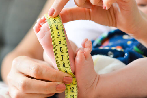
Photo source: Getty images
Babies are adorable and sometimes look a little too much like adults in appearance. Modern dresses, designer shoes, and "little adult" is the world. But in reality, babies are more different by a certain age than it might seem at first glance. If you have a baby at home or are expecting one, it's good to know these differences. Why is it important?
Article content
- In and out of mommy's tummy.
- First breath and further development of the respiratory system
- How does the blood circulation change after childbirth?
- Digestive system and organs of the abdominal cavity
- The oral cavity is adapted at an early age to suck milk
- The chubby cheeks are not only beautiful, but also have their significance
- From reflex sucking to targeted biting - the development of teeth
- The pharynx allows reflex swallowing of food
- Esophagus and stomach
- The oversized liver of young children fills most of the abdominal space
- Did you know that a child has a longer intestine than an adult?
- The excretory system develops rapidly
- The immature bone system
- Skin and thermoregulation
- The child's nervous system and senses
A new life begins with the union of a male and a female sex cell. This is the fusion of a sperm and an egg in the fallopian tube, called fertilisation. After fertilisation, the egg begins to divide (grow). It travels through the fallopian tube and enters the uterus as an embryo consisting of several cells.
Interesting:
If you thought you were Daddy's fastest sperm, you were very wrong. Sperm move at about 5 mm per minute. That's slower than a snail, which travels about 13 mm per second.
Their movement is aided by the uterine contractions during a woman's orgasm.
When the sperm reach the egg, only one of them penetrates the egg, but it is not the first.
The exact mechanism is still a mystery to us.
If all goes well, the embryo nests in the uterus and grows for nine months. Every day it becomes more and more human-like. After birth, we see a little person who is no different from an adult.
But the opposite is true. In fact, some organs and systems take years to develop.
In and out of mommy's tummy.
Life inside the womb is carefree and completely different from after birth. From the dark, damp environment where the fetus receives nutrition and oxygen through the placenta, birth brings it into the light of day.
With the first breath, the baby undergoes many changes to which it must adapt.
Why do we like being in mummy's tummy?
The uterus (Latin for uterus) is the organ in which the baby's growth and development takes place. It creates a mechanical barrier and thermal insulation to give it maximum protection during this vulnerable period. The mother's circulation provides heat regulation.
The supply of oxygen and nutrients occurs even though the blood circulation of the mother and the embryo is separated. This is done through the placenta. The placenta, or even the amniotic sac, is a temporary organ at the top of the uterus. After the birth of the baby, the placenta is also delivered.
Childbirth represents a major adaptive change for the baby
Pregnancy ends with childbirth.
Pregnancy ends with childbirth. A parent is alerted to impending labour by mild pain in the lower abdomen, spotting or light bleeding, irregular contractions of lesser intensity that may occur a few hours to days before birth, and pressure in the sacrum and rectum (pressure like stool).
Table of stages of labour:
| The first period of labour | Opening phase | Opening of the gate under the onslaught of fetal and sheath pressure |
| Second stage of labour | expulsion phase | the actual delivery of the baby |
| Third period of labour | final stage | delivery of the placenta |
First breath and further development of the respiratory system
Just before birth, the respiratory system of the fetus is sufficiently developed to be capable of taking its first breath after birth. In the womb, although the baby does not breathe, it already performs rapid and irregular breathing movements, resembling respiration.
In spite of these movements, no amniotic fluid or only a very negligible amount enters the lungs of the fetus. This is forced out by pressure on the chest during labour.
Subsequently, with the first breath, the lungs begin to fill with air and the newborn begins to breathe.
Complete aeration of the lungs occurs within a few minutes.
Why do I need to be careful with a full nose?
The oral and nasal cavities are very small. Most of the oral cavity is filled by the tongue and pharyngeal tonsils. This makes it impossible for the newborn to breathe through the mouth. The baby breathes mainly through the nose.
Therefore, the nasal cavity is very important in the process of breathing. A baby's stuffy nose can cause an obstruction in breathing, which the mouth cannot compensate for adequately.
For this reason, it is very important to prevent infections and in case of nasal discharge, the baby should be suctioned properly.
The newborn has a short and high larynx. The size and position of the larynx allows it to take in food and breathe at the same time.
Trachea, bronchi and lungs. What's different, what's the same?
The adult human has a trachea located about in the middle, in the throat area. Two main bronchi of equal length emerge from the trachea.
In a baby, the trachea is ovoid and extends mostly down the right side of the neck. The right bronchus is a kind of continuation of the right bronchus. The left bronchus bends at a greater angle and is longer.
Interesting:
The right bronchus is a continuation of the trachea. It has the same slope and is about half a centimetre shorter than the left. This fact is important especially when choking on a foreign body (beans, toy parts).In children, due to the slope of the left bronchus, it is very unlikely to be blocked. The body almost always enters from the right.
The last part is the lungs, which are bilaterally the same size in the adult despite the unequal number of lung lobes on the right and left sides. The fetal lungs are wider and higher up.
After birth, they enlarge and expand with each breath.
A baby's gland is an organ you won't find in an adult
The infant gland (Lat. thymus) is a temporary glandular organ that is located behind the sternum, in front of the trachea after birth. It reaches its largest size during this period and extends to the neck area.
The thymus is part of the lymphatic system and plays an important role in the immune system, maturing white blood cells, specifically T-lymphocytes, which are important in fighting infections.
It begins to disappear around the third year of life, but does not completely disappear until puberty.
In adults, the infant gland is absent, but its non-functional remnants can persist in connective tissue into old age in some people. They are of no significance.
How does the blood circulation change after childbirth?
Already in the first month of pregnancy, the foundation of the heart is formed.
However, it is far from being the organ we know from cardiology. The growth and functionality of the heart is a complex process that, despite its early start, is not fully formed until the age of 12.
The first breath closes the holes and closes the shunts in the fetal heart
The newborn goes from fetal to normal circulation at birth. The differences in fetal and adult circulation are enormous. One fact to remember: the fetal pulmonary circulation is non-existent. The lungs receive a minimum of blood.
The blood from the inferior vena cava flows directly into the right atrium, from where it passes into the left atrium through an opening called the foramen ovale. This opening closes with the first breath of the newborn, when it simultaneously switches to normal circulation.
In some cases, there is no closure and the blood of the atria mixes. This developmental defect is called an atrial septal defect. There is no septum between the ventricles. It forms later. If it does not form completely, there is a ventricular septal defect.
In addition to holes, there are shunts in the fetal heart on the blood vessels that provide fetal circulation. The most famous is the ductus botalli, which connects the pulmonary trunk to the main heart artery, the aorta. It also disappears after birth.
There are a large number of congenital heart defects related to the transition to normal circulation and failure of development, so they are not listed.
The heart is as big as its owner's fist
The heart is generally the size of a clenched fist. That is, its size corresponds to the size of the body.
- However, the shape of the heart in a newborn is spherical. The chambers are larger than the ventricles. The small volume of pumped blood and the thin musculature of the heart result in its faster action. A newborn therefore has a heart rate of around 140 beats per minute.
- By rapidly growing in length, the heart becomes oval-shaped around the third year of life. The muscles become massive and the heart rate is around 100 beats per minute.
- The typical ventricular shape does not develop until around the age of twelve, when the heart action is at adult levels of 80 beats per minute.
With age, the heart's ears and valvular apparatus also develop into the resulting structures of an adult. The heart muscle (especially the chambers of the heart) becomes massive. Already around the seventh year, the chambers are larger than the atria.
The position of the child's heart up to the age of one is transverse. It gradually turns oblique. It becomes fully oblique at the age of six.
Digestive system and organs of the abdominal cavity
The alimentary canal and organs of the digestive tract develop from an originally straight and primitive digestive tube consisting of three basic compartments.
The anterior compartment is the basis of the oral cavity, pharynx, oesophagus, stomach, duodenum, liver and pancreas. The middle compartment is the precursor of the small intestine, appendix and part of the large intestine. The posterior compartment gives rise to the remaining part of the large intestine and the rectum.
Interesting fact: From the blind and impassable fetal tube, a passage organ is formed, beginning in the oral cavity and ending in the rectum. In some cases, the openings do not form as they should, and congenital defects of impassability arise.
The oral cavity is adapted at an early age to suck milk
The oral cavity of a newborn is very narrow in relation to its overall weight and length. It is externally lined by lips with substantially thick musculature, which allows the baby to latch on firmly to the breast. The gums, which protrude into a sharp ridge, also help the nipple to grip.
The tongue is relatively wide compared to the oral cavity and its bulk practically fills the entire mouth by about 3 months of age.
The tip of the tongue is short and not very mobile. It often sticks out of the mouth (comical tongue protrusion). The child spits out and ejects solid food through it.
The root, on the other hand, is larger and thicker, allowing reflex swallowing of liquids in the first half of life. Its surface is relatively dry (lower saliva production) and often forms a white coating.
Interesting fact: The oral and nasal cavities form a single unit during intrauterine development. With the formation of the palate, the nose and mouth are divided into two distinct parts. In the case of palatal developmental disorders, cleft palate often occurs.
The chubby cheeks are not only beautiful, but also have their significance
It is not only the sucking reflex and the strong lips and pointed gums that are involved in breastfeeding. The chubby cheeks are also involved, as their structure forms a kind of soft cushion (mostly made up of cheek fat).
This prevents them from collapsing during active sucking, thus maintaining the correct vacuum in the oral cavity for breastfeeding.
From reflex sucking to targeted biting - the development of teeth
The foundations of the milk teeth are already formed in the womb during pregnancy, around the 6th and 7th week. They are deposited inside the upper jaw before we are born.
By 6 months, only the gums are visible externally, arranged in a pointed ridge. Around 6 to 8 months of age, the ridge disappears and the baby teeth begin to erupt through the flattened gums.
The final deciduous dentition is visible around the age of 2. It consists of 20 teeth, namely 8 incisors, 4 eye teeth and 8 molars.
Teething allows the child to transition from sucking to biting and chewing, which is extremely important for food intake. In addition to the teeth, the chewing muscles, tongue and salivary glands are also involved in this process.
The amount of saliva increases with the age of the child. In the neonatal period, it is poor. Infants produce about 100 ml of saliva per day, which is 10 times less than in an adult.
These teeth gradually fall out and are replaced by the permanent dentition. This happens at the age of 6 to 7 years. The permanent dentition has a total of 32 teeth (8 incisors, 4 eye teeth, 8 molars and 12 molars).
The pharynx allows reflex swallowing of food
Like everything, a baby's pharynx is smaller in size. In a newborn, it reaches about 4 cm, which is only one-third that of an adult.
In the neonatal period, it is positioned lower than in an adult and allows for swallowing of food. It lengthens with age and reaches its final dimensions, along with the nasopharynx, around the age of 15.
The size of the pharynx would not be unusual, but its lateral wall is dominated by the pharyngeal tonsil, which paradoxically grows to a larger size. Normally this is not a problem.
Esophagus and stomach
The oesophagus of the newborn reaches 11 to 15 cm, and about 40 cm in adulthood. At this age it forms a straight tube extending directly from the pharynx to the stomach. It grows very rapidly (faster than the thoracic spine running in parallel).
It is the speed of growth that causes it to bend forward axially at the neck. Final bending occurs by 2 years of age, which increases its flexibility.
The end of the oesophagus attaches to the stomach. The stomach is an organ that varies considerably in the neonatal period. It is mainly in terms of shape (tubular shape) that it begins to take on the basic features of its final shape just a few days after birth.
The lining of the baby's stomach is thicker, made up of deeper folds. The folds cause its functional surface area to be larger than that of an adult, despite its smaller capacity.
Its capacity is only 8 ml of fluid. After about a week, its capacity increases up to tenfold. Thereafter, the stomach's capacity increases by about 20 to 25 ml of fluid each month. In the first year, it is almost 300 ml, doubling by the age of three.
The stomach musculature is relatively weak despite its thickness, resulting in frequent vomiting of stomach contents back into the oesophagus and mouth - retching.
The oversized liver of young children fills most of the abdominal space
The size of the liver in newborns and young children is approximately 5% of the body weight. As we age, this organ shrinks and by adulthood it makes up only 2.5% of the total weight. Ultimately, this means that it is up to twice as large in children.
In an adult, the liver is located in the upper right part of the abdomen and is covered and protected by the right rib arch. Anatomically, it consists of two lobes. The right lobe is considerably larger than the left.
In children, the liver is not protected by the ribs. In the newborn, it even overlaps the rib arches on both the right and left sides.
The lobes are the same size, sometimes the left one can be even larger. Their size extends to the umbilicus. Deep in the liver is the gallbladder, which is practically immobile because of its size.
The development of the liver's function does not end until around 10 years of age. The remodelling mainly concerns changes in the liver's vasculature. It is only after 10 years that the structure of the liver lobules is formed.
Did you know that a child has a longer intestine than an adult?
The digestive tube of the fetus is sterile. During or shortly after birth, it is initially colonized by bacteria. Microorganisms enter the intestine through the mouth, nose and rectum.
This colonisation is important for the natural intestinal microflora that is part of our body. It starts 2 to 3 days after birth. It is slow until the colon is completely emptied.
A newborn's small intestine measures about 40 cm. By the age of three, it grows to a length of 1.5 to 2 m, which is a fairly rapid increase.
Compared to an adult, it is too large, resulting in a disproportionately larger baby tummy. The overall growth of the body flattens the originally bulging tummy.
Its curved villi press directly against the abdominal wall and are not protected by the peritoneum until about 3 years of age. The peritoneum develops, but is too short to cover such a long intestine.
Interesting:
When a child is choking on a foreign body, the Heimlich maneuver should not be performed until age 3.
The reason is the fragility of the intestine, which is not covered by the peritoneum (risk of intestinal rupture and spillage of intestinal contents - inflammation, sepsis, death). Unlike an adult, it has a larger blood supply (risk of haemorrhage, death) and in addition, the liver fills almost the entire part of the cavity from the navel upwards, including the intestines (liver rupture, haemorrhage, death).
The small intestine is almost the same at age 3 as in an adult. This cannot be said for the large intestine, which develops throughout childhood.
The large intestine of a newborn is at fetal level, reaching about 65 cm.
It is only after half a year that the adult-like protrusions begin to form. Until then, they are not present. The mucous membranes, algae and villi begin to take shape at about 3 years of age.
However, it is still developing during the following years. It reaches its normal length at about the 7th year of life. Its position in the abdominal wall is constantly changing until adulthood.
Interesting fact: Because of the immaturity and constantly changing position of the colon in newborns and smaller children, it can easily bulge out through the rectum. This is most often the case with stool. It is not a serious condition. The colon usually withdraws spontaneously.
The appendix differs mainly in size. Its size plays a role in the location of the worm-like appendix, which is much higher up in the right side of the abdominal cavity.
Normally, the appendix is insignificant. Around the age of 3 years, the appendix moves downwards. It does not reach its final topographical position in the right hypogastrium until the age of 14 years.
Interesting fact: With appendicitis, the child may not have pain in the right lower abdomen. The appendix often insists on the right kidney, sometimes even on the lower part of the liver.
The newborn has a naturally small rectum.
Because of its size, it is relatively long and stretchable, despite the not very strong musculature of its sphincters. The weak musculature causes reflex defecation, which is not influenced by will.
The excretory system develops rapidly
Already around the second month of pregnancy, the kidneys and the urinary tract as such begin to develop. This is an organ system that is one of the first to form. A fluid that is already very similar to urine is excreted into the amniotic fluid.
The high demands on the kidneys are evidenced by wet diapers
After birth, the kidneys and urinary tract are anatomically fully developed.
They are much larger in mass and are stored relatively low in infancy. Functionally, however, they have their gaps. Despite their functional immaturity, up to twice the load is placed on them.
Why?
The diet of the newborn and infant is predominantly liquid. It contains a high amount of water, which is mainly excreted through the urinary tract. This means that the child urinates much more than the adult, about 70 ml/kg.
The lower efficiency and lower filtering capacity of the kidneys causes the urine to be less concentrated. This is reflected externally by its paler colour.
The development of the urinary tract gives us the answer to why children wet the bed
Two ureters with weak musculature protrude from both kidneys. Their rapid growth, weak wall and low positioning of the urinary organs cause them to temporarily wave-shaped.
This shape is the reason for frequent urinary tract infections in children.
The ureters empty into the bladder. The bladder is also composed of a fine layer of muscle that only thickens over the years. It is not at the adult level until around the age of 6.
In young children, especially in the neonatal and infant period, a full bladder is visible to the naked eye. Its filling objectively enlarges the tummy, giving observant mothers a chance to prevent minor accidents during the diaper withdrawal period.
We see will-controlled urination in about two-year-olds. The frequency decreases to 10 times a day, but the bladder filling is greater.
Interesting:
Surely you are no stranger to the image of a doctor or nurse holding a freshly born newborn and he pees right in his face. This is to be expected due to the change in thermal stimuli and mechanical pressure on the abdominal wall as it passes through the birth canal. The good news is that he subsequently stops peeing for one full day, which is to be savored.
Why?
After this quiet period, the baby urinates about 30 times a day.
The immature bone system
The bone structure of newborns and young children is very immature. However, this weakness is compensated extremely well by their strong skeleton (periosteum), which protects the fragile bone skeleton.
The newborn's head is proportionally larger than the rest of the body
The structure of the skull of a newborn and an adult is very different. The most obvious difference is its size, compared to the rest of the body. In addition, the facial part is considerably smaller than the brain part.
The bones that make up the cranial vault are thin but flexible. They have loose joints, which is important at birth. At the top of the head - the site of future joints - there is a large fontanelle and at the back of the head a small fontanelle. The fontanelles (called scapulae) are formed by ligaments at the sites of future bone sutures.
The bones of the skull do not have a spongy structure like those of the adult. This begins to form around the sixth month and formation ends around the second year.
The child also lacks cavities (maxillary, nasal, frontal and wedge). Their development is very slow. The foundations of these cavities are not seen until children are 2 years old.
The spine makes up to 40% of the child's total length
Despite the relatively long spine, the vertebral bodies of newborns are short. The length of the spine is compensated for by the taller intervertebral discs.
The spine is more pliable and does not yet have the typical curvature, although there are signs of this already in the fetus. Therefore, its shape adapts to the base. The spine does not become stable until around the sixth year.
The curvature of the cervical spine occurs when the baby is able to hold its head upright. The sacral curvature occurs when the baby is able to stand on its feet. These curvatures result in thoracic kyphosis.
Large head, large trunk and short limbs
Short arms and legs are not a defect. Compared to the skull and torso, they grow much faster. The greatest growth is seen in children under four years of age, the period when they are learning to crawl and walk.
Changes in the size, shape or growth of bones and skin protrusions take place throughout childhood and end in adulthood. Joints are much more mobile in childhood. As for their shapes, this is very individual for each individual. They are also determined by external factors (load, weight, mobility).
Skin and thermoregulation
The skin of the fetus is made up of only one layer of cells at the beginning of pregnancy. The multi-layered epithelium is formed around the third month of pregnancy. The folds and skin lines are completely absent. This makes the skin of the fetus completely smooth and soft.
Skin furrows appear around the 4th month of fetal development (the bases of the unique fingerprints). The cornified layer appears sporadically only in the 7th month of pregnancy.
The softness of the baby's skin hides the high water content
In the womb, the fetus is protected from the effects of amniotic fluid by a whitish, oily mass composed of epithelial cells, skin cells, lanugo and fat.
This layer also protects the baby from injury during and shortly after birth. Subsequently, it thins and disappears completely.
Hair is produced in the womb. After birth, the body is continuously covered with fine hairs (lanugo), except on the hands and feet, which are smooth. Most of this hair falls out before birth.
At the same time, definitive pubic hair is formed. The nails are also formed before birth, have white stripes and grow rapidly.
The skin of the child and the adult does not differ in anatomical structure after the protective layer has disappeared. It is made up of skin cells. The superficial cornified layer is weaker and thinner, and therefore often peels off.
The softness and suppleness of children's skin, despite the smaller number of elastic fibres, is due to its high water content - almost 80%. It is even so soft that the vascular pattern shines through.
The reaction of newborn skin is alkaline. It changes to acidic after a few weeks. The acidic reaction is important as a defence against infection, which our babies are exposed to every day through constant skin contact with urine or faeces.
Interesting fact: For the first 3 days after birth, the baby does not sweat at all. The sweat glands start to function around the 4th day of life. The sweat is alkaline and the final acidic pH occurs at the end of the first month.
Even a black baby is born white. How is that possible?
The colour of the skin of newborns is the same in every human being, regardless of race and genetic predisposition. It contains no pigments. Even in the case of the black race, a newborn baby is pale pink.
But it doesn't take long for differences to become apparent. Skin pigments begin to form immediately after birth. The subsequent colouring of the baby is influenced by the genetic make-up it has encoded in it.
Body temperature regulation
In the mother's tummy, the baby is protected from external influences. In addition, the mother's body temperature influences and regulates the body temperature of the fetus while the baby's thermoregulatory system is in the developmental stage.
In newborn babies, the subcutaneous connective tissue is only 2 mm. The layer of subcutaneous fat is also thin. Therefore, especially in the newborn period, but also in infancy, the baby easily loses body temperature. This can lead to general hypothermia even though the body temperature regulation is fully functional.
Interesting fact: Most of the body heat escapes through the head, which is proportionally larger in relation to the baby's body. Therefore, it is important not to forget the caps, which are of great importance in the prevention of hypothermia and colds.
We should also be vigilant throughout the toddler period. Colds are the cause of frequent colds in children.
The child's nervous system and senses
The nervous system, despite its complexity, is the most mature system of the newborn. It must be so because it is essential for survival itself. The nervous system could be said to be complete and only improves during life. The nerve cells mature fully between 3 and 8 years.
Interesting.
Brain and spinal cord
The forebrain is the largest part of the brain, and the same is true for children. In a newborn, the brain weighs up to 390 g. In a six-month-old child, its weight doubles. At one year, it weighs a staggering 1,200 g.
The frontal lobe of the brain is the smallest, and it grows rapidly. The whorls are formed, only their shape and position relative to the fontanelle in the skull changes.
The spinal cord is longer than that of an adult, and in a newborn it extends to the third lumbar vertebra. In fact, it shortens because it filled the entire spine during intrauterine development.
Basic senses - how's your little one?
A baby develops all its senses right after birth. But everything takes its time. That means they mature and become more refined with age.
Table with the basic senses of a person:
| Visual receptors |
|
| Auditory receptors |
|
| Olfactory receptors |
|
| Taste receptors |
|
| Touch receptors |
|
Interesting resources
Related

Bc. Lucia Sýkorová
Healthcare worker
I graduated from the Secondary Medical School and the Faculty of Medicine in Emergency Medicine. Since 2008 I have worked for 10 years in the emergency medical service as a paramedic. I am still involved in urgent pre-hospital care and emergencies as an emergency medical service operator on the emergency call line 155.
View all articles by the same authorThe aim of the portal and content is not to replace professional
examination. The content is for informational and non-binding purposes
only, not advisory. In case of health problems, we recommend seeking
professional help, visiting or contacting a doctor or pharmacist.


