Retinal and vitreous diseases involve various problems that affect these two closely adjacent structures. Risk of eye damage. Early diagnosis is important.
Retinal and vitreous disorders involve a variety of problems. Their origin may be in the gradual aging of the body, but also as a result of other diseases. They occur long-term (chronically), however, the second group are those that occur acutely, ie suddenly.
Even in this group of diseases, a preventive professional examination is important.
Early diagnosis and detection of difficulties helps with effective treatment. Which can significantly affect the extent of eye damage.
What is the retina and vitreous?
These two structures are close together, neighbors together.
Anatomy of the eye - anterior pole of the lens and posterior pole of the retina. Photo: Getty images
Retina
It is one of the most important parts of the eye. Its task is to capture light rays. Light-sensitive cells, such as rods and suppositories, help her with this.
These capture light.
We know these light-sensitive retinal cells :
rods are light-sensitive cells that process light of lower intensity
they do not recognize the color
suppositories are used to capture the light of different wavelengths, and therefore colors
they recognize colors, intensity, color saturation
ensure visual acuity
the highest number is in the central hole of the yellow spot, the so-called fovea centralis
there are approximately 6 million suppositories on the retina
Obraz zachytený sietnicou sa následne zrakovými nervami prenáša do zrakového centra v mozgu. Tu sa zrakový vnem ďalej spracuje a vytvára sa obraz, ktorý aktuálne vidíme.
There is an important macula lutea on the retina, a yellow spot about 5 mm in size. The macula consists of neuroreceptors, mainly suppositories, and at the edge of the rods. It also contains a xanthophilic pigment, which has a typical color, thanks to carotenoids and vitamin A.
Glass
The vitreous makes up 80% of the volume of the eyeball (bulbus oculi) and is, therefore, the largest part of the inside of the eye. In the front, it touches the surface of the lens case. It is clamped at the posterior pole of the eye at the outlet of the optic nerve.
Vitreous is made up of components:
water - 98% content
collagen, a structural protein
hyaluronic acid, a glycoprotein, gives the eye a gel-like consistency
chondroitin sulfate
Vitreous, technically also Corpus vitreum, is a transparent clear, colorless jelly-like mass. Its main task is to maintain the intraocular pressure, and thus the shape of the eye, plus it is part of the optical system of the eye.
The site where the vitreous urges the retina is referred to as the vitreoretinal interface .
The vitreous and retina are normally connected in only a few places. And these are:
around the target of the optic nerve
at the site of retinal vessels
in the vitreous base area
The rest of the vitreous is only loosely attached to the retina.
The most common diseases of the retina and vitreous
The retina and vitreous work closely together. Both can affect different diseases, which in some cases have a common connection. They cause visual impairment.
The table shows the most common diseases of the retina and vitreous
occlusions (closures) with ischemia (non-bleeding)
Causes
We know various diseases of the retina and vitreous. These two structures are closely adjacent and interact with each other. In the following section, we list some of the diseases that affect them.
Macular degeneration
It is also referred to as age-related macular degeneration or VPDM for short. The disease affects the central part of the retina, and thus the macula, the yellow spot.
The name itself means that old age is a risk factor. However, the exact cause has not yet been fully revealed.
Other risk factors such as genetic predisposition, familial occurrence, the effects of sunlight, smoking, alcoholism, diabetes, and high blood pressure are also attributed to the outbreak of difficulties. The risk of occurrence is also increased by the already present refractive error, especially farsightedness.
The disease itself is further divided into dry or wet forms.
The dry form represents approximately 90% of cases. It arises as a result of the accumulation of waste products of metabolism in cells. It is manifested by small yellow dots in the retina, which are visible during the examination of the ocular background.
Disability leads to visual impairment and decreased visual acuity. Blurred vision, impaired vision at dusk, and in low light conditions. In the last phase until the loss of vision.
The wet form is less common in approximately 10% of VPDM cases. It is caused by new blood vessels and peeling of the retina.
Newly formed blood vessels damage the retina, causing bleeding and swelling of the retina. This type occurs suddenly. It causes image distortion and significantly impairs visual acuity.
It arises as a complication of diabetes. Diabetes in the long run results in various difficulties throughout the human body.
In the case of the eye, this is a negative for the small vessels of the retina. These are damaged, which in the worst case leads to blindness.
It can also occur with uncomplicated diabetes, which is mild.
The vessels of the retina are affected by various changes that cause bleeding, swelling, or heart attack, and thus bloodlessness. The most severe form is proliferative diabetic retinopathy. It is characterized by the formation of new, however, pathological vessels.
Pathological vessels often bleed into the retina or vitreous. As a result, retinal detachment can also occur.
Like diabetes, high blood pressure has a negative effect on the whole body. In addition to the risk of stroke and heart attack, it also causes other problems.
The problem is especially untreated, poorly or insufficiently treated high blood pressure. At high values of blood pressure, spasms occur - contractions of blood vessels and also the transfer of fluid into space outside the vessels, and thus into the retina or vitreous.
Similarly, in this case, there is bleeding, swelling, or non- bleeding. The damage occurs gradually. As time goes on, difficulties such as deterioration in visual acuity and loss of field of vision gradually add up.
Retinal vascular diseases
There are more common eye diseases that occur acutely and cause significant vision problems.
They are divided into artery or vein occlusions. Arteries are vessels that carry oxygenated blood and veins, which are not oxygenated.
The mechanism of arterial occlusion is the occlusion of small vessels by the embolus. An embolus is a loose blood clot that travels in the body's bloodstream and clogs the smallest blood vessels.
An ischemic stroke, for example, also occurs.
Atherosclerosis is also important for the development of these problems. This results in a narrowing of the lumen of the vessel. The main risk factors involved in the development are hypertension, diabetes, high blood fat, obesity, smoking, and thus a bad lifestyle.
Another mechanism is due to venous occlusions that result from narrowing or complete occlusion of the vessel. The main cause is atherosclerosis of the vessels. Blood accumulates behind this blockage, resulting in swelling and bleeding into the retina.
We know that they arise due to various risk factors, such as:
hypertension
hypercholesterolemia
old age, over 65, affects up to 50% of the elderly population
thermocoagulation disorders
diabetes
increased intraocular pressure
Retinopathy of prematurity
It is listed as the most common cause of blindness in children.
It occurs in newborns who are born before 32 weeks of pregnancy or in those whose birth weight was less than 1,500 grams.
These children have insufficient breathing and are therefore placed in an incubator for a long time. There they are given oxygen therapy. Where the oxygen concentration is high, above 40%.
During this period, the newborn's eyes are not fully developed, not even the retina or blood vessels. The high concentration of oxygen in the incubator results in the eye getting used to this increased value of the partial pressure of oxygen in the ambient air.
Consequently, outside the incubator, the normal value of the oxygen content in the air is no longer sufficient. There is an increased formation of new blood vessels to ensure a sufficient supply of oxygen.
These new blood vessels grow through the retina, can pass into the vitreous, and there is a risk of retinal detachment. Similar to macular degeneration.
Retinal detachment
The retina separates from the pigment layer, on which it normally attaches freely. As a result, fluid penetrates between the retina and the pigment layer.
It can be caused by an eye injury, but it is also a complication of other diseases, such as diabetes. In particular, a higher degree of myopia requires increased attention.
Symptoms that occur in this case:
flashes of light, phosphenes (flickering before the eyes)
floating flies in the field of view - as if threads, tufts
The vitreous loosely urges the retina, it is as if attached.
Under normal circumstances, the two structures merge in only three places. And this is the target of the optic nerve, in parts of the course of the retinal vessels and the area of the vitreous base.
The site where the vitreous urges the retina is referred to as the vitreoretinal interface.
As the body ages, changes occur in the area. The vitreous collapses, peeling from the retina.
If the condition is not further complicated, there are only milder difficulties.
There are difficulties such as flying flies in the field of view, also referred to as muches volantes. Possibly occasional flashes in front of the eyes.
The opposite is the case when complications occur.
If a stronger connection is formed between the vitreous and the retina, the collapsing and peeling vitreous pulls the retina. The retina crouches, lifts.
The result is image distortion, ie metamorphosis, which arises as a result of the pull of photoreceptors.
The retina is the thinnest in the macular area.
In the most severe case, a macular hole is formed.
The macular hole is a defect that affects the entire thickness of the neural layer of the retina, in the macular area. Manifestations are complications and visual disturbances, ie image deformation - metamorphosis, but also reduced visual acuity or loss of visual field, in the central part (in the middle).
It is actually an opening in the area of the yellow spot, through which the lower structures are visible. The size of the defect - the hole gradually increases.
However, its cause is not only old age. It occurs as a single disease or as a complication of other eye problems. An example is diabetic retinopathy or a post-traumatic condition.
The overall difficulties that affect a person depend on several factors, such as:
localization
the extent of the defect
duration, ie the time since its inception
Epiretinal membrane
The difficulty is due to the state of an epiretinal membrane (EM), that is, the surface membrane of the retina film with time becomes thicker. The result is its opacity, wrinkling.
The retina under the membrane deforms and deforms.
In a more severe stage, the pull persists and a macular hole is formed.
Manifestations are :
decrease in visual acuity
primarily into the distance
later also a problem with reading
image distortion - wavy lines
significant decrease in vision
up to a failure of the central field of view
Other vitreous changes, vitreous opacities or eye flies
As we already know, the vitreous makes up 80% of the intraocular environment. It reaches a volume of approximately 4 milliliters. From 98% it consists of water. The rest consists of collagen, hyaluronic acid, and chondroitin sulfate.
The vitreous insists on the lens at the front and the retina at the back. Towards the edges of the vitreous, its gel-like mass is denser. This part is also referred to as the base, it has a front and a back part.
The higher density is formed by the increased concentration of hyalocytes, which are cells that makeup collagen and hyaluronic acid.
Because the vitreous and retina are closely related, pathological changes in the vitreous also affect the retina. As above.
The vitreous itself can be affected by turbidity, for various reasons :
due to age, ie involutional changes
degenerative changes
bleeding
inflammation
1. Age-related vitreous changes
The vitreous has a homogeneous, uniform structure from birth. However, changes in its structure gradually begin in the second decade of life.
The table shows some age-related changes in vitreous
Title
Description
Syneresis
structural changes that occur after the second decade of life
liquefaction of the middle part of the vitreous
collagen-hyaluronic acid-binding disorder
Posterior vitreous membrane ablation
detachment of the posterior vitreous membrane
after 70 years of life up to 70% of the population
the thickened posterior surface is detached from the retina
2. Degenerative vitreous changes
This category includes those changes that are not caused by the aging of the body, and therefore the eye.
The table shows the degenerative changes of the vitreous
Title
Description
Asteroid hyalinosis
vitreous cataracts
small yellow - pale color
reflective and fixed (fixed)
non - liquefied vitreous
content of calcium, phosphorus, fats
after 60 years of life
predisposition in diabetes
mostly unilaterally
Synchisis scintilans
also small
flat
reflecting cholesterol crystals in the vitreous
golden brown color
they are freely movable
due to vitreous hemorrhage
they sit on the bottom of the cavity
Amyloidosis
the haze is in the form of granules or cotton wool
in the area of vitreous cortex
family occurrence
after inflammation or tumors
3. Vitreous blood flow
Vitreous hemorrhage is caused by various causes. It's not just after an accident.
Causes of vitreous hemorrhage:
after the injury, however, the eye may not be directly injured
in retinal vascular diseases, such as diabetic retinopathy, retinal vessel occlusion
in other general diseases
high blood pressure
leukemia
blood clotting disorders
during long-term treatment with anticoagulants, ie anticoagulants
There are various difficulties in bleeding into the vitreous. For example, seeing falling shadows in the field of view, decreased visual acuity, or the feeling of a veil in front of the eye.
4. Inflammatory changes of the vitreous
In inflammation of the vitreous, inflammatory cells as well as proteins from the blood penetrate.
Inflammation is caused by standard bacteria, viruses, fungi, fungi or parasites.
Inflammation is in most cases from the external environment after the injury, but also after eye surgery. Intraocular inflammation is uncommon. The reason for this type is usually blood transmission, the so-called hematogenous spread in other infectious diseases.
The vitreous is turbid by various particles, which may be pale or yellow. Which depends mainly on the cause of the infection. Inflammation is usually fast, the worst complication is loss of vision, ie blindness.
Eye floaters - muches volantes
This refers to small floating features in the field of view, which are visible especially on a bright, pale surface. For example, when reading or driving. They have the shape of dots, threads, or tufts.
Flies, such as vitreous turbidity, can be the cause of increasing age, they also occur in myopia, injury, inflammation, cataract surgery or diabetes.
However, the reason for their occurrence may be m
Symptoms
How the individual diseases manifest themselves is influenced by the place of disability, the extent, and duration. The difficulties concern the eye, and therefore the vision.
Here are some of the symptoms that are part of them:
decrease in visual acuity
the problem may be initially with remote image viewing
reading also makes it a problem later
eye floaters, ie muches volantes
floating formations in the field of view
they have different shape and size
better visible on a white wall or sky
they change position when moving the eyes
dots
nite
cobwebs
clumps
spots
flashes
falling soot
shadow or aperture, shielding of the field of view
visual field failure
deformation of lines and image, metamorphopsia
Deformation of the observed image - metamorphosis. Photo: Getty images
Diagnostics
Today, various diagnostic options are available. An ophthalmologist, ie an ophthalmologist, examines visual acuity, the back of the eye. The anamnesis and clinical difficulties described by man are important.
If necessary, an examination of the image deformation on the Amsler grid is added, photo documentation, OCT is created.
OCT, an ocular tomography, but also optical coherence tomography, is a non-invasive non-contact examination. More information about the examination can be found on Wikipedia.
This method is suitable mainly because it shows the individual structures of the eye in detail. The team is involved in the diagnosis of various eye problems. Distinguishes stage, development, and the exact type of difficulties.
Methods such as the Watzke-Allen test, the laser beam test or autofluorescence, microperimetry are also known for the examination of these diseases. Or the Gass hole classification.
It is an important differential diagnosis and determines the exact cause of the problem.
In particular, people with other general illnesses should have an eye examination at least once a year. Others should not forget about important preventive check-ups.
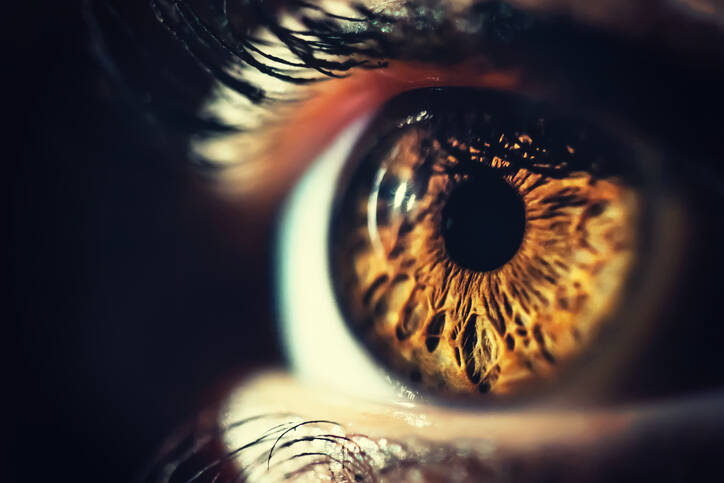
What is seen in the retinal examination. Photo: Getty images
Course
A wide range of diseases can have a diverse course. It is important to remember that if the problem occurs suddenly, acutely, ie quickly from full health, an immediate examination by a specialist is necessary. In this case, it is an ophthalmologist, ie an ophthalmologist.
Beware of the rapid onset of difficulties!
How it is treated: Retinal and vitreous diseases
Treatment of retinal and vitreous diseases: medications and surgery
"Facts About Retinitis Pigmentosa". National Eye Institute. May 2014. Retrieved 18 April 2020.
Openshaw, Amanda (Feb 2008). Understanding Retinitis Pigmentosa (PDF). University of Michigan Kellogg Eye Center.
Shintani, Kelly; Shechtman, Diana L.; Gurwood, Andrew S. (2009). "Review and update: Current treatment trends for patients with retinitis pigmentosa". Optometry. 80 (7): 384–401.
"Facts About Age-Related Macular Degeneration". National Eye Institute. June 2015. Archived from the original on 22 December 2015. Retrieved 21 December 2015.
Apte, Rajendra S. (5 August 2021). "Age-Related Macular Degeneration". New England Journal of Medicine. 385 (6): 539–547. doi:10.1056/NEJMcp2102061. PMID 34347954. S2CID 236926930.
"Retinal detachment". MedlinePlus Medical Encyclopedia. National Institutes of Health. 2005. Retrieved 2006-07-18.
Shukla Manoj, Ahuja OP, Jamal Nasir (1986). "Epidemiological study of nontraumatic phakic rhegmatogenous retinal detachment". Indian J Ophthalmol. 34: 29–32.
Ivanisević M, Bojić L, Eterović D (2000). "Epidemiological study of nontraumatic phakic rhegmatogenous retinal detachment". Ophthalmic Res. 32 (5): 237–9.
American Cancer Society (2003). "Chapter 85. Neoplasms of the Eye". Cancer Medicine. Hamilton, Ontario: BC Decker Inc. ISBN 978-1-55009-213-4.
Ryan SJ, Schachat AP, Wilkinson CP, Hinton DR, Sadda SR, Wiedemann P (2012-11-01). Retina. Elsevier Health Sciences. p. 2105. ISBN 978-1455737802.
"Retinoblastoma - Symptoms and causes". Mayo Clinic
Martin–Doyle, J. L. C. (22 October 2013). A Synopsis of Ophthalmology: Volume 4. Butterworth-Heinemann. ISBN 978-1-4832-8490-3.
D, Chaudhury; R, Meenakshi; R, Ramakrishnan; A, Mitra; S, Raju (2015-10-02). "Lipaemia Retinalis". The Official Scientific Journal of Delhi Ophthalmological Society. 26 (2): 107–110
Quesnel, NM; Lovasik, JV; Ferremi, C; Boileau, M; Ieraci, C (Jun 2004). "Laser in situ keratomileusis for myopia and the contrast sensitivity function". Journal of Cataract and Refractive Surgery. 30 (6): 1209–18.
Goldman, Lee (2012). Goldman's Cecil Medicine (24th ed.). Philadelphia: Elsevier Saunders. pp. 2429. ISBN 978-1-4377-2788-3.
The Sailor's Word-Book, Admiral W.H. Smyth, p. 483; Conway Maritime Press, UK, 1991. ISBN 0-85177-972-7
The secondary medical school in Nitra gave me the basis for my career in the field of health and diseases. Thanks to it, I worked for 2 years in the traumatology clinic and outpatient clinic at the Nitra Hospital. Since 2006 I was employed in the emergency medical service, where I stayed until 2017.
I completed my bachelor's degree at the University of Constantine the Philosopher in Nitra in the field of emergency health care. The bachelor's degree allowed me to continue my mission as a paramedic. In the meantime, I got a job at the emergency line 155. I have been working in pre-hospital health care until today.
I had an interest in people, health and even diseases in my childhood, which gave me the prerequisite to pursue this topic in adulthood. Studying and acquiring new information in practice provided me with a great basis for writing professional texts, in the form of articles that can be understood by ordinary people. Thus, my interest in the Health Portal has a solid foundation in years of practice and personal interest. Similarly, I am also interested in healthy eating, nutrition and overall healthy lifestyle. I fill my free time with family and sports.


.jpg "What is seen in the retinal examination. Photo: Getty images")


