Retinal and choroidal disorders
Retinal and choroidal disorders include

Diabetic retinopathy
Diabetic retinopathy is a disease that affects the ability to see. It can manifest in varying degrees of visual impairment and even blindness. It is related to diabetes.

Macular degeneration is a disease that affects the retina of the eye. The exact cause is not revealed. The genetic influence and action of internal and external factors, as well as lifestyle, are assumed.
Retinal detachment
Retinal detachment is a serious eye disease that can lead to poor vision and permanent loss of sight, i.e. blindness. Early treatment may be helpful.
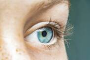
Diseases of the retina and choroid affect the most important part of the eye, where the light-sensitive visual cells, i.e. the cones and rods, are located, which is why it is important not to underestimate these diseases. Whether it is various inflammations, retinal detachment and detachment problems, or problems of a vascular nature or retinal nutrition, several diseases can also permanently damage vision. Some retinal and choroidal diseases can only be partially addressed, and such therapy is mainly in the form of laser treatment.
On the outer layer there is a layer of rods and cones at the back of the retina, while on the inner layer there is a layer of multipolar cells. In between there is still a layer of bipolar cells. At the front of the retina and in the choroid there are pigment cells which send out projections just between the rods and cones, these cells are also called photopigments and transform light into chemical impulses of the nerve type. The choroid is located between the white of the eye and the retina and contains many blood vessels that are used to supply nutrition and oxygen to the eye.
Inflammations of the retina and choroid
Retinitis and choroiditis most often occur together and are not so much manifested by pain as by a deterioration in the quality or acuity of vision. They are most often caused by various microbial infections that enter the retina from surrounding tissues or through the bloodstream from other parts of the body. Often the inflammations are asymptomatic, with a decrease in visual acuity only being a signal if the macula is also affected. However, early diagnosis as well as treatment can prevent permanent visual impairment.
Inflammations include focal chorioretinal inflammation, which has several forms such as retinitis, retinochoroiditis, chorioretinitis or choroiditis, also diffuse chorioretinal inflammation with the same forms, inflammation called Pars planitis, inflammatory Harada's disease or other unspecified inflammations. The most common inflammation is the common retinitis, or acute inflammation of the retina and choroid, also called professionally retinitis. In our country, two forms are the most common, namely retinitis pigmentosa and retinitis cytomegalovirus.
Retinitis pigmentosa is a disease directly affecting the light-sensitive rods and cones located at the back of the retina, with the rods that provide peripheral and night vision being affected more than the cones responsible for central and colour vision. This is why the disease is most often manifested by so-called night blindness and impaired visual field or side vision. Over time, the disease steadily worsens and treatment should be started as soon as possible.
Retinitis cytomegalovirus is an inflammation caused by cytomegalovirus, when this virus attacks the cells in the retina. In the initial stages, this type of inflammation hardly manifests itself at all, possibly with only partial disturbances of peripheral vision, similar to the previous disease, and at first only one eye is affected, but later the other eye is affected as well. Symptoms do not include, on the other hand, increased sensitivity to light or pain. In the case of this inflammation, too, it is true that if left untreated it can lead to loss of vision.
Retinal detachments and retinal tears
Retinal detachments, retinal detachments and retinal tears are among the most serious diseases that can affect this part of the eye and must be treated exclusively by surgery. They can arise from a variety of causes and accordingly there are several types of these diseases. These include retinal detachment with retinal tear, retinal detachment with retinal tear, serous retinal detachment without or with retinal tear, retinal detachment with traction, such as proliferative vitreoretinopathy with retinal detachment.
In addition to retinal detachment, various forms of retinal tears can also form on the retina without retinal detachment. There are also several forms of the disease and these are Horseshoe retinal tear without detachment, Round hole retinal tear without detachment, Operculum retinal tear without detachment or non-specific retinal tear. In addition, retinoschisis and retinal cysts, parasomatous retinal cyst, retinal pseudocyst and ora serrata retinae cyst also cause problems with retinal tear or damage.
Retinal detachment is a disease that can cause irreversible visual impairment in the later stages in some forms. The sensory part detaches from the pigmented part, which remains fixed to the choroid. The detachment may be idiopathic, i.e. a tear is formed, or without a tear, which is usually the result of an infectious disease in the eye. In retinal detachment, the rods and cones, which are important photoreceptors, are also severely disrupted.
The retina as such is nourished indirectly through the richly vascularized choroid, so that detachment and detachment of the retina from the choroid means disruption of this nourishment and subsequent death of the light-sensitive cells, which very quickly begins to manifest itself in vision and visual disturbances. The disease usually progresses very rapidly and arises from either inflammatory or post-traumatic causes. It must be treated surgically and, depending on the type of detachment, either by conventional or laser treatment.
Vascular occlusion
Diseases affecting the retina include vascular occlusions. There are several types of occlusions, for example, transient central retinal artery occlusion, centralis retinalis occlusion, retinal vein occlusion or other central retinal artery occlusions. Occlusion of the retinal vein may be incipient, partial, tributary or central. There are also several types of disease in central retinal artery occlusions, such as Hollenhorst plaques, arterial occlusion, partial, branch occlusion, or microembolism.
Occlusion is the closure of the stellate artery and vein, and can be temporary or permanent. While arteries carry oxygenated blood to the eye, veins carry deoxygenated blood out of the eye. Occlusion is manifested by sudden loss of vision and usually affects only one eye, or loss of central vision in a gradual form if the veins and not the arteries are affected. Occlusion occurs, for example, when a clot forms, embolization from another site, or as a result of certain inflammatory diseases of the blood vessels.
Occlusion most commonly affects men over the age of 60 and may also be related to diabetes, smoking or high blood pressure. Depending on the type of occlusion, it is addressed either by removing the obstruction in the blood vessels or by creating conditions to bypass the obstruction. The most common form of central retinal vein occlusion is combined retinal vein occlusion, but these cases of combined central retinal vein occlusion are rare in all retinal diseases.
In some cases, there may be simultaneous damage to the eye by occlusion and hearing loss or nerve inflammation, such as in the localised form of Wegener's granulomatosis. Overall, however, up to half of all cases of severe changes in the eye and vision are manifested by occlusion of the ophthalmic or orbital arteries, with the most frequent outbreak of vascular occlusion occurring at night. The cessation of nutrition is immediately manifested by unilateral loss of vision.
Other diseases of the retina
In addition to inflammation and problems with retinal detachment or detachment of the retina from the choroid, the retina is also affected by other diseases that may be related to the nervous system or may be degenerative or other changes directly on the retina. These diseases include background retinopathies and retinal vascular changes, such as Retinal Vascular Changes, Retinal Microaneurysms, Neovascularization, Perivasculitis, Vasculitis or Varixes and various retinopathies.
Within retinopathic retinal diseases, Coats retinopathy, exudative or hypertensive retinopathy, proliferative vitreo-retinopathy and retrolental fibroplasia affect the eye. The retina is also affected by various types of dystrophies, retinal hemorrhages, retinal pigment epithelium detachment, central serous chorioretinopathy, peripheral retinal degeneration reticular, microcystic, palisade, squamous or reticular, and also degenerations of the macula and posterior pole of the eye, such as various cysts, holes, wrinkles, drusen, Kuhnt-Junius degeneration and atrophic senile macular degeneration.
Other diseases of the choroid
In addition to the above-mentioned diseases and disorders, some other diseases, degenerations or deformities also affect the coil separately. These may include, for example, chorioretinal scars, which include post-inflammatory and post-traumatic posterior pole scarring and solar retinopathy, or choroidal degenerations such as choroidal atrophy or choroidal sclerosis. Coil rupture and subsequent bleeding, such as expulsive or complete choroidal detachment, is also dangerous.
Among the degenerative and destructive diseases occurring on the choroid is hereditary choroidal dystrophy. This is a group of diseases comprising different types of dystrophies such as choroideremia, choroidal gyrata dystrophy or central areolar dystrophy, generalized choroidal dystrophy and peripapillary choroidal dystrophy. These dystrophies disrupt the choroid, causing disturbances in its oxygen and nutrient supply and, consequently, disturbances in the nourishment of the retina itself.
Atrophy is manifested in the choroid by underdeveloped capillaries, which means a decrease in the nourishment of the choroid. Problems with the choroid can also occur as a result of a systemic disease, such as liver disease. In choroidal degenerations, there is a significant thinning of the choroid and various scarring scars also appear. In particular, scarring occurs at the edges in the pigment cells, resulting in epithelial scarring that can lead to visual disturbances.
Both retinal and choroidal disorders also occur as a result of traumatic events, such as injuries to the eye. Most commonly, choroidal disorders occur with blunt force trauma to the eye, which causes a concussion of the eyeball and can result in either a concussion or a direct tear of both the retina and choroid. This results in haemorrhage, while externally the haemorrhage may not be apparent and only visual disturbances occur. Also, as a result of some scleritis, there is an interruption of retinal nourishment by the choroid, causing degeneration.


