- solen.cz - Age-related macular degeneration - in pdf
- solen.sk - Macular degeneration - natural treatment and prevention - in pdf
- encyklopedia.akv.sk - about the disease at AKV
- nspskalica.sk - pdf os information about VPMD
Macular degeneration: What is it and how does the damaged retina manifest?

Macular degeneration is a disease that affects the retina of the eye. The exact cause is not revealed. The genetic influence and action of internal and external factors, as well as lifestyle, are assumed.
Most common symptoms
- Twinkles before the eyes
- Blindness in one eye
- Blindness
- Loss of field of vision
- Deterioration of vision
Characteristics
Macular degeneration, otherwise age-related macular degeneration ARMD, is a disease that affects the retina of the eye. As the name implies, more precisely macula.
The exact cause of macular degeneration is not clear.
The disease has a multifactorial basis and various risk factors contribute to its development. These include genetic predisposition, the influence of internal or external factors, which also include high blood pressure, diabetes, smoking, and overall lifestyle.
Most often one is interested in:
What is macular degeneration?
How does the damaged retina of the eye manifest itself?
What is the macula of the eye?
How is it treated and can it be treated naturally?
Macular degeneration is the world's most common visual disease leading to blindness, in developed countries. It affects approximately 30% of the population over 75 years of age.
Degeneration in this case affects the macula. The macula is an area of yellow spots. It is located in the central part of the retina.
Degeneration is an acquired disorder that is associated with impaired metabolism, leading to an undesirable change in the appearance and function of cells.

Complex pathogenesis in a nutshell and simply
In short, the retina contains two main layers.
1. a neurosensory layer that is inner and 2. an outer layer, which is a layer of retinal pigment epithelium - RPE.
Retinal pigment epithelium has a variety of important functions. Such as maintaining vitamin A metabolism or reducing light scattering.
It is stated that the accumulation of waste products in the RPE layer is mainly the cause of the disease. This leads to a gradual disruption of function to complete damage to the photoreceptors, which are the cells that process light.
In old age, waste products, known as drusen, accumulate in metabolically active cells. Also important is lipofuscin, which is normally broken down at a young age and removed by blood vessels. Its transformation is ensured by lysosomes.
Druse are round yellow bearings , which are divided into hard or soft.
Hard drusen are small , rounded - they are formed by accumulated lipids.
Soft are larger , softer in appearance, yellow - gray.
They do not have a precise boundary, they cluster.
With aging, this process is suppressed, and this leads to an undesirable accumulation of waste substances, and thus to a disruption of cell function.
Light itself also has a role to play in the negative influence. This turns into electrical stimuli on the retina. However, not all energy is consumed in this process.
Excess energy then causes oxidative stress. At the same time, free radicals are released, which promotes degenerative damage.
The vasogenic factor, VEGF, also plays an important role. It is responsible for the formation of new vessels that have a negative effect on one form of VPDM.
VPDM is divided into ...
Macular degeneration is divided into two important forms.
- dry atrophic
- moist exudative
The table shows the difference between dry and wet form
| Form DM | Difference |
| Dry |
|
| Wet |
|
What is a macula?
The macula is the central part of the yellow spot.
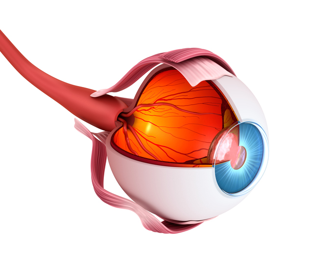
The yellow spot is located on the retina and is the place of sharpest vision. In this part is the highest concentration of visual cells. These cells are sensitive to light and color.
There is a small depression in the back of the eye, referred to as the central hole, professionally the fovea centralis. Here is the mentioned yellow spot, ie macula lutea. In the middle is a central hole, the foveola centralis, which is the place of sharpest vision.
The fovey area is called the macula.
The retina is located on the inside of the eyeball. It is the sensory visual organ to which the light rays passing through the optical system of the eye are directed.
In the retina, light is converted into electrical impulses, which then go to the brain, and thus to the visual center. There, the signals are processed and the overall picture is formed.
Causes
The exact cause of macular degeneration is not yet known
Both multifactorial and environmental effects are expected
Risk factors are known to be involved in the development of age-related macular degeneration.
Risk factors :
- age, from 65 to 75 years of age affects about 10% of the population, over 75 years, even more than 30%
- genetic predisposition
- Gender - Women are at higher risk after menopause for reduced estrogen levels
- familial occurrence t, the risk increases to 50% in familial occurrence
- race,
- the Caucasian race has the highest risk
- blacks more melanin and lower risk
- iris color - paler colors for less melanin - increased risk
- sunlight
- in case of excessive sun exposure and exposure to sunlight
- blue light from monitors or mobile phones also increases the risk
- reduced content of antioxidants in the diet - vitamins A, C and E - insufficient elimination of free radicals
- smoking - increases the risk up to 4x
- alcohol
- hypertension
- diabetes
- obesity
- hypercholesterolaemia - increased cholesterol in the blood
- refractive error - increased risk of farsightedness
- cataract - gray cataract
Symptoms
The retina is the most important part of the vision. The rays of light, and thus the observed image, is projected onto the cells of the retina, which is deposited on the posterior pole of the eye. Subsequently, the light on the retina is converted into an electrical signal, which travels on to the brain, the visual center.
There is a macula in the central part of the retina. This is important for the fact that it provides sharp vision. This is especially important when reading, writing, driving.
In macular degeneration, changes occur that lead to visual impairment.

There is a decrease in visual acuity. Both eyes are affected, but
Gray shadows or spots appear in the center of the observed. One is not able to read, the problem is also the perception of shape and contours, recognition of colors or faces. The image is distorted
The table shows the main differences between the dry and wet forms
| Dry form | Wet form |
| more frequent occurrence - 90% | rarer occurrence - 10% |
| slower onset of difficulties | rapid visual impairment and a more dramatic course |
| blurred vision | retinal bleeding and swelling |
| impaired vision at dusk and dusk | gray to black shadows and spots in the center of the field of view |
| inability to focus | peripheral vision is usually impaired |
| total visual field loss at a later stage | inability to read, write, drive |
| and therefore blindness | image deformation - metamorphopsia |
| inability to recognize shape and color |
Pain does not occur in any of the forms of macular degeneration.
Watch for warning signs:
- problem with reading, driving
- blurred text
- problem watching tv
- the lines are uneven, deformed
- dark spots in the middle of the field of view - gradually increasing
- loss of the ability to recognize colors, shapes, faces
Diagnostics
. The subjective symptoms that the affected person describes leading to the diagnosis of macular degeneration.
In the foreground is mainly reduced visual acuity, distortion, and deformation of shapes and objects. They also describe dark spots.
An early examination by a specialist is important. And thorough differential diagnostics.
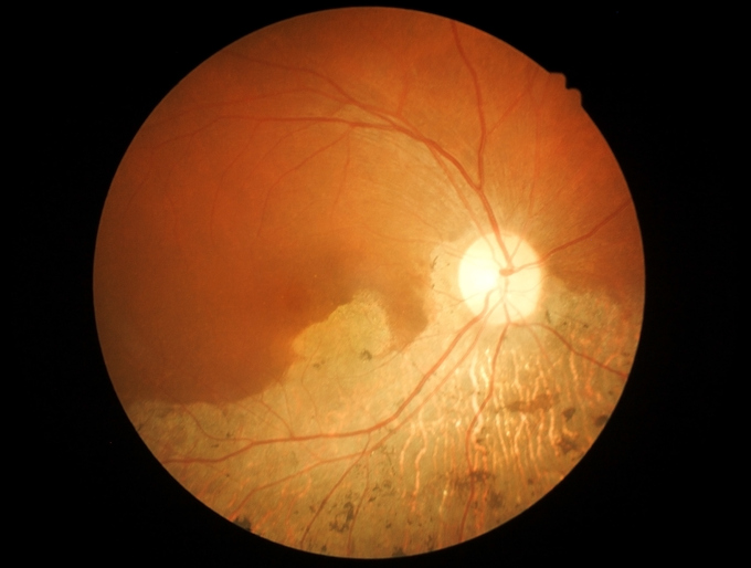
An ophthalmologist looks for signs of a dry or wet form of degeneration on the retina of the eye during an ophthalmoscopic examination.

The table shows the main diagnostic differences between the dry and wet forms
| Dry form | Wet form |
| the atrophic central part of the retina | lifting of the central part of the retina |
druse
| retinal swelling |
| transfer of pigment cells | bleeding present |
It is performed fluorescein angiography, which is used to diagnose the creation of new and surplus vessels that grow through the retina. Macular atrophy can also be assessed.
Using OCT - optical coherent topography
A simple test reveals macular degeneration
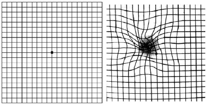
Even a simple test can help you suspect macular degeneration. Home self-examination is based on the Amsler test.
How to test for macular degeneration?
- primarily with both eyes
- then individually, with one eye covered
- do you use prescription glasses or lenses? Keep them
- from a distance of 30 cm
- you are looking at the grid
- there is a dark spot in the middle to focus on
- you also notice the area around the point, the whole grid
- all grid lines should be straight
- and they should be clear as well

The warning signal is if you see:
- bending lines into ripples
- image distortion
- image loss
- color changes
- increased attention, especially in the age over 50 years
Course
The course of the disease depends on whether the form is dry or wet. It also depends on the extent of macular involvement with atrophy and the formation of pathological vessels - neovascularization.
The dry has a long course, on the contrary, the wet form starts quickly and behaves destructively when there is a severe loss of the central visa.
It also has an impact on the quality of life.
The disease affects both eyes, however, the course is asymmetric. When the other eye has half of the affected already after five years and after ten years all with DM.
How it is treated:
Video for more information about the disease
is treated by
Other names
Interesting resources


