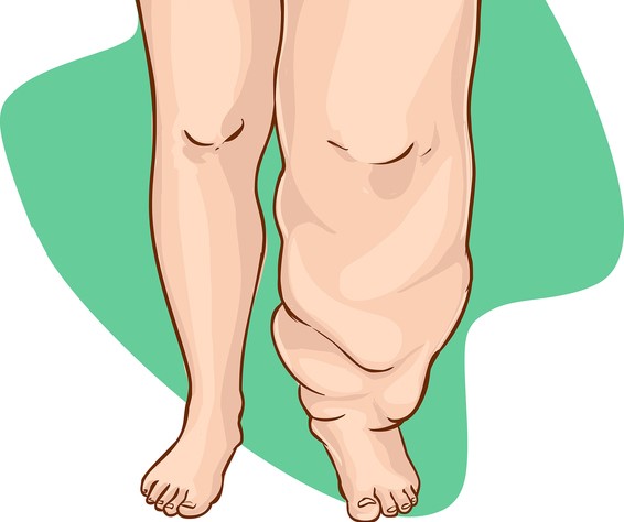
Lymphedema: What is it and how does it manifest? + Treatment
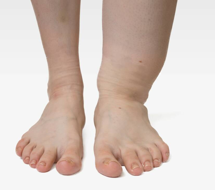
Lymphedema: What is it and how does it manifest? + Treatment
Photo source: Getty images

Lymphedema is a long-term disease that worsens over time or progresses. It is divided into primary and secondary. In the latter case, it is necessary to look for the cause of another disease. It also occurs as a result of impaired function of the lymphatic system. It causes the accumulation of fluid in the tissues and subsequent swelling.

Characteristics

Lymphedema is a chronic disease that worsens over time or progresses. It is the result of an imbalance between the formation and removal of lymph. As a result, there is an accumulation of pulmonary fluid in the tissues, ie lymphocytosis.

The swelling results from an increased content of proteins. It is complicated by frequent inflammation and fibrotic changes of the subcutaneous tissue and tissues. It can be primary or secondary. It arises primarily as a developmental error, but also for the hereditary factor. Secondary as a result of another disease or damage to the pulmonary system.

The lymphatic system or the lymph system  is designed to transport lymph, ie lymph fluid in the body. Especially between the blood and the intercellular space. The pulmonary system also plays an important role in human immunity .

What is the lymphatic system?

The lymphatic system, including the pulp system, is a large system in the human body. It plays an important role in the body's immune processes. It includes the pulp fluid, ie lymph, lymphatic vessels, nodes, thymus, and, for example, the spleen. An important part also includes the digestive system or bone marrow, lungs, and liver.

The formation of lymph occurs at the level of blood vessels, where the fluid from them is filtered into the extravascular, so-called interstitial space. Approximately 2-4 liters of this fluid are formed during the day. It contains various products of metabolism, but also, for example, salts or proteins.

Lymph is a colorless fluid and is formed as a product of cell metabolism, but also as a filtrate from blood vessels. It contains ingredients such as:

  • blood plasma
  • protein
  • in the area of ​​the intestines also fats, which are then transferred to the liver
  • lymphocytes

The pulp is further transported in the pulp vessels. Lymphatic vessels begin blindly in the interstitial space, ie in the interstitium. This small tangle of blood vessels combines into large pulp collectors. The wall of the pulmonary vessels is very thin, unlike the blood vessels.

In one day, 20 liters of tissue fluid are produced.
10%, or about 2-4 liters, is lymph.
90% of this fluid is reabsorbed into the blood vessels.

Lymphatic vessels are highly permeable. Bacteria that do not pass through the wall of blood vessels also penetrate into them. The pulp then travels to the lymph nodes. Lymph nodes play a filtering role and play an important role in immune responses.

Lymph nodes are distributed throughout the body. Their most common storage is near large vessels. Their filtering ability allows the capture of various foreign substances. These are, for example, the mentioned bacteria, but also other pathogenic germs or tumor cells. Also for this property, the nodules enlarge in various inflammations and diseases. There are lymphocytes in the nodes that are part of the immune system.

The pulmonary system serves as a drainage system for lymph, which is formed as a metabolite of cells or a filtrate of blood fluid.
It has an important role in the body's immune processes.
Lymphatic vessels are a common route for the transmission of tumor cells in the body, also known as metastases.

Causes

The cause of lymphedema is an insufficient function of the lymphatic system. It cannot remove excess fluid from the tissues. Subsequently, they accumulate locally in various places, causing swelling.

The causes of lymphedema are various

There can be various causes of lymphedema. An example is a damage to the structure of lymphatic vessels or their functional damage at the valve level. The reason may also be a clogged vessel. The result is the accumulation of pulp, and thus the other substances it contains. Such as proteins, metabolites, or foreign substances.

Accumulation causes swelling. The complication is inflammation, but also binding of the subcutaneous tissue. Inflammation can also be non-infectious. Reddened skin. The swelling worsens over time.

Lymphedema is divided into primary and secondary, with:

  • it is primarily due to congenital malformations of the lymphatic system or heredity
  • secondary occurs as a result of damage to the pulmonary system and as a symptom of another disease, such as:
    • infection
    • inflammation (non-infectious)
    • vascular disease
    • oncological disease
    • injury, trauma, damage during surgery
    • long - term immobilization

The primary cause is developmental anomalies in the lymphatic system. For example, lymphangiodysplasia. The cause can also be an inherited factor, and therefore a familial occurrence. It mainly affects women.

Lymphedema occurs congenitally, but can also occur during adolescence. When the cause is unknown. The most common areas of origin are the lower parts of the limbs, especially the lower limbs. It then spreads upwards.

The secondary cause is often due to damage to the lymphatic system. As a result of clogging or blockage of the river. The result is, for example, surgery, especially radically large-scale surgical solutions to cancer. But also inflammation or injury.

Swelling occurs where drainage is limited. And then it spreads downwards. It affects women and men alike. In our republic, the most common lymphedema of the upper limb is after surgery in the breast area, or after mastectomy in breast cancer.

Symptoms

The most characteristic symptom of lymphedema is swelling. However, this does not occur as the first symptom. According to available sources, lymphedema is divided into 4 basic stages.

The table describes the 4 stages of lymphedema

Title Description
Stage I latent, ie hidden when an objective finding is not yet present,
however, it can be proved by scintigraphic examination the
transport function is still sufficient
subjective difficulties occur, such as:
  • feeling of tension in the affected area
  • pain
  • limb mining
  • limb fatigue
Stage II spontaneously reversible, ie reversible at this stage, the subjective difficulties increase
swelling occurs after a full day of exercise
helps the positioning of the limb
is marked by the spongy character of the tissue
after pushing the finger does not leave a hole
Stage III irreversible, which means irreversible now the tissue is already solid
after compression at the site of swelling, the hole remains
fibrosis of the subcutaneous tissue
limb enlarges (unilaterally)
Stage IV lymphostatic elephantiasis change in the shape of the limb
deformation of the limb
restriction of the mobility of the joints the
tissue is completely fibrotically changed

Another form is the division of lymphedema into degrees, as shown in the table

Title Description
level 1 slight swelling the circumference of the affected limb by less than 4 cm
does not affect the affected limb in its entirety
level 2 moderate swelling when the difference in the circumference of a healthy limb is 4 - 6 cm the
swelling affects the whole limb
after pushing on the skin the hole remains in the swelling
level 3a severe swelling the difference is more than 6 cm
in addition to the limb, the swelling is also affected by the corresponding quadrant of the body
there are also skin changes
level 3b the swelling affects two or more limbs
level 4 elephantiasis deformation of the limb is associated

In the case of primary lymphatic edema, the characteristic edema begins, which begins in the lower limb. Subsequently, this swelling spreads upwards to the limb. In secondary pulmonary edema, the procedure is different.

Secondary lymphedema often occurs after surgery, chemotherapy, and radiotherapy. Most often within 1 year after oncological treatment. The swelling occurs in a place under the damage of the pulp system, then spreads downwards from this place. Also referred to as early edema.

The late-type develops over a lifetime. However, lymph swelling has not yet been detected. However, its trigger can be an injury or even an insect bite, and even a blood draw. Another example is malignant edema, which is caused by the aggravation of the underlying disease. Its manifestation is, for example, significant pain.

Other symptoms include:

  • pale skin
  • sweaty skin
  • increased skin temperature
  • loss of visibility of the vascular drawing, especially in the area of ​​the back of the hand
  • skin hyperkeratosis ie increased keratinization
  • superficial damage to ulceration, ie ulceration of the skin
  • on the contrary, with severe fibrosis of the subcutaneous tissue, the limb may be reduced, stiff with restricted movement
  • hair loss

The complication is a significant risk of exacerbation

Complications are common in lymphedema. These pose a significant risk of worsening, and thus progression. These include, for example:

  • Inflammation of the skin, ie dermatitis, especially dangerous is infectious inflammation, most often the presence of bacteria such as staphylococci and streptococci
  • eczema
  • inflammatory - rheumatic disease
  • mycosis, especially as a complication of a humid environment in the swollen tissue and its incisions
  • impaired joint mobility
  • restriction of daily activities
  • Steward - Treves syndrome, ie lymphangiosarcoma, is a rare occurrence

Diagnostics

A thorough and correct history is sufficient to prove the disease. A history of injuries, operations, chemotherapy, radiotherapy, and other diseases is sought. Subsequent evaluation of the clinical picture. And so it is important to know the duration, and how the difficulties develop.

Finger pressing on the skin and assessing whether a hole remains in place is also used in diagnosis. Subsequently, the circumference of the limbs is measured. It is important to evaluate skin changes. The so-called Kaposi - Stemmer sign is used, which determines the possibility of skin algae formation on the II. toes. This sign is especially evaluated in the case of primary swelling.

Imaging methods are also used in lymphedema. Such as lymphography, scintigraphy (lymphoscintigraphy). CT, MRI, soft tissue X-ray, USG, also known as SONO are used. If cancer is suspected, a sample is taken by biopsy. Laboratory tests include examination of thyroid hormones, blood sugar levels, blood counts, FW, CRP, creatinine, urea, albumin, and proteins.

TIP: Information on the various causes of edema  in the Swelling symptom.

Course

Lymphedema can occur at an early stage in secret. And so without the limb being swollen, and therefore enlarged. The first symptom may be a feeling of pressure, weakness, or fatigue of the limb. Subsequently, edema develops during a full day of exercise. However, at this point, the positioning, ie the elevated position of the limb, helps.

The third stage is characterized by persistent swelling. This is also evident in that the hole remains in the skin after being pushed with a finger. It will disappear over time. However, this condition persists for a shorter time. In the case of a greater extent of fibrotic tissue damage, the skin is stiff. And even this pressure test is not demonstrable.

The swelling is noticeable during this period, the limb is enlarged. Paradoxically, with extensive fibrotic tissue changes, limb shrinkage occurs. The skin is stiff, movement in the joint is limited. Skin inflammation may also be associated. And even in the form of surface damage or significant ulceration. When deep ulcers develop on the surface of the skin

The late-stage is characterized by deformities of the limb, also referred to as elephantiasis. In case of complications, worsening of lymphedema is known. In the case of primary lymphedema, swelling begins to develop in the lower limbs and progresses upwards. In the case of secondary edema, it is in the area under the disability, downwards.

lower limb lymphedema, elephantiasis
Lymphedema, elephantiasis of the lower limb. Photo source: Getty images

How it is treated: Lymphedema

Lymphedema and its treatment: lifestyle interventions and medications

Show more

What is a practical life like with lymphedema?

fshare on Facebook

Interesting resources

  • Joos E, Bourgeois P, Famaey JP (June 1993). "Lymphatic disorders in rheumatoid arthritis". Seminars in Arthritis and Rheumatism. Elsevier BV. 22 (6): 392–398. 
  • Grada AA, Phillips TJ (December 2017). "Lymphedema: Diagnostic workup and management". Journal of the American Academy of Dermatology77 (6): 995–1006. 
  • Sleigh BC, Manna B (January 2020). "Lymphedema". StatPearls [Internet]. Treasure Island (FL): StatPearls Publishing. 
  • Grada AA, Phillips TJ (December 2017). "Lymphedema: Pathophysiology and clinical manifestations". Journal of the American Academy of Dermatology77 (6): 1009–1020. 
  • Martin MB, Kon ND, Kawamoto EH, Myers RT, Sterchi JM (October 1984). "Postmastectomy angiosarcoma". The American Surgeon50 (10): 541–545. 
  • Chopra S, Ors F, Bergin D (December 2007). "MRI of angiosarcoma associated with chronic lymphoedema: Stewart Treves syndrome". The British Journal of Radiology80 (960)
  • Requena L, Sangueza OP (February 1998). "Cutaneous vascular proliferations. Part III. Malignant neoplasms, other cutaneous neoplasms with significant vascular component, and disorders erroneously considered as vascular neoplasms". Journal of the American Academy of Dermatology38 (2 Pt 1): 143–75, quiz 176–8. 
  • Publishing, Licorn (2009-10-28). "Body image and quality of life in secondary lymphedema of the upper limb". Servier - Phlebolymphology
  • "Oncology Fact Sheet" (PDF). aota.org. Retrieved 2019-09-03.
  • "A Visual Guide to Lymphedema". WebMD
  • Shenoy, R. K. (2008-09-20). "Clinical and Pathological Aspects of Filarial Lymphedema and Its Management". The Korean Journal of Parasitology46 (3): 119–125. 
  • "Lymphoedema - Causes". nhs.uk. 2017-10-20. 
  • Jeannie Burt; Gwen White (1 January 2005). Lymphedema: A Breast Cancer Patient's Guide to Prevention and Healing. Hunter House. pp. 9. ISBN 978-0-89793-458-9.
  • Choices, NHS. "IPS retired". nhs.uk
  • Brorson H, Ohlin K, Olsson G, Svensson B, Svensson H (June 2008). "Controlled compression and liposuction treatment for lower extremity lymphedema". Lymphology41 (2): 52–63.