- Nienaber, CA; Clough, RE (28 February 2015). "Management of acute aortic dissection". Lancet. 385 (9970): 800–11. ndell, J; Crowe, J (2013). "Acute aortic emergencies – part 2: aortic dissections". Advanced Emergency Nursing Journal. 35 (1): 28–52.
- Criado FJ (2011). "Aortic dissection: a 250-year perspective". Tex Heart Inst J. 38 (6): 694–700. PMC 3233335. PMID 22199439.
- Arima D, Suematsu Y, Kurahashi K, Nishi S, Yoshimoto A. Use of coagulation-fibrinolysis markers for prognostication of Stanford type A acute aortic dissection JRSM Cardiovascular Disease. 2021 Jan;10. PMCID: PMC8613881
- Robert O. Bonow; Douglas L. Mann; Douglas P. Zipes; Peter Libby (2011). Braunwald's Heart Disease E-Book: A Textbook of Cardiovascular Medicine. Elsevier Health Sciences. p. 1321. ISBN 978-1-4377-2770-8.
- John Marx; Ron Walls; Robert Hockberger (2013). Rosen's Emergency Medicine - Concepts and Clinical Practice E-Book. Elsevier Health Sciences. p. 1125. ISBN 978-1-4557-4987-4.
- John Elefteriades (2007). Acute Aortic Disease. CRC Press. p. 31. ISBN 978-1-4200-1976-6.
- Isselbacher E.M.; Cigarroa J.E.; Eagle K.A. (1994). "Cardiac Tamponade Complicating Proximal Aortic Dissection. Is Pericardiocentesis Harmful?". Circulation. 90 (5): 2375–78.
- Sheikh, AS; Ali, K; Mazhar, S (Sep 3, 2013). "Acute aortic syndrome". Circulation. 128 (10): 1122–27.
- Cassoobhoy, A (30 June 2020). "Aortic Dissection". WebMD. Retrieved 29 June 2021.
- Orihashi K (May 2016). "Mesenteric ischemia in acute aortic dissection". Surg. Today. 46 (5): 509–16.
- Kamman AV, Yang B, Kim KM, Williams DM, Michael Deeb G, Patel HJ (2017). "Visceral Malperfusion in Aortic Dissection: The Michigan Experience". Semin. Thorac. Cardiovasc. Surg. 29 (2): 173–78.
- Alfson, DB; Ham, SW (August 2017). "Type B Aortic Dissections: Current Guidelines for Treatment". Cardiology Clinics. 35 (3): 387–410.
- Di Tullio, Marco R.; Homma, Shunichi (2016-01-01), Grotta, James C.; Albers, Gregory W.; Broderick, Joseph P.; Kasner, Scott E. (eds.), "33 - Atherosclerotic Disease of the Proximal Aorta", Stroke (Sixth Edition), London: Elsevier, pp. 576–590,
- Rahimi, A; Geiger, Z (2021). "Anatomy, Thorax, Subclavian Arteries". National Center for Biotechnology Information, U.S. National Library of Medicine
Aortic dissection: What are the causes of arterial rupture and symptoms? + Risks and treatment
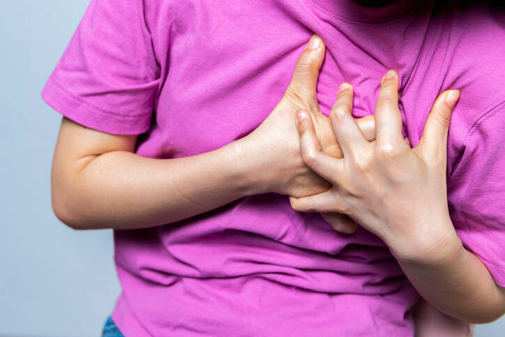
Photo source: Getty images
During aortic dissection, the vascular wall ruptures, with blood flowing between its inner layers. The extent may be small, but also extensive and affecting the entire aorta.
Most common symptoms
- Shoulder Blade Pain
- Malaise
- Speech disorders
- Chest pain
- Abdominal Pain
- Headache
- Limb pain
- Leg Pain
- Pain that Radiates into the Shoulder
- Groin Pain
- Back Pain
- Spirituality
- Bleeding
- Blue leather
- Nausea
- Low blood pressure
- Defence
- Swelling of the limbs
- The Island
- Erectile dysfunction
- Disorders of consciousness
- Slowed heartbeat
- Muscle weakness
- Pressure on the chest
- Head spinning
- Fatigue
- Vomiting
- High blood pressure
- Stuck winds - stopping the outflow of gases
- Urinary retention - anuria/retentiveness
- Confusion
- Accelerated heart rate
Show more symptoms ᐯ
Characteristics
Aortic dissection is one of the less common but even more serious diseases. This is due to the anatomical nature of the largest artery of the human body and its function.
Dissection = tearing, cracking.
The dissecting is tearing, dividing.
Aorta = heart.
During dissection of the aorta, the vascular wall ruptures and ruptures. Through the damaged inner layer, blood under pressure penetrates between the vascular layers.
A new, so-called false lumen of the vessel is formed. This false course of the artery is often larger than the true course of the vessel.
The lumen is the inner part of a hollow vessel.
This mechanism of dividing the vessel into several layers is the basis of various difficulties. For better illustration, we also provide brief information about the aorta.
Aorta - the largest artery of the human body
The heart, or aorta, is the largest vessel in the human body and therefore the artery.
We know several types of vessels, and one form of division divides them into arteries and veins. In short, veins are those vessels that carry blood to the heart. Arteries, on the other hand, draw blood from the heart.
Oxygenated blood usually flows in the veins and mostly oxygenated in the arteries.
There is an exception for the lungs and pulmonary veins.
Artery = vessel
Vein = ducts.
Venules= capillaries.
The vessels generally contain three layers:
- internal - tunica intima, formed by endothelial cells
- middle - tunica media, mostly muscle cells, reticular and elastin fibers
- outer - tunica adventitia, formed by collagen ligament and elastic fibers
Smaller vessels are nourished through diffusion, ie the transfer of nutrients and oxygen to vascular cells. Larger vessels, and thus vessels over 1 mm, have vessels in the vessel wall.
Vascular vessels = vasculature.
The aorta is a massive vessel that protrudes from the left half of the heart and, more precisely, from the left ventricle. The heart pumps into it - it expels blood under high pressure.
This pressure acts on the aortic wall and burdens it for life.
The heart further distributes this blood, transmitting it to other parts of the human body.
The aorta is divided into several sections:
- aortic root
- thoracic aorta - thoracic aorta
- ascending part - ascending aorta
- aortic arch - arcus aortae
- descending, aorta descendens
- abdominal aorta - abdominal aorta
The thoracic and abdominal sections are separated by a diaphragm.
The branches of smaller arteries emanate from the aorta. The first include two coronary arteries - the right and left coronary arteries ( coronary artery dextra et sinistra).
In the part of the arch there are: the
shoulder-head trunk (truncus brachiocephalicus), the
left common cervix (arteria carotis communis sinistra) and the
left subclavian artery (arteria subclavia sinistra).
Various branches of the arteries protrude from the abdominal aorta, which further branch and go to the abdominal cavity and its organs. The final section of the abdominal aorta passes and branches to the artery of the iliac communis dextra et sinistra, ie the right and left lumbar arteries.
+
The aorta is the body's largest artery.
The aorta has a diameter of about 3 up to 4 centimeter.
More precisely, it is stated that:
For men it is 3.63 - 3.91 cm on average and
for women 3.50 - 3.72 cm.
The role of the aorta is to distribute blood from the heart to the whole body. And thus the supply of vital and all other organs, tissues, cells of the body.
In addition, it has other roles.
The main tasks of the heart :
- distribution of blood - to other parts of the body
- control and management of systemic vascular resistance - aortic pressure control controls vascular resistance
- heart rate control - aortic pressure control affects heart rate
- elastic function - vascular pump, similar to the heart muscle
Want to know more about aortic dissection?
About what causes it?
What are his symptoms?
How is it treated?
Read with us.
What is aortic dissection?
Aortic dissection is a rare but even more dangerous disease. It can be typical, but also atypical, when it resembles another disease.
It usually has an acute, ie sudden course. He is in dire trouble. And it doesn't always end happily, it can be deadly.
It affects men + people over the age of 60 to a greater extent.
The dissection is a rupture of the aortic wall.
The expelled blood from the heart penetrates through the damaged vascular wall due to the pressure.
It gradually passes between the layers of the heart.
The pumped blood further separates the individual layers from each other by pressure.
The outer layer of the artery, the tunica adventitia, is not disturbed.
There is no blood leakage between the bloodstream.
As other arteries leave the aorta, blood does not flow into these branches as a result of damage to the artery. The false lumen of the vessel suppresses the original course of the aorta and the distances of other arteries. This causes bloodlessness of organs, other structures and tissues, and thus their ischemia.
Ischemia = non-blood supply to a specific organ or tissue.
Lack of nutrients and oxygen will cause damage and even death.
Plus, harmful metabolic products accumulate in a given place.
The risk of dissection is also the rupture of the artery across its entire width. This leads to massive bleeding. This condition is called aortic rupture and is mostly fatal.
+
The blood penetrates through a crack in the inner layer of the vessel, through the intima.
The blood flow does not stop, but continues.
The blood thus progresses between the intima, the medium or the Advent.
A so-called false channel is being created. It is a division of the vascular lumen - the inner channel of the vessel - into two cavities.
Right canal = aortic canal that normally exists.
+
False channel = newly formed channel in the vascular space.
It happens that this new fake channel is bigger than the original and oppresses it. As a result, the already mentioned restriction of blood flow in the aorta, and thus also blood flow to the target organs behind the damaged section, is already mentioned.
During dissection:
- entry opening
- fake channel
- the complete dissection also contains the output channel - reentry
- this returns the blood to the right lumen of the artery
- incomplete dissection does not contain an output reentry channel
- a separate part causes narrowing of the right aortic canal
Dissection can occur in any section of the aorta. For example in the place apart from the heart and the aortic valve in the affected section spacing coronary artery associating hypohemia heart - heart attack .
In the case of a section of arteries leading to the head and brain, the manifestations of cerebral anemia and stroke are associated. Similarly in any section, bloodlessness of the intestines, kidneys and other abdominal organs or lower limbs.
By size.
There are small dissections a few millimeters. When they form a small intramural hematoma (IMH), and thus blood clot in the artery wall.
The opposite is ...
Extensive dissections can extend the entire length of the aorta to the lumbar artery.
The rupture of the vascular wall usually has a spiral course. Tearing of the vessel wall can also spread to the protruding branches of the arteries.
The incidence of approximately 5-30 cases out of one million is reported.
There are approximately 25-150 cases in Slovakia.
Acute aortic dissection is one of the acute aortic syndromes - abbreviated AAS. More is also written in the article on aortic diseases.
Division into two types.
According to the dissection site, it is divided into two types, according to the Stanford classification :
- Type A - ascending aorta, when the section of the aortic distance is affected and mostly just above the aortic valve - ascending aorta
- proximal dissections - proximal = closer to the head
- two thirds of cases
- Type B - indicates the involvement of the descending aorta and usually spreads to the abdominal aorta to the arteries of the lower extremities - descending aorta
- distal dissection - distal = distant from the trunk - lower
This division is of practical importance from the point of view of the necessary method of treatment. While for dissection type A it is always operating surgical approach.
Stanford classification according to Daily and co-workers from 1970.
The type B approach is initially conservative in order to lower blood pressure. The evaluation may be followed by endovascular treatment. Then the catheter is inserted into a larger artery, most often the femoral artery, and continues to the affected area.
Another form of division ...
DeBakye classification as early as 1965, distinguishes dissection according to location and extent.
DeBakey Classification:
- DeBakey I. - involvement of the entire aorta
- DeBakey II. - involvement only in the initial section of the aorta - the ascending aorta
- DeBakey III. - involvement of the aorta below the certainty, from the artery of the subclavian sinistra - descending aorta
65% involvement of the ascending - ascending aorta,
20% involvement of the descending - descending aorta,
10% in the arch section.
We also know the division in terms of time :
- Acute aortic syndrome - 14 days after the onset of the problem
- Subacute aortic syndrome - 15 to 90 days of difficulty
- Chronic aortic syndrome - over 90 days
Dangerous complications
First of all, it is about the non-blood supply of tissues and organs. They are supplied by blood vessels leaving or behind the dissection site.
The cause for concern is the ischemia of vital organs such as the heart or brain. These are very sensitive to blood flow and their ischemia is characterized by a rapid onset of difficulties.
An example is the non-congestion of the spinal cord, which causes paralysis of the lower limbs. A kidney from ischemia started renal failure.
+
The second serious complication is a rupture of the aorta.
In this case, a complete defect in the aortic wall occurs and blood escapes from the bloodstream. Major internal bleeding usually ends in death. In this case, the location and extent of arterial damage are equally important.
About 1/6 of patients are reported to die before aortic rupture surgery.
Causes
Thus, dissection is tearing of the aortic wall. It causes blood to penetrate between the leaves of the vessel wall. The pressure of the flowing blood increases the tear, thus prolonging the damaged section of the aorta.
Decreased resistance of the vessel wall is damage and blood penetration between the layers of the vessel wall = one of the factors of origin.
You ask:
Why does this happen?
The cause is multifactorial, which means that several risk factors are involved in the development of cardiac dissection.
Risk factors for aortic dissection:
- genetic predisposition
- familial forms - familial occurrence
- cystic medionecrosis
- congenital - congenital connective tissue diseases
- Marfan syndrome
- Turner syndrome
- Ehlers-Danlos syndrome
- aortic coarctation
- hypertension - high blood pressure
- untreated hypertension in up to 72% of cases
- injury
- in traffic accidents and falls from great heights
- deceleration, ie a sudden stop
- blunt injuries
- iootrogenic damage during another medical procedure
- atherosclerosis
- atherosclerotic ulceration, ie rupture of the atherosclerotic plaque of the aorta
- PAU - penetrating aortic ulcus
- atherosclerotic ulceration, ie rupture of the atherosclerotic plaque of the aorta
- infectious cause - rare today (syphilis and other diseases)
- Gender - Men are affected more often, up to twice as often as women
- autoimmune disease
- age over 40 years, the highest incidence rate after the 60th to 70th year of life
- untreated hypertension significantly reduces the incidence period
- obesity
- pre-existing aortic disease
- aortic valve disease
- disorders of fat metabolism
- smoking
- stimulant drugs such as cocaine or methamphetamine
Aneurysm + aortic dissection
How do these two diseases relate to each other?
An aneurysm is a dilation of a blood vessel. Even in the case of a bulge, a multifactorial effect is reported. The altered structure of the vessel wall causes the weakened vessel wall to not maintain pressure in the artery.
It can take place for a long time undetected, without any symptoms present.
We know several types of bulges, and they are described in more detail in the article Aneurysm. A right bulge is one in which the entire width of the artery is weakened and affects all three layers of the vessel.
80% of right bulges are said to affect the aneurysm.
Such a weakened vascular wall is risky in terms of two complications.
One is a rupture of the aorta. During rupture, the entire width of the aortic wall ruptures, causing massive bleeding.
The second is dissection. It is called a dissecting aortic aneurysm - aneurysm dissecans aortae. With the weakened and damaged inner layer of the vessel, blood passes between the layers of the artery.
Aortic rupture
Rupture, ie rupture of the heart = damage to the aortic wall with its opening.
She is accompanied by massive bleeding. Blood flows through the aorta under high pressure, as well as escapes from the damaged artery.
The tear point may be different.
According to the area of aortic damage:
- bleeding into the pericardium , which leads to cardiac tamponade
- blood accumulates in the heart sac
- accumulated blood oppresses the heart
- in diastole, relaxation of the heart muscle is not possible enough filling the heart
- started hypotension, reduction of cardiac output, a reduction in blood supply to organs and tissues to shock-like state
- thoracic haemorrhage - hemothorax and respiratory failure
- abdominal bleeding - hemoperitoneum
Massive bleeding + hemorrhagic shock = sudden death.
A closed form may also occur.
In this case, the damaged vascular wall is closed by nearby structures, such as the pericardium, the pleura, or another urgent organ on the artery wall.
Symptoms
Symptoms of dissection may be typical, but there are cases where difficulties are present that indicate another disease.
Plus...
Depending on the location and extent, symptoms of non-blood supply to tissues and organs are also associated. An example is the co-dissection of dissection with heart failure, myocardial infarction, or stroke.
Characteristic manifestations of dissection:
- sharp, strong, cruel, shocking, intense pain
- sharp, jerky, stabbing
- the worst pain in life
- does not respond to analgesics - painkillers
- throbbing pain
- up to 95% of cases
- sudden onset
- chest pain - associated with damage to the ascending aorta
- pain between the scapulae - descending aorta
- abdominal pain - abdominal aorta
- + the pain spreads
- to the back - between the shoulder blades
- to the abdomen - abdominal pain
- the spread of pain is along the advancing dissection
- the pain sometimes subsides, either temporarily or permanently
Other possible difficulties such as:
- weakness
- dizziness
- difficulty breathing, shortness of breath, shortness of breath
- heartbeat
- high blood pressure to hypertensive crisis
- or, conversely, low blood pressure, heart failure, bleeding
- fear of death
- pallor
- neurological disorders in the brain
- indigestion - abdominal area, like a sudden abdominal event
- kidneys - oliguria, ie reduced urine production to anuria - complete cessation of urination
- upper or lower limb bleeding - limb ischemia, limb weakness and pain
- pain in the groin
- paralysis of the lower limbs - damage to the spinal cord
- apostasy, syncope, collapse, short-term loss of consciousness
- unconsciousness, shock
- sudden death, extensive damage or rupture of the aorta
It is reported that only 5% of cases go chronic.
As many as 95% of untreated patients end in death .
Plus, the following phenomenon can occur during dissection:
Paradoxical pulse and a large, noticeable difference in the measurement of blood pressure on the two upper limbs.
Paradoxical pulse = pulsus paradoxus refers to a condition in which the heartbeat is present on one upper limb and intangible or difficult to feel on the other.
It's similar with blood pressure. When measuring the pressure on both upper limbs and comparing it, the difference will be too noticeable and large.
Pressure difference higher than 20 mmHg .
In this case, it is necessary to think about aortic dissection.
Diagnostics
Diagnosis plays a very important role in the occurrence of acute problems that indicate dissection. As the risk of serious complications and death increases every hour, which is not revealed.
However, making a correct diagnosis may be complicated by an atypical course or the occurrence of other associated symptoms.
A history is a matter of course, which includes personal and family history. And thus other diseases, such as mainly hypertension and familial occurrence of serious illnesses or deaths.
The clinical picture is evaluated, ie how the difficulties go, when and how they started. Plus physical examination and evaluation of vital functions such as blood pressure, pulse, respiration and more. It complements the laboratory blood test.
Imaging methods play an important role :
- X-ray
- CT - 90 to 100% success rate of diagnosis of dissection
- ECHO
- transesophageal ECHO - TEE , via esophagus
- esophageal echocardiography
- endoscopy + ultrasound probe
- similarity with endoscopic examination of the stomach and esophagus by gastrofibroscopy
- 85-99% success rate of diagnosis of dissection
- transthoracic TTE, across the chest wall, lower sensitivity
- transesophageal ECHO - TEE , via esophagus
- ECG - differentiation of ACS, acute coronary syndrome, heart attack
- SONO
- MRI
- PET
- aortography
Course
The course of aortic dissection is usually typical , but in some cases it may resemble another disease .
Or, the initial difficulties are alleviated and the symptoms of associated non-congestion of the target organ, organ system or tissue come to the fore.
About 95% of cases are marked by a sudden and abrupt onset. Present is intense and severe pain. The sufferer can describe it as the worst pain in life.
Pain can be characterized by several properties.
An example is that a high intensity and does not take the medication soothing pain - analgesics. It can be pulsating.
With this type of chest pain, it is assumed that the initial part of the heart - the ascending aorta - will be affected.
If it is located in the area of the back between the scapulae, it is probably a damaged arch of the heart - arcus aortae.
Abdominal pain and possible symptoms of an abdominal event occur when the abdominal section is affected - the abdominal aorta.
+
The pain may initially come from the chest area. However, as the dissection expands and progresses to the lower parts of the artery, so does the pain.
The pain spreads during dissection.
In some cases, the intensity of the pain subsides or subsides completely. Which may be one of the reasons for viewing a dissection. Therefore, it is necessary to think about this life-threatening condition during the initial difficulties.
Complications and consequences can be fatal.
If the not treated :
Pitch mortality by 1 - 2% for each hour during the first 24 hours .
21% die within 24 hours.
49% within 4 days.
74% within two weeks.
93% within one year.
Survival at a well-established diagnosis and appropriate treatment is 74-92% !
Heavy physical exertion can prevent the problem. There are also reports in the literature where a person has leaned forward to tie his strings and feel a rupture. The cause was not the plate, but the dissection.
+
Symptoms may mimic another health problem.
In addition to the pain, there are other complications that result from the site of the aortic lesion. And here either heart, neurological, abdominal or kidney problems, or as with a spine injury.
Manifestations are difficulties such as:
- spine ache
- radiation between the blades
- pain in the thoracic and lumbar spine to the lower back
- abdominal pain , sudden abdominal event, gallbladder colic and others
- radiation under the right rib arch and under the right scapula
- nausea on the stomach
- vomiting
- renal symptoms
- kidney stones, renal colic
- oliguria to anuria, ie reduced to stopped urine production
- pain and weakness in the limbs - upper and lower
- torment
- noticeable weakness of the limbs
- inability to walk and move
- immobility of the lower limbs - paraparesis to plegia - paralysis due to spinal cord blood
+ Their mutual combination.
Accompanying phenomena can be hypertension, accelerated hypertension (sudden to sudden rise in blood pressure, causes damage to organs if left untreated) to a hypertensive crisis.
The table shows the occurrence of less typical dissection difficulties
| Symptom | % |
| Apostasy, collapse, syncope | 4% |
| Neurological difficulties |
|
| Heart attack | 1 - 2% |
| A sudden abdominal event | 3 - 5% |
| Acute renal failure | 5 - 8% |
| Acute limb ischemia | 12% |
How it is treated: Aortic dissection
How is aortic dissection treated? Conservatively and surgically
Show moreAortic dissection is treated by
Aortic dissection is examined by
Interesting resources

Bc. Lukáš Tóth
Healthcare worker
The secondary medical school in Nitra gave me the basis for my career in the field of health and diseases. Thanks to it, I worked for 2 years in the traumatology clinic and outpatient clinic at the Nitra Hospital. Since 2006 I was employed in the emergency medical service, where I stayed until 2017.
I completed my bachelor's degree at the University of Constantine the Philosopher in Nitra in the field of emergency health care. The bachelor's degree allowed me to continue my mission as a paramedic. In the meantime, I got a job at the emergency line 155. I have been working in pre-hospital health care until today.
I had an interest in people, health and even diseases in my childhood, which gave me the prerequisite to pursue this topic in adulthood. Studying and acquiring new information in practice provided me with a great basis for writing professional texts, in the form of articles that can be understood by ordinary people. Thus, my interest in the Health Portal has a solid foundation in years of practice and personal interest. Similarly, I am also interested in healthy eating, nutrition and overall healthy lifestyle. I fill my free time with family and sports.
View all articles by the same author


