- Maton, Anthea; Jean Hopkins; Charles William McLaughlin; Susan Johnson; Maryanna Quon Warner; David LaHart; Jill D. Wright (1995). Human Biology Health. Englewood Cliffs, New Jersey: Prentice Hall. ISBN 978-0-13-981176-0.
- Tortora, Gerard J. (1995). Principles of Human Anatomy (Seventh ed.). Harper Collins. pp. 341, 367, 369. ISBN 978-0-673-99075-4.
- Tortora, Gerard J.; Grabowski, Sandra Reynolds (1996). Principles of Anatomy and Physiology (Eighth ed.). Harper Collins. p. 634. ISBN 978-0-673-99355-7.
- Hole, John W. Jr.; Koos, Karen A. (1994). Human Anatomy (Second ed.). Wm. C. Brown. p. 479. ISBN 978-0-697-12252-0.
- De Graaff, Van (1998). Human Anatomy (Fifth ed.). WCB McGraw-Hill. pp. 548–549. ISBN 978-0-697-28413-6.
- Putz, R.; Pabst, R., eds. (2006). Atlas van de menselijke anatomie (Translated from German (Atlas der Anatomie des Menschen)) (in Dutch) (3rd ed.). Bohn Stafleu van Loghum. ISBN 978-90-313-4712-4.
- Drake, Richard L.; Vogl, Wayn A.; Mitchell, Adam W. M. (2010). Gray's Anatomy for Students (2nd ed.). Churchill Livingstone (Elsevier). ISBN 978-0-443-06952-9.
- Netter, Frank H. (2003). Atlas of Human Anatomy (3rd ed.). ICON Learning Systems. ISBN 978-1-929007-21-9.
- Drake, Richard L.; Vogl, Wayne; Tibbitts, Adam W.M. Mitchell; illustrations by Richard; Richardson, Paul (2005). Gray's anatomy for students. Philadelphia: Elsevier/Churchill Livingstone
- cedars-sinai.edu - Aortic Infection
- suscch.eu - Guidelines for the Diagnosis and Management of Patients With Thoracic Aortic Disease 2010
- sks.sk - 2014 ESC Guidelines on the diagnosis andtreatment of aortic diseases
Aortic diseases
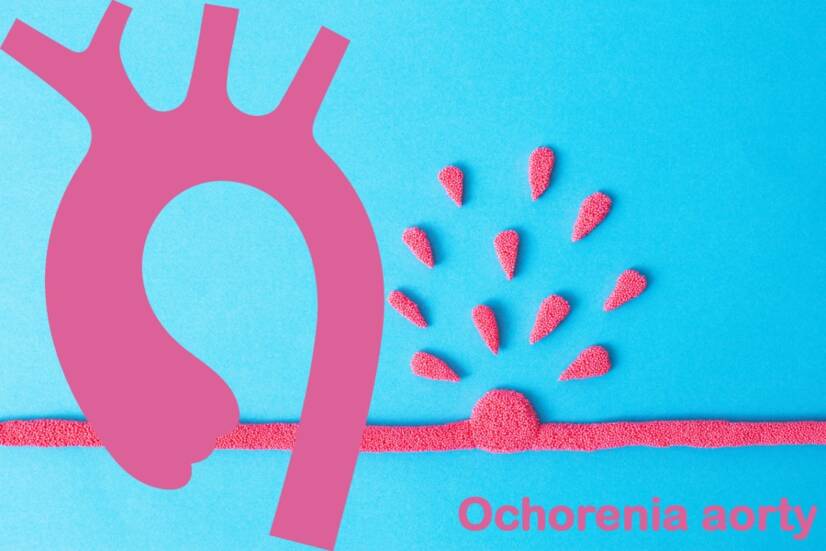
Photo source: Getty images
Aortic disorders are not common but are all the more serious. They can take place very quickly and acutely with life-threatening but also chronically.
Most common symptoms
- Shoulder Blade Pain
- Malaise
- Chest pain
- Stomach pain after a meal
- Abdominal Pain
- Groin Pain
- Spirituality
- Fever
- Pain that Radiates into the Shoulder
- Nausea
- Head spinning
- Blue leather
- Low blood pressure
- Defence
- Lung Island
- Swelling of the limbs
- Tingling
- Erectile dysfunction
- Disorders of consciousness
- Back Pain
- Dry cough
- Pressure on the chest
- Fatigue
- Coughing up blood
- High blood pressure
- Accelerated heart rate
Show more symptoms ᐯ
Characteristics
Aortic diseases are less common, but their course may be all the more serious. In terms of speed, therefore, they are acute, created suddenly, which endanger a person's life and chronic in the long run.
The aorta is the largest vessel in the human body. We also know it as heartworm.
Blood is expelled from the heart under high pressure by the heart muscle into the aorta.
From the aorta, blood is then directed to other parts of the body and to target organs, muscles, or other tissues. In the first section, two coronary arteries protrude from it, which nourish the heart.
Furthermore, blood flows to the head, brain, upper limbs, chest, abdomen, and lower limbs.
The left ventricle creates muscle pressure when pumping blood into the body. The aorta is adapted to this by its size and composition of the vascular wall.
The left ventricle and aorta are separated by a valve.
This prevents the blood from flowing back into the left heart chamber.
This is at the time of diastole, when it is released again and filled with blood.
More about the aorta ...
The aorta is a large elastic, or flexible, artery through which approximately 200 million liters of blood flow during a person's life.
From the anatomical point of view, it is divided into several sections.
The aorta is divided into the following parts:
- Aorta ascendens - ascending aorta
- emanates from the left ventricle of the heart
- is separated from the ventricle of the left heart by an aortic valve
- the arterial supply of the heart recedes from it
- right and left coronary arteries
- The Arcus aorta is the aortic arch, in which the aorta turns left
- the receding arteries carry blood to the brain and upper limbs, the muscles of the neck or shoulders, the larynx, and the chest wall
- Aorta descendens - descending aorta, which is divided by a diaphragm into two parts, namely:
- thoracic aorta - the thoracic aorta - approximately at the height of the vertebrae Th3 to Th 12
- aorta abdominalis - the abdominal aorta - from the diaphragm and Th12
During the course of the heart, other sentences of the arteries withdraw from it. These lead the blood to target organs, muscles, and other tissues.
In its final section, the abdominal aorta branches into the artery of the ilica communis dextra et sinistra, ie the lumbar arteries - right and left.
The vessel wall has three layers.
The vascular wall of the aorta also has three layers.
The inner one is called the tunica intima.
It consists of endothelial cells.
The middle layer, ie tunica media, is the inner thick layer.
It consists of elastin, collagen fibers.
Plus it consists of smooth muscle - muscle.
The outer layer - tunica adventitia is composed mainly of collagen.
The vessels of the vessels are located in this layer.
Vessels of vessels - ie vessels that nourish large vessels.
Large arteries and veins have a thick vascular wall, such as an artery muscle layer. It must be sufficiently congested, nourished for what the blood vessels are used for.
Small and tiny blood vessels are nourished through the diffusion of oxygen and nutrients directly from the blood.
The vessels of the vessels are also referred to as vasa vasorum .
Vessel vessels are divided into three types :
- vasa vasorum internae - receding from the aortic lumen and branching to the vessel wall
- vasa vasorum externae - receding from the branches of the aorta, then directed back to the vessel wall
- venous vasa vasorae - from the vascular wall of the mouth into the lumen of the aorta or into a parallel vein
Back to the heart.
The main role of the aorta is the distribution of blood, ie its conduction to other parts, to other arteries, and to other parts of the human body.
The role of the aorta is:
- blood distribution - directing to other parts of the bloodstream
- control and management of systemic vascular resistance
- heart rate control
- band function - vascular pump, similar to the heart muscle
As the aortic pressure increases, systemic vascular resistance and heart rate decrease.
Vice versa...
Decreased aortic pressure leads to the output of vascular resistance and acceleration of heart rate.
Pressure control and management is provided by pressure receptors - presorecetrors in the ascending part of the aorta and in the aortic arch.
The aorta is the largest artery in the human body.
Its diameter is approximately 3 - 4 centimeters. The overall anatomy is influenced by several factors, such as age, gender, height, and body weight.
The dimensions of the aortic root in adults are approximately:
in men 3.63 - 3.91 cm ,
in women 3.50 - 3.72 .
Its diameter decreases downwards.
Over time, as the body ages, there is a slight increase in the diameter of the heart. It is reported to be approximately 0.9 mm for men and 0.7 mm for women every 10 years.
Want to know more about aortic disease ?
What is sclerosis or aortic dilatation?
What is the cause of them ?
How do they work ?
What is their treatment ?
Read on.
.
What aortic diseases do we know?
In general, vascular diseases cause a variety of problems. It's the same with the aorta. At the same time, various disease states arise, according to which they are divided.
They are among the cardiovascular diseases.
The severity of the disease is given by several contexts.
The aorta emerges from the heart, withstands a large pressure load during cardiac activity.
It is the largest artery and has a lot of blood flowing in it.
+
It is stored deep in the chest, which makes surgical access difficult. In the abdominal cavity, it passes between the intestines and the spine.
We know congenital and acquired diseases .
With innate ones, a person is born. They occur during intrauterine development, such as aortic coarctation - a narrowing of the aorta.
The acquired ones begin during a person's life.
The table lists some aortic diseases
| Title | Description |
| Acute aortic syndromes |
they are divided into:
|
| Aortic aneurysms |
|
| Genetic diseases of the aorta |
|
| Atherosclerotic disease of the aorta |
|
| Aortitis |
|
| Aortic tumors |
|
Causes
The causes of aortic disease are diverse. One example does not show that this is to blame.
An example is the genetic and familial errors that a person is born with as a child. This is also the case with a narrowing of the aorta, which manifests itself immediately after birth.
The weakening of the vascular wall also has a broad basis. And why an aortic bulge is not caused by a single factor in each case.
According to the recommendations of the ESC - European Society of Cardiology, aortic diseases are divided into three basic groups, namely:
- Acute aortic syndrome - CAAS - 14 days after the onset of symptoms
- Subacute aortic syndrome - CSAS - 15 to 90 days of difficulty
- Chronic aortic syndrome - CCAS - over 90 days
Acute aortic syndrome
The grouping of these diseases under one main category has its practical significance.
The exact cause may vary, but the course and clinical manifestations have common features.
Plus.
The problem is a delayed or incorrect diagnosis. As these are acute conditions, delay inappropriate treatment can lead to complications, endanger health and life, and even the risk of death.
The possibility is confusion with heart attack.
Similar symptoms.
Different diagnosis.
Different treatment.
Delay = risk of death.
Therefore, in the case of this category of diseases, determining the correct diagnosis is paramount and most important.
The CAAS group is listed in the table
| Disease | Description |
| Aortic dissection |
dissection - dissecting - dissecans = division, rupture, bifurcation, tearing
|
| IMH - intramural hematoma |
|
| PAU - penetrating aortic ulcus |
|
| Rapidly expanding aortic aneurysm |
|
| Traumatic injury |
|
Aortic aneurysm
It is an aortic aneurysm. In this case, the weakened vascular wall leans out of the normal course of the aorta.
Weakening occurs for various reasons.
An example is the involvement of the vasa vasorum. Ischemia occurs ie blood supply to the wall of a large vessel - the aorta. This bloodlessness of the aortic wall weakens.
Another mechanism is the deposition of inappropriate substances in the vessel wall, as in atherosclerosis. It also causes impaired vascular function, elasticity, and strength.
An inflammatory process that can have an infectious or non-infectious origin as well.
Even in aortic aneurysms, the congenital forms that cause a genetic defect are different.
The opposite is ...
Acquired aneurysms. In their case, various internal or external factors are mentioned. Their multifactorial action supports the development of the disease.
There are three basic forms of the aneurysm, namely:
- right bulge - aneurysm verum which is a bulge of the whole vascular wall, and therefore all 3 layers
- dissecting aneurysm - aneurysm disecans
- a rupture of the vessel wall is added to the bulge
- pseudoaneurysm - false aneurysm professionally also aneurysm falsum
- blood flows into the bulge during cardiac systole
- during diastole, the heart drains
- the risk is rupture, rupture of the vessel
An aneurysm can affect any part of the aorta.
This is stated to be an approximate redistribution by frequency, to:
- 60% affects the aortic root + ascending aorta (dilatation of the ascending aorta)
- about 40% descending aorta (dilation of the descending aorta)
- 10% of the aortic arch and thoracic - abdominal part
We also provide general information in the article Aneurysm .
Rupture of the aortic aneurysm
Rupture = rupture.
Tearing of the aorta, ie damage to the aortic wall with its opening, will cause massive bleeding . As blood flows through the aorta under high pressure.
Depending on the place of rupture, it may then be:
- bleeding into the pericardium, which leads to cardiac tamponade
- blood accumulates around the heart, in the bag in which it is stored
- accumulated blood oppresses the heart
- in diastole - the relaxation of the heart muscle, the heart is not able to expand enough and suck blood
- hypotension, reduction of cardiac output, reduction of blood flow to organs and tissues to shock occur
- thoracic hemorrhage - hemothorax and respiratory failure
- abdominal bleeding - hemoperitoneum
Bleeding deepens into hemorrhagic shock and can result in sudden death.
During rupture, a closed-form can also occur.
In this case, the damaged vascular wall is closed by nearby structures, such as the pericardium, the pleura, or the urgent organ.
Other ...
What other conditions does the group of aortic diseases include?
Other causes:
- genetic diseases and rare diseases
- atherosclerotic disease of the aorta
- inflammatory disease of the aorta - aortitis
- aortic tumors
Genetically determined diseases of the aorta
There are several diseases in this group, which are also associated with aortic damage and in which they must be taken into account.
Genetically determined and rare diseases.
Marfan syndrome is a congenital disease affecting connective tissue. It is a mutation affecting the FBN1 gene. It encodes the production of the protein fibrillin.
Fibrillin is synergistic in its formation and is the basis of various types of tissues and organs. The disease affects the bones, blood vessels, heart, lungs, eyes, and also the spinal cord.
This includes several diseases and syndromes.
Others include Turner syndrome, Loeys-Dietz syndrome, Fabry disease, aortic coarctation, and others.
= diseases caused by a gene error.
They manifest in various ways, depending on the topic, with the present involvement of the cardiovascular system and also the aorta.
Atherosclerosis of the aorta
Atherosclerosis is a long-term and progressive process in which the vascular wall is affected.
More specifically, atherosclerosis affects medium and large arteries, and therefore also the thoracic or abdominal aorta.
Then we talk about sclerosis - more precisely atherosclerosis of the aorta.
It affects the blood vessels anywhere in the body. From the brain through the aorta to the smaller arteries of the lower extremities.
The damage concerns the vessel wall, with substances that do not normally belong to the disturbed artery penetrate.
These are mainly fats + other blood components.
Over time, the inner surface of the vessel narrows. This change has a negative effect on blood flow, which in turn contributes to the hardening process.
Plus ...
This process also negatively affects the elasticity of the vessel, in this case, the aorta.
An atherosclerotic lesion forms in the vascular wall. The complication is thus the mentioned reduction of the space for blood flow.
Narrowed aorta is not the only problem ...
However, a steeper course occurs in the event of rupture of the atherosclerotic lesion - plaque.
Platelets and other components of hemostasis are placed on the damaged vessel wall.
A thrombus, a blood clot, forms. This in turn contributes to the narrowing of the space inside the vessel and restricts blood flow.
Acute aortic occlusion syndrome.
Acute aortic occlusion syndrome occurs.
Occlusion = closure, closure, obstacle.
This impediment to blood flow will cause the target organ or tissue, the limb, not to bleed.
According to the field of occlusion passes the risk of loss of blood supply , such as when a lower limb him necrosis and gangrene . Sudden death in the higher parts of the aorta .
If the blood supply to the target tissue or organ through the collateral circulation is maintained, no signs may be present and it is asymptomatic.
Collateral = lateral, secondary, surrounding.
Alternatively, there is only a partial malfunction. Which depends on the state of the lateral blood supply.
+
Thrombus = risk of tearing = embolism in another part of the body.
More information in the article Atherosclerosis .
Inflammatory diseases of the aorta - Aortitis
Two forms are described, infectious and non-infectious.
- infectious form - if left untreated, it is a risk for the development of a condition such as:
- thromboembolism - formation of blood clots with their embolization
- rupture - rupture of the aorta
- until death
- it is caused by an infectious mass - septic embolus originating from the heart, this happens, for example, in infectious endocarditis
- risk of syphilis, salmonellosis
- the non-infectious form combines several basic diseases, such as:
- rheumatoid arthritis
- spondyloarthropathy such as ankylosing spondylitis
- systemic lupus erythematosus
- Behcet's syndrome
- idiopathic aortitis
- large cell - giant cell arteritis
- Takayasu's arteritis
Main risk factors
As with other diseases, common multifactorial risk factors have been reported for aortic damage.
They work together to form a disability. And in various combinations.
Risk factors are divided into internal and external.
Internal groups group those that are unaffected. This means that we can not change them by our actions.
Vice versa.
We know the external influences.
Internal = endogenous / external = exogenous.
The set of risk factors consists of, for example:
- age - the wear and tear of the vessels increases, which conditions the atherosclerotic process + the ratio of fibers in the vessel wall changes
- gender - mainly men and women after menopause
- genetic predisposition and familial occurrence
- smoking
- disorder of fat metabolism
- hypertension
- diabetes
- metabolic syndrome
- overweight and obesity
- injuries
- mainly chest injuries
- in traffic accidents
- falls from a great height
- lifting a heavy load
- lack of exercise
- poor eating
- alcohol
- drugs, cocaine
- stimulants
- thrombotic condition
- hyperuricemia
- inflammation
Interesting information in the article :
Stenosis of the carotid artery
disorders of cerebral vascular
peripheral vascular disease,
valvular heart disease
Ischemic heart disease
Myocardial heart muscle
Heart failure
Cardiogenic shock
Symptoms
The symptoms that develop in aortic disease depend on the exact cause, the location of the problem, and the extent to which it has been damaged.
Of course, they also take place depending on the time horizon during which the difficulties occur.
The occurrence of manifestations is related to the blood supply and insufficient blood supply to the target organs, or to the failure of function, in a wide range.
There is an imbalance in blood supply and demand.
The aorta passes through the thoracic and abdominal cavities, around various structures. The branches of the arteries, which are responsible for the congestion of many parts of the body, whether it is the brain, the organs of the chest or abdomen, tissues, muscles, limbs, depart from it.
In the following section, we present a summary of possible symptoms that are described in connection with the disease of the largest artery of the human body.
Among the symptoms may include the following:
- pain, vague feeling - discomfort
- chest pain
- back pain
- stomach ache
- sore throat and jaw
- pain in the lower abdomen, in the groin up to the lower limbs
- pain is characterized by several properties:
- sudden start
- it is fierce, intense to cruel
- sharp, rusty , jerky
- throbbing pain
- the pain is permanent and does not take analgesics (painkillers)
- pain spreading the direction dissecting aneurysm
- pallor
- hypertension or hypotension
- the difference in measured blood pressure when measured on two limbs
- intangible pulse on one upper limb
- paradox pulse - pulse paradoxus
- dizziness
- shortness of breath - difficulty breathing
- heartbeat
- a cough
- fear of death
- fatigue
- weakness and loss of performance
- swallowing disorder due to esophageal oppression
- painful swallowing
- the feeling of a foreign body in the throat
- hoarseness when oppressing the vocal cord nerve
- indigestion and abdominal pain after eating
- feeling full and without eating
- the feeling of pulsations in the abdomen - palpable pulsating resistance in the abdomen
- fast weight loss
- renal failure
- erectile dysfunction
- weakness in the limbs
- numbness of the limb - mostly on one side of the body
- septic condition, fever, weakness, and general symptoms - infectious form
- loss of consciousness
- neurological deficit - CNS dysfunction, symptoms of CMP
- reduction in blood pressure to heart or breathing failure
- multiorgan failure - fails the function of several vital organs - respiration, CNS, heart, kidneys, and subsequently the GIT and others
- shock
- until death
Diagnostics
Diagnosis is very important. In principle, the rule is that the disease should be considered in the event of these difficulties and symptoms.
However, the disease can be confused with another cardiovascular disease. And then the risk of complications increases.
As the dissection of the thoracic aorta can also occur as asymptomatic, it is difficult to diagnose it. Therefore, available examination methods must be performed in the differential diagnosis of the difficulties encountered.
The basis includes a history, which includes the difficulties encountered, the period before their occurrence, previous diseases, and also the family occurrence.
Clinical and physical examination from blood pressure, pulse, respiration, among others. Blood collection and its laboratory parameters.
Getting arterial stiffness - measuring the pulse wave velocity - P uls Wa of W elocity PWV .
Imaging methods are important :
- X-ray
- CT
- MRI
- ECHO, transthoracic or transesophageal
- plus ECG
- SONO, USG
- PET
- Aortography
- monitoring of blood pressure (BP) and pulse rate (PF, SF)
If aortic dissection is not diagnosed and treated in time, the risk of death increases as follows:
- 21% after 24 hours
- 49% after 4 days
- 74% after two weeks
- 93% after a year
Remember to aortic disease in case of:
- severe and sudden pain - 74 - 90% of cases
- Pain is spread and is well-located
- 90% pain in the front of the chest - ascending aorta - 90% of cases
- pain between the scapulae - descending aorta - 90% of cases
- can occur after great physical exertion, lifting a load
Course
The course of the disease is variable and depends on the exact cause.
In some cases, it happens that the disease is hidden without symptoms - asymptomatically. It may be discovered at random during another examination.
For example, atherosclerosis also has a chronic and progressive course. One does not know about it until it gets acutely worse. This happens when a sclerotic plaque ruptures in the vessel wall.
It is similar to an aneurysm, ie swelling of a vessel anywhere in the body. The concave vascular wall may be hidden and persist without symptoms.
However, the risk of complications still persists.
An example is an aortic rupture. Even in this case, it depends on where and to what extent it occurs.
Subsequently, the clinical picture and symptoms of the acute exacerbated disease develop accordingly.
Disabilities indicate a sudden onset and severe, sharp pain. Its location is on the chest, in the back between the shoulder blades, or in the abdomen.
Aortic damage by section and symptoms:
Heart failure and signs of heart attack = ascending, ascending aorta.
Sore throat and jaw = aortic arch.
Pain between the shoulder blades up to the abdomen = descending part of the aorta / thoracic - abdominal part of the aorta.
The pain may shift. In the case of aortic dissection, it is along the rupture - a crack in the vessel wall.
Painkillers do not work.
In some cases, it may temporarily alleviate the difficulties. Which can make it harder to look for aortic disease.
Most serious = most acute.
The most serious group consists of the most acute forms that endanger a person's life. Massive bleeding, acute vascular occlusion or other acute syndromes.
Their incorrect and late diagnosis or neglect leads to a shocking state up to sudden death.
On the other hand, the opposite of severe pain is a course during which pain does not occur.
Rarely, aortic dissection may not be manifested by pain, but rather by a collapse - syncope, impaired consciousness, or neurological deficit such as stroke, or heart failure.
This is especially true for a group of older people.
Plus.
In addition to these symptoms, any symptoms of failure of the function of the target organ, tissue or limb, which suffers from bloodlessness - ischemia, may also be added.
How it is treated: Aortic diseases
How is aortic disease treated? Medication and surgery
Show moreAortic diseases is treated by
Aortic diseases is examined by
Interesting resources

Bc. Lukáš Tóth
Healthcare worker
The secondary medical school in Nitra gave me the basis for my career in the field of health and diseases. Thanks to it, I worked for 2 years in the traumatology clinic and outpatient clinic at the Nitra Hospital. Since 2006 I was employed in the emergency medical service, where I stayed until 2017.
I completed my bachelor's degree at the University of Constantine the Philosopher in Nitra in the field of emergency health care. The bachelor's degree allowed me to continue my mission as a paramedic. In the meantime, I got a job at the emergency line 155. I have been working in pre-hospital health care until today.
I had an interest in people, health and even diseases in my childhood, which gave me the prerequisite to pursue this topic in adulthood. Studying and acquiring new information in practice provided me with a great basis for writing professional texts, in the form of articles that can be understood by ordinary people. Thus, my interest in the Health Portal has a solid foundation in years of practice and personal interest. Similarly, I am also interested in healthy eating, nutrition and overall healthy lifestyle. I fill my free time with family and sports.
View all articles by the same author


