- solen.cz - Aortic coarctation in adulthood. Solen. Daniela Žáková a spol.
- solen.cz - Aortic coarctation as a rare cause of secondary hypertension. Solen. MUDr. Zdeněk Štekl
- casopisvnitrnilekarstvi.cz - Acute and chronic diseases of the thoracic and abdominal aorta in adults. Journal of Internal Medicine. Peter Gavornik et al.
- heart.org - Coarctation of the aorta (CoA). Symptoms of heart attack and stroke online
- cdc.gov - Facts about aortic coarctation
- cincinnatichildrens.org - Coarctation of the aorta
Aortic coarctation: causes and symptoms of aortic narrowing?

Coarctation of the aorta is a congenital heart defect characterized by narrowing of a major heart vessel. It accounts for nearly one-tenth of congenital heart defects. How does this developmental cardiovascular disease arise?
Most common symptoms
- Malaise
- Sweating
- Headache
- Muscle Pain
- Sensitivity to light
- Spirituality
- Tinnitus
- Blue leather
- Low blood pressure
- Cold extremities
- Muscle weakness
- Fatigue
- High blood pressure
- Accelerated heart rate
Characteristics
The etiology, symptoms, diagnosis and current treatment options can be found in the article.
Cardiac aorta in a nutshell
The aorta, also referred to as the heart tube, is the largest blood vessel in the human body.
It originates from the left ventricle of the heart upward, forms a characteristic arch and curls downward. It continues into the abdominal and pelvic region of the individual. Along the way, it forms direct vascular branches that supply the organs with blood and nutrients.
The aorta is anatomically divided into 3 basic parts:
- The ascending portion (aorta ascendens), which originates from the left ventricle and progressively passes around the aortic arch.
- The arch of the aorta (arcus aortae ) is the arched part which then curves downwards. The vascular branches supplying the brain also emerge from the arch.
- The descending part (aorta descendens ) is the descending part of the blood vessel downwards into the abdominal and pelvic cavities of the body.
What is aortic coarctation?
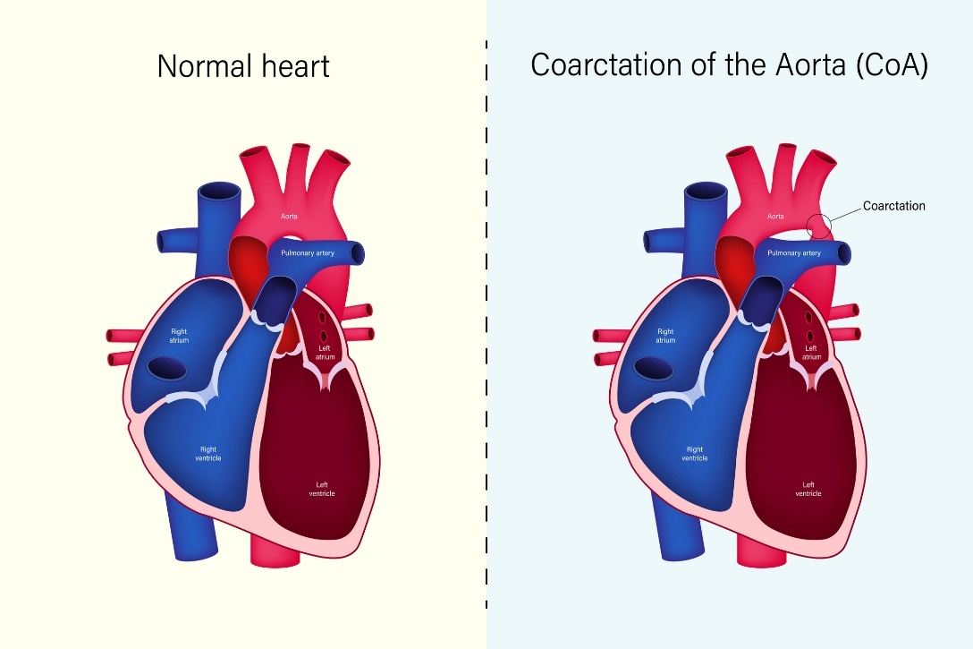
Coarctation (congenital narrowing) of the aorta is a developmental generalized disease of the human cardiovascular system. It is also referred to as CoA. It accounts for approximately 8% of all congenital heart defects.
The disease affects men 2 to 3 times more often.
The main feature is a congenital narrowing of part (most commonly the descending part) of the aortic arch. The exact location and extent of the narrowing of the vessel is individual and determines the severity of the disease itself.
The pathophysiology of the disease is obstruction of the outflow of blood from the left ventricle of the heart. The consequence is the development of hypertension (high blood pressure) and hypotension (low blood pressure) in the areas after coarctation.
Left ventricular hypertrophy (enlargement) and dysfunction, congestion of the heart, development of accelerated atherosclerosis and impaired coronary blood flow occur.
In most cases, aortic coarctation is diagnosed and operated/treated in childhood. In rare cases, the diagnosis is not made until the individual is an adult.
Late indications for surgery mainly include hypertension (high blood pressure), recoarctation and aortic pseudoaneurysm.
The main health risks of aortic coarctation are hypertension, atherosclerosis and the subsequent development of vascular (ischemic) stroke.
Causes
Botall's duct is the vascular connection between the aorta and the main pulmonary vessel. Its pathology is one of the common causes of aortic coarctation.
In healthy newborns, the arterial connection between the aorta and the pulmonary artery is closed. This ensures adequate blood flow throughout the body and prevents backflow of blood into the lungs.
In coarctation of the aorta, this closure does not occur and a narrowing of some part of the aorta occurs. There is a significant flow of blood from the aorta to the pulmonary vessels and at the same time a reduction in blood flow to the rest of the lower body.
The consequence is insufficient blood supply to the lower body and excessive congestion of the heart. However, the blood supply to the upper body is not impaired.
Depending on the exact location of the narrowing of the vessel, the clinical symptoms and health risks to the patient depend on the narrowing itself.
Most commonly, the descending part of the aorta is narrowed.
In rare cases, it can also occur in the ascending portion or in the aortic arch itself.
Coarctation of the aorta is an isolated defect, but it can also occur with associated lesions and heart defects. The most common associated defects include bicuspid aortic valve, ventricular septal defect or aortic stenosis.
In intrauterine and postnatal life, underdevelopment of part of the aorta, changes in intrauterine blood flow, premature narrowing of the arteries, and other cardiac disorders may occur.
Coarctation of the aorta is also more common in people who have other genetic disorders present, such as Turner syndrome.
CoA can also be part of complex congenital heart defects such as transposition of the great arteries with ventricular septal defect or hypoplastic left heart syndrome.
Genetic factor plays an important role in the development of aortic coarctation and heart defects.

Symptoms
The main symptom is shortness of breath due to decompensated hypertension and vascular difficulties in the lower extremities due to insufficient blood supply to the area. The patient may experience weakness and fatigue of the lower extremities or a feeling of cold feet.
Headaches, tinnitus (ringing in the ears) and sinus bleeding may be present.
Objective symptoms may include hypertrophy(enlargement/cardiomyopathy) of the left ventricle, systolic murmur over the site of aortic constriction or a weak femoral pulse in the lower limbs.
High blood pressure is present in the upper half of the body and conversely low blood pressure in the lower half of the body.
Possible manifestations of coarctation of the aorta:
- Shortness of breath (breathing difficulties, dyspnoea)
- Headache
- Tinnitus (ringing and whistling in the ears)
- Dizziness
- Increased sweating
- Nosebleeds
- Feeling of cold feet
- Muscle weakness in the lower limbs
- Increased fatigue

Diagnostics
Most cases of aortic coarctation are diagnosed in early childhood.
The basic diagnosis consists in measuring blood pressure, which varies in the upper and lower half of the patient's body.
The basic diagnosis includes instrumental echocardiography, which senses the electrical impulses of the heart muscle. The result is an ECG curve that determines the exact waves, oscillations and intervals of the heartbeat.
In coarctation of the aorta, left ventricular hypertrophy can be observed. A corrected gradient is measured.
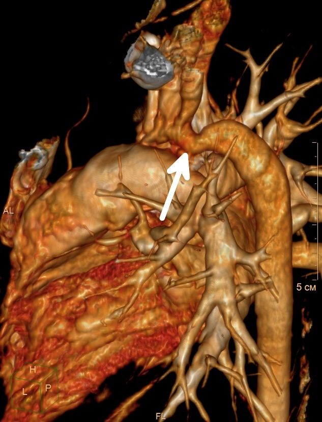
Angiography, X-ray, computed tomography CT or magnetic resonance imaging MRI may be indicated to view the internal structures in detail, to determine the exact site of pathology and the segment of aortic narrowing itself.
The diagnostic program also includes regular Holter blood pressure monitoring to determine and monitor the patient's hypertension.
Prognosis after treatment of aortic coarctation
Today, the prognosis of treated patients is much better than in the past due to early surgery in childhood and the development of surgical techniques. Approximately 84% of operated patients survive and of these, 11% undergo aortic reoperation.
Accelerated atherosclerosis in the vasculature before coarctation is the cause of coronary events and sudden cardiac death, which is the most common cause of death in patients with aortic coarctation. The average age of fatal myocardial infarction is 48 years.
Patients after aortic coarctation surgery should be followed up by a cardiologist for life.
Aortic coarctation and pregnancy in women
Pregnancy in a woman with an unoperated aortic coarctation or recoarctation is high-risk because of the possibility of aortic dissection.
Pregnancy in women after successful aortic coarctation surgery is well tolerated. The likelihood of miscarriage is approximately 16%.
Due to hypertension, frequent monitoring by both a cardiologist and gynecologist and adherence to medical precautions are necessary.
Prevention of cardiovascular health
Diseases of the cardiovascular system are among the most serious causes of disability worldwide and are also one of the leading causes of hospitalization.
Preventive factors:
- Regular preventive checkups with a cardiologist
- Seeing a doctor early when symptoms worsen
- Elimination of stress factor
- Healthy and nutritious diet
- Elimination of fatty and greasy foods
- Control of optimal weight
- Limiting tobacco products
- Limiting alcohol consumption
- Regular adequate exercise as part of the diagnosis
- Quality regular sleep
- Home blood pressure measurement
Read also: How to prevent cardiovascular disease?
How it is treated: Coarctation of the aorta
Treatment of aortic coarctation: surgery/angioplasty is the first option
Show moreCoarctation of the aorta is treated by
Coarctation of the aorta is examined by
Other names
Interesting resources
Related


