Cardiomyopathy refers to a group of diseases that affect the heart muscle, the myocardium. We know several forms, they have different symptoms and also different treatments.
Cardiomyopathy is the name for a group of diseases affecting the heart muscle.
Myocardium = heart muscle. Cardio = heart related, cardiac. Myo = related to muscle, muscles. Patia = disease ending.
These diseases are divided into several forms. Each may have a different cause, manifested by different symptoms or necessary treatment.
In cardiomyopathy, the heart enlarges.
This results in a malfunction of the muscle. The roughened fabric has different properties and its elasticity does not meet the necessary requirements.
Disease changes are also affected by the conductive system of the heart, which results in heart rhythm disorders. We also know these as arrhythmias.
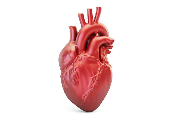
The heart is a pump that pumps blood to the human body for life.
In the case of a blood supply disorder, the brain and the heart itself mainly suffer. Of course, so do other cells, tissues, and organs. With the sudden interruption of blood circulation and the failure of the heart as a pump, unconsciousness, respiratory arrest, and, after a few minutes, death.
Most often you are interested in: What is cardiomyopathy and why does it occur? What is the hypertrophic, dilated, restrictive, stressed or alcoholic form? How does it manifest and heal? Answers to these questions and other interesting information in the article.
Myocardium = heart muscle
The heart pumps blood to the whole body. And without a break for life.
This function is provided by the heart muscle. This is the thickest layer of the heart wall. Muscle with regular movements ensures the expulsion of blood into the body and its suction back.
Upon withdrawal, blood is expelled into the aorta. This phase is also called systole.
The weakening of the heart muscle then ensures the return of blood from the large veins to the heart. And this phase is called diastole.
The thickest layer of muscle is located in the left ventricle. The left ventricle blows blood into the aorta , and thus into the whole body. It must therefore overcome the highest pressure. Thanks to which oxygenated blood from the heart travels to the body.
The heart muscle is made up of muscle cells called cardiomyocytes. There is a combination of transversely striated and smooth muscle.
Although it contains a type of transversely striated muscle fibers, its function cannot be controlled by will. Its influence has an autonomic nervous system, and frequency.
The sympathetic nerve accelerates and the parasympathetic nerve slows down heart activity.
The very control of contraction, ie contractions and muscle weakness, is ensured by the heart's own transmission system.Disruption of this system results in various cardiac arrhythmias.
Like all cells in the human body, heart cells must be oxygenated and supplied with nutrients. Blood flows to them through blood vessels, also known as coronary arteries or coronary arteries.
This ensures trouble-free function and operation of the heart like a pump. If the supply of oxygen and nutrients is disrupted, then, depending on the extent of restriction or blockage of blood flow to the muscle, ischemic heart disease or myocardial infarction of the heart muscle occurs.
Layers and envelope of the heart : The inner layer of the heart = endocardium - the inner lining of the heart cavities and also forms the heart valves. Middle layer - myocardium. The outer layer of the heart = epicardium. Pericardium = a bag in which the heart is stored.
+
The heart has 4 cavities: the right atrium - the entry of large veins and the inflow of deoxygenated blood from the body. The right ventricle - the outflow of blood to the lungs, where it is re-oxygenated. Left atrium - entry of oxygenated blood back into the heart. Left ventricle - expels oxygenated blood into the aorta, and thus into the whole organism.
The left ventricle has approximately three times the muscle mass of the right ventricle.
In the rest of the article, you'll learn: What is cardiomyopathy and how it divides . Sudden deaths in athletes and young people. Why cardiomyopathy occurs . What are the symptoms and treatment available?
A closer look at cardiomyopathy
Cardiomyopathies are diseases of the heart muscle. The basis is a change in the size of the heart.
This disease change results in a change in the function of the heart muscle and transmission system. This is negatively reflected on the work of the heart as a pump and also on the heart rhythm.
In addition to cardiac arrhythmia, there is a risk of developing heart failure.
From a broader point of view, it is divided into primary or secondary.
Primary = has no known cause. Secondary = specific disease with a known cause.
Cardiomyopathy is also referred to as KMP.
Cardiomyopathies are divided into several types, which are listed in the table
Type of cardiomyopathy
Description
1. Dilated cardiomyopathy
dilatation = diseased enlargement, increase in volume, dilation
pathological enlargement of the heart = dilation of the heart compartments
the most common type
it affects the left ventricle to a greater extent
blood pumping disorder - reduction of the ejection fraction of the heart below 40%
various causes
ischemia and massive myocardial infarction
unknown cause
viral disease - myocarditis = inflammation of the heart muscle
alcohol
heredity or impaired immunity and others
2. Hypertrophic cardiomyopathy
hypertrophy = increase in cell, tissue, organ volume
enlargement of the heart muscle cells - especially the left ventricle and septum, ventricular compartments
the heart cavities do not expand
the expulsion of blood is not as disturbed as its filling into the ventricles of the heart
slowing of the filling of the heart chamber
increase in pressure inside the chamber
consequently, the pressure in the lungs also increases
obstructive form = disruption of filling and expulsion of the chamber
a common cause of sudden death in young people and athletes
genetic defects and hereditary factor
3. Restriction cardiomyopathy
rare form
the heart is hardened - rigid
behind the formation are infiltration mechanisms = pathological deposition of substances in the wall of the heart
for example in diseases:
Amloidosis
Hemochromatosis
Sarcoidosis
the pumping and expulsion of blood is impaired
4. Arrhythmogenic dysplasia of the right ventricle
ADPK
congenital genetic disease
fibrous damage of the right ventricle + fatty tissue remodeling
normal tissue is morbidly replaced by non-valuable adipose and fibrous tissue
change of electrical properties and creation of electrical excitations, which control the activity of the heart, come out and run through the area of the right atrium and ventricle
often asymptomatic
possibly with the first symptoms in young people
palpitations = palpitations
apostasy and syncope = short-term disorder of consciousness
sudden death in a hitherto healthy person
already in people under 35 years of age
genetic basis
risk of malignant lethal arrhythmias
is not the cause of chronic heart failure
Other rare forms
Tako Tsubo Cardiomyopathy - TTK
stress cardiomyopathy - stress KMP
you may have heard of broken heart syndrome
especially in women
named after the pitchers in which Japanese fishermen catch squid
these are called Takotsubo
the cause is excessive stress such as:
death in the family
divorce
or severe chronic mental stress
change the shape and size of the left ventricle
+ disorder of the kinetic properties of the apex and base of the left ventricle
the condition usually resolves after a few weeks
however, during this time there is an increased risk of heart rhythm disorders
maybe confused with myocardial infarction
postpartum cardiomyopathy
non-compact myocardium
diabetic cardiomyopathy
and other
Sudden death in young people and athletes
Death of a young person may be the first sign of a previously unspecified heart defect.
The fact that a now healthy person is experiencing a change in health leading to death is devastating.
This is largely due to unrecognized hypertrophic cardiomyopathy or arrhythmogenic dysplasia of the right ventricle of the heart. And also other causes.
Severe - malignant heart rhythm disorder= ventricular tachycardia → progresses to ventricular fibrillation .
These two arrhythmias cause the blood to fail to fill the heart and pump blood from the heart.
There is heart failure as with a pump. Death occurs without immediate assistance, defibrillation and resuscitation.
Therefore, it is important to pay attention to every dropout and collapse in young people, especially during sports activities or increased physical activity.
Important: Early detection of warning - alarming signs.
Alarming symptoms:
loss of consciousness, collapse, syncope, apostasy
about 18% have a cardiac cause
quick return to consciousness within 20 seconds
in uncomplicated syncope, a good sign
persistence of impaired consciousness - unconsciousness
when one does not take consciousness
body cramps in a person who is not being treated for epilepsy
onset of convulsions until loss of consciousness
in epilepsy, mostly with loss of consciousness
difficult to absent breathing
growl
catching breathing - fish breathing
irregular and ineffective breathing
discoloration of the skin of the face and lips
Gray color
bluish to purple color
beware heart disorder
Warning! Pulse measurement may have an incorrect result.The rescuer can feel his own pulse! And not the pulse of the affected person.
You ask:
How to proceed?
We proceed as follows:
secure access
control of consciousness - by voice, touch
does not answer?
call for help 155 / in case of accident 112
the emergency operator asks questions
answer
will advise you on what to do in a situation immediately threatening your health and life
respiratory control
not breathing normally?
his chest and abdomen are not raised
airway clearance
head tilt
still not breathing normally?
revival
chest compression 5 - 6 cm to depth
100 times per minute
center of chest
does the rescuer have a first aid course?
+ mouth-to-mouth breathing
ratio of 30 chest compressions and 2 breaths - 30:2
inhalation takes about 1 second
if the rescuer does not have a first aid course, he only performs chest compressions!
children have their first breaths - anyway!
5 introductory breaths
subsequently compressing the chest to approximately one-third of the height
in children, the most common respiratory (respiratory) cause is circulatory arrest
Remember: In children, 5 first breaths, then chest compressions. Don't have a first aid course? = "ONLY" COMPRESS your chest.100 * 1 minanimation - resuscitation will not hurt. Broken ribs heal.Death is irreversible. Therefore, even imperfect resuscitation is BETTER than DOING NOTHING!
Early defibrillation is important in heart rhythm problems. It could be described as resetting the heart's transmission system. And it should restore normal heart rhythm function.
The defibrillation public are designed automatic external defibrillators abbreviation AED.
They are available in some public places :stations,stadiums,sports halls, shopping mallsor workplaces. They are marked: White heart with lightning on a green field.
AED marking. Photo: Getty images
BUT...
In some cases, the error is detected accidentally even before the first symptoms occur. Or the first symptoms are less severe, such as palpitations, or palpitations.
In this case, preventive implantation of a cardioverter-defibrillator in a person at risk may be chosen. Another method is the treatment method, and thus catheter ablation, which interrupts the pathological conduction of stimuli through the heart and prevents the onset of arrhythmias.
Causes
The causes of cardiomyopathies are diverse. In some cases, the exact reason will never be found. This is referred to as primary cardiomyopathy.
The second group is secondary cardiomyopathies. These are specific forms of the disease, as listed in the table above. Their causes are usually different.
The table shows the distribution of causes according to the form of KMP
Form
Causes
Dilatation KMP
ischemia - decreased myocardial blood flow
massive myocardial infarction
unknown cause
myocarditis = inflammation of the heart muscle
viral disease
protozoa
drugs
alcohol
heredity and genetic factors
immune system disorder and autoimmune causes
pregnancy
Beri-Beri, a vitamin B1 deficiency disease
Hypertrophic KMP
HKMP
in most cases the genetic basis
and thus also the familial occurrence
up to 50% of cases
approximately 150 gene mutations are known to which it is associated
Fabry disease , Danon's disease, and others
Restriction KMP
RKMP
based on the pathological deposition of substances in the heart in other diseases
Amyloidosis - metabolic disease, storage of protein, amyloid in organs, lungs, heart, liver, joints
Hemochromatosis - a disorder of iron metabolism, the storage of iron in organs and their subsequent damage, in addition to the heart, such as the liver and other
Sarcoidosis , also the deposition of small nodules in organs, is an unknown cause
post radiation damage
unknown cause
the heart is stiff, rigid
Arrhythmogenic dysplasia of the right ventricle
ADPK
genetically determined disease
fibrous damage of the right ventricle and fat remodeling
physiological tissue is replaced by non-functional adipose and fibrous tissue
this will cause a change in electrical properties and the generation of electrical excitations
Some other causes
rheumatoid arthritis
scleroderma
heart valve defects
muscular dystrophy
thyrotoxicosis
diabetic KMP
in case of insufficient nutrition
for chronic tachycardia
toxic causes
long-term and severe stress
unknown cause
and other
Interesting: Normally, the left ventricle has a muscle thickness of approximately 12 millimeters. In hypertrophic cardiomyopathy, it can be as much as 60 millimeters.
You ask: At what age does it develop?
The age category is not precisely limited, as it can develop in children, young and adults.
Symptoms
Each form of cardiomyopathy may have different symptoms. However, it happens that it takes place asymptomatically, ie asymptomatically.
In some cases, serious heart problems occur first. An example is a heart rhythm disorder characterized by palpitations.
Sudden death may also be the most serious first symptom. The information resonates especially in the case of the death of a young athlete.
In this case, mainly two forms of KMP are considered:
arrhythmogenic right ventricular dysplasia
hypertrophic cardiomyopathy
Until then, they run asymptomatic, are not diagnosed and the trigger is physical exertion.
Thus, the symptoms of cardiomyopathies cannot be precisely specified in one set. Sometimes KMP does not show up at all, other times it has typical symptoms of a cardiac problem.
The conditions with loss of consciousness and respiratory disorder are especially dramatic, as described at the beginning of the article in the section on the Death of young people and athletes.
From general symptoms may occur such as:
fatigue and weakness
fainting, collapse, syncope
increased fatigue and reduced performance
dizziness
swelling, first of the feet, ankles and later higher
Initially, symptoms can only occur with more severe physical activity. Depending on the degree and extent, the required load rate decreases. Severe myocardial damage is manifested by minimal effort.
+
The disproportion in the size of the heart cavities results in the formation of blood clots (thrombi). Similar to cardiac arrhythmia.
The basis is disruption of blood flow. The risk is the expulsion of clots from the heart to the body. A serious complication is a stroke.
Thrombus in the heart → after its release → embolus → risk of clogging of a vessel in another part of the body → embolization.
A common cause of brain non-blood flow is a thrombus expelled from the heart. Examples are atrial fibrillation, valve defects, and others.
Athlete's heart
The heart can adapt to long-term sports load and activity. Therefore, it is common for athletes, and especially in elite sports, for the heart to enlarge to some extent.
It is also hypertrophy.
This is usually an increase in chamber thickness to 13 millimeters.
Myocardial thickness has been reported to be up to 12 millimeters under normal circumstances.
Possibly.
The so-called gray zone is also mentioned. It is a thickening of the muscle up to 15 mm. In this case, further examinations and condition monitoring are required.
Diagnostics
Diagnosis focuses on the anamnesis and clinical picture, ie the manifestations of the disease. At a higher stage of the development of the disease, the symptoms that lead to the heart cause are usually present.
Of course, in addition to the physical examination, other specific examinations are also required.
An example is the ECG, which records the electrical activity of the heart. Possible arrhythmias are also detected here, and a specific ECG image is determined. A 24-hour recording, ie an ECG Holter, can be helpful.
It complements the ECHO. During echocardiography, ie the examination of the heart by ultrasound, the dimensions of the heart, its cavities and the thickening of the heart muscle wall, the enlargement of the heart sections, the condition of the valves, and other parameters can be determined.
MRI - magnetic resonance imaging can be performed to exclude structural changes. And to recognize coronary artery disease and atherosclerosis, coronary angiography is added.
The basic diagnostics also include X-rays, CT, lungs and heart - chest, blood collection for laboratory examination, of course, blood pressure, pulse frequency, and its regularity are measured. Possibly genetic testing.
Course
Enlargement of the heart can be asymptomatic. And especially in the initial period. The asymptomatic course is interrupted by occasional difficulties, especially during exercise.
Examples are palpitations, increased tiredness or dizziness, and a feeling of falling away.
Higher levels of physical exertion and provocative mechanisms initially cause milder difficulties. However, over time, symptoms of the disease may occur even after mild exercise.
Heart failure develops.
A high level of danger to health and life is conditions when a full acute deterioration occurs in full health. Rhythm disorders are mainly risky, Which can lead to unconsciousness, respiratory disorders, and even death.
The table lists some of the symptoms that occur during dilated or hypertrophic KMP
Dilatation KMP
Hypertrophic KMP
gradual heart failure
arrhythmia
leads to cardiac decompensation
chest pain
to swelling of the lungs
exertional dyspnea
commonly occurring:
palpitations
arrhythmia
to orthopnoea
palpitations - palpitations
there are syncopations - falls off, especially during exercise
chest pain - angina pectoris, but with a normal finding in the coronary arteries
or milder pre-collapse states - a feeling of falling off
shortness of breath - dyspnoea, first after exertion
+
the chamber of the heart fills worse due to its increased muscle mass, the expulsion of blood into the body circulation is not disturbed. The chamber fills more slowly and increased pressure is present
a cough
+
the left ventricle is normally filled, but there is a disorder of its emptying = expulsion of blood into the aorta
And ...
There is also swelling of the lower limbs, which develops over time and according to the degree of heart involvement. It progresses higher, from the legs, through the ankles, and to the forelegs to the thighs and abdomen. Thus, it may be a symptom of chronic heart failure.
People diagnosed with KMP are also at risk for developing malignant arrhythmia = a serious heart rhythm disorder. Such as ventricular tachycardia and fibrillation.
How it is treated: Cardiomyopathy
Treatment of cardiomyopathy - medications and healthy lifestyle
"What Are the Signs and Symptoms of Cardiomyopathy?". NHLBI. 22 June 2016.
"Who Is at Risk for Cardiomyopathy?". NHLBI. 22 June 2016.
"Types of Cardiomyopathy". NHLBI. 22 June 2016.
"What Causes Cardiomyopathy?". NHLBI. 22 June 2016.
"How Is Cardiomyopathy Treated?". NHLBI. 22 June 2016.
GBD 2015 Disease and Injury Incidence and Prevalence, Collaborators.
"What Are the Signs and Symptoms of Cardiomyopathy? - NHLBI, NIH". www.nhlbi.nih.gov.
Bakalakos, Athanasios; Ritsatos, Konstantinos; Anastasakis, Aris (1 September 2018). "Current perspectives on the diagnosis and management of dilated cardiomyopathy Beyond heart failure: a Cardiomyopathy Clinic Doctor's point of view". Hellenic Journal of Cardiology. 59 (5): 254–261.
Rath, Anika; Weintraub, Robert (23 July 2021). "Overview of Cardiomyopathies in Childhood". Frontiers in Pediatrics. 9: 708732.
Gorla, Sudheer; Raja, Kishore; Garg, Ashish; Barbouth, Deborah; Rusconi, Paolo (December 2018). "Infantile Onset Hypertrophic Cardiomyopathy Secondary to PRKAG2 Gene Mutation is Associated with Poor Prognosis". Journal of Pediatric Genetics. 07 (04): 180–184.
^ Law, Michelle L.; Cohen, Houda; Martin, Ashley A.; Angulski, Addeli Bez Batti; Metzger, Joseph M. (February 2020). "Dysregulation of Calcium Handling in Duchenne Muscular Dystrophy-Associated Dilated Cardiomyopathy: Mechanisms and Experimental Therapeutic Strategies". Journal of Clinical Medicine. 9 (2): 520.
The secondary medical school in Nitra gave me the basis for my career in the field of health and diseases. Thanks to it, I worked for 2 years in the traumatology clinic and outpatient clinic at the Nitra Hospital. Since 2006 I was employed in the emergency medical service, where I stayed until 2017.
I completed my bachelor's degree at the University of Constantine the Philosopher in Nitra in the field of emergency health care. The bachelor's degree allowed me to continue my mission as a paramedic. In the meantime, I got a job at the emergency line 155. I have been working in pre-hospital health care until today.
I had an interest in people, health and even diseases in my childhood, which gave me the prerequisite to pursue this topic in adulthood. Studying and acquiring new information in practice provided me with a great basis for writing professional texts, in the form of articles that can be understood by ordinary people. Thus, my interest in the Health Portal has a solid foundation in years of practice and personal interest. Similarly, I am also interested in healthy eating, nutrition and overall healthy lifestyle. I fill my free time with family and sports.