- ospalecek.cz/ - Paleček Association
- solen.sk - Achondroplasia. Solen. MUDr. Ľubica Tichá, PhD
- genetickesyndromy.sk - Achondroplasia. Genetic syndromes.sk
- vzacna-onemocneni.cz - Czech Association of Rare Diseases
- pubmed.ncbi.nlm.nih.gov - Achondroplasia: a comprehensive clinical review. Richard M Pauli
Achondroplasia: What are the causes, symptoms of congenital bone disorder?

Achondroplasia refers to a developmental genetic disease of the bones and cartilage. Why does this congenital disorder occur and what are its characteristic manifestations?
Characteristics
Achondroplasia is a genetic disorder of cartilage development in which there is a defect in bone growth, improper development and ossification of articular cartilage. The disproportion is already present in newborns and is genetically determined.
The etiology, symptoms, treatment options and much more can be found in this article.
What is achondroplasia?
Achondroplasia refers to a condition of impaired cartilage growth. It is caused by a disruption in the process of echondral ossification of bone and results in abnormal bone growth.
The primary problem, however, is not the undeveloped cartilage, but the process of ossification itself - the conversion of cartilage to bone during development. It is particularly evident in the upper and lower limbs.
The cause of the disease was first described by Dr. John J. Washmuth and his research team in 1994. The disease is rare, occurring in approximately one in 15-20 thousand births.
The genetic disease is caused by a mutation in a growth factor receptor gene called FGFR3.
This is a mutation in the gene that codes for the receptor for fibroblast growth factor. This factor is responsible for regulating linear bone growth.
The characteristic manifestation of achondroplasia is a defective body structure in terms of short stature, short bones and a head that is disproportionately large compared to the body.
Causes
The basic cause of achondroplasia is a genetic disorder on chromosome 4, specifically the aforementioned FGFR3 gene.
Achondroplasia is autosomal dominant in nature. Autosomal means that the pathological gene is not on the sex chromosome. Therefore, the disease also affects both sexes - male and female.
Dominant type inheritance means that the newborn inherits the disease from one of the parents with achondroplasia. However, approximately 80% of people with achondroplasia have parents of normal stature without the diagnosis.
In most patients, there is no obvious objective factor of family history. However, the older age of the father (over 35 years) may be a definite risk factor in cases of this rare disease of achondroplasia.
The chance of conceiving a child with achondroplasia in both healthy parents is very small.
An individual with achondroplasia who has a partner of physiological stature has approximately a 50% chance of having a healthy child. However, if both parents have achondroplasia, the chance of having a healthy child is approximately 25%.
There is also a 25% chance that the child will have a homozygous mutation that is incompatible with life.
Symptoms
The main characteristic symptom due to abnormal bone growth is a small, short stature (dwarf build) with short arms and legs, average torso size and a larger head compared to the body.
Hydrocephalus (accumulation of fluid in the brain cavities) is also common.
The average height for adult males with achondroplasia is approximately 131-136 cm and for adult females 124-130 cm.
In most cases, individuals are shorter than 130 cm.
Within the face, the frontal region and the undeveloped root of the nose are particularly dominant. The elongated, pear-shaped skull is characteristic. The mandible and supraorbital arches are also enlarged. Dental and oral health problems are also common.
However, life expectancy may be slightly reduced due to respiratory problems (risk of sleep apnoea), cardiovascular disorders, non-physiological spinal cord position or recurrent infections.
Low blood pressure is often present in childhood.
Excess body weight (obesity), increased lordosis and kyphosis of the spine, hunching, joint instability and muscle imbalances and spinal pain are common.
Obesity is one of the main problems in achondroplasia. It leads to increased morbidity and overloading of the joints and spine. Obesity is associated with lack of exercise, increased appetite and psychological instability of the individual.
Another characteristic feature is the unphysiological position of the knees (legs in an imaginary letter O).
Macrocephalic skull in infants, short neck, short limbs and muscular hypotonus together negatively affect the motor development of the patient.
Characteristic features of achondroplasia:
- Short upper and lower limbs
- Large head against the body
- Short, dwarf stature
- Prominent forehead and supraorbital arches
- Defective posture
- Lordosis and kyphosis of the spine
- O-shaped knees

Diagnostics
Diagnosis is primarily based on characteristic clinical features such as the individual's overall height, head circumference, skull structure, limb length, and the proportion of upper and lower body segments.
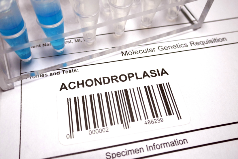
Diagnosis is primarily confirmed by molecular genetic testing for the FGFR3 gene mutation.
Prenatal diagnosis is possible during prenatal ultrasound examination in the 3rd trimester of pregnancy. Shortening of the long bones can be detected.
Confirmation of the diagnosis is also possible by genetic testing (amniotic fluid/peripheral blood) of fetal DNA for the presence of the FGFR3 mutation at position 380 (G380R) in almost all patients with achondroplasia.
Developmental skeletal defects of achondroplasia can be seen on ultrasound from approximately 22-24 weeks of gestation.
However, the biggest problem in prenatal diagnosis is that in some cases the changes are only observable in the third trimester. Sometimes the disease is even detected only postnatally.
On a radiograph of the total skeleton, shortened long bones with thickened metaphyses (the forelimb of the bone) of irregular shape are visible.
The lateral skull X-ray shows enlargement of the frontal segment, facial hypoplasia and narrowing of the foramen magnum (opening of the cranial cavity).
The foramen magnum requires an MRI scan, which may show cervicomedullary compression, myelomalacia or ventriculomegaly and other disorders.
How it is treated: Achondroplasia
Treatment of achondroplasia: Can it be cured?
Show moreAchondroplasia is treated by
Achondroplasia is examined by
Other names
Interesting resources


