- solen.sk - STEREOTACTIC RADIOCHIRERY IN THE TREATMENT OF HEAD AND NECK DISEASES
- gponline.com - CLINICAL REVIEW ACUSTIC NEUROMA
- emedicine.medscape.com - acoustic neuroma
- mayoclinic.org - Mayo Clinic Acoustic Neurinoma
What is schwannoma, neurinoma of the statoacoustic nerve and its symptoms?
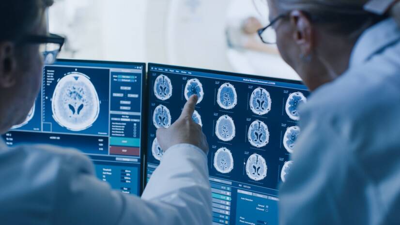
Photo source: Getty images
Acoustic neurinoma and schwannoma are synonyms for one and the same benign tumor.
Most common symptoms
Show more symptoms ᐯ
Characteristics
Acoustic neurinoma, schwannom...
The tumor affects the cranial nerve, which comes directly from the brain. It's responsible for hearing and balance, so those two functions will be most severely affected.
It's not malignant. It's not cancer in the true sense of the word. But it grows and behaves expansively. In the confined space of the skull, important brain centres are suppressed.
The statoacoustic nerve (VIIIth cranial nerve, Latin nervus vestibulocochlearis) is a nerve that runs at the base of the skull towards the inner ear.
It is responsible for the perception of sound (hearing) and also for the perception of body and head position. It serves the organ of balance of the inner ear.
All cranial nerves have a sheath on their surface that serves to protect, nourish and guide the fibres. This sheath is called the myelin sheath. It is made up of cells called Schwann cells. They are named after the physiologist Theodor Schwann.
They wrap around the nerve ending like a roll of toilet paper. One Schwann cell thus forms a sheath for only one nerve ending, one axon.
When Schwann cells begin to grow uncontrollably, they form a tumor-like formation called a schwannoma. This tumor grows expansively, does not overgrow and is not invasive to surrounding tissues. It is therefore a benign process.
Schwannoma accounts for about 8% of all brain tumours. The highest incidence is between 40 and 60 years of age. Women are affected up to 2 times more often than men.
Schwannomas can affect any of the twelve cranial nerves, but most commonly the statoacoustic nerve. The trigeminal nerve is the second most common.
Causes
The direct cause of vestibular schwannoma is not yet known. No risk factors are apparent in most patients.
Epidemiological studies have investigated the effect of mobile phone use, but the results have not shown a direct association between wireless phone use and the incidence of vestibular schwannoma.
Other studies have linked the incidence of schwannoma to chickenpox or exposure to multiple X-rays of the skull as a potential risk factor.
So far, the only established predisposing factor for neurinoma is an inherited disease called neurofibromatosis type II.
Neurofibromatosis type II occurs in patients who have a defective tumor suppressor gene located on chromosome 22q12.2. The disrupted protein produced by the mutated gene is called merlin or schwannomain.
The main clinical features of neurofibromatosis type II are bilateral acoustic tumours.
Other manifestations include peripheral neurofibromas, meningiomas of the brain and spinal cord, and gliomas.
Patients are usually diagnosed in late adolescence or early adulthood.
Rarely, they may be caught later in the fifth to seventh decade. In these individuals, the tumors grow very slowly, and so symptoms from oppression become apparent later.
Symptoms
As a benign tumour, schwannoma grows very slowly. It is usually present in the brain for several years before neurological symptoms start to appear.
A small tumour usually shows no symptoms. If it grows faster or is larger, it will start to show gradually and subtly.
It does not show any particularly specific symptoms. It manifests itself very similarly to other common neurological diseases, which may not even be related to pathological processes in the brain.
For example, dizziness and balance problems may not be related to the tumour, but to stiff neck and cervical spine problems. However, some symptoms deserve attention, especially if they are long-lasting, do not improve, but get progressively worse.
The most common symptoms of schwannomas are:
- Gradual hearing loss
Approximately 80-90% of people who are later diagnosed with acoustic neuroma suffer hearing loss at various levels.
The patient is not initially aware of the hearing loss and may sometimes downplay it. For example, he or she notices that he or she can hear better in one ear when talking on the phone.
The worsening situation is only brought to his attention by deafness in normal interpersonal conversation, which interferes with the individual's social life.
Hearing loss caused by neurine is called perceptual (sensorineural) deafness. It is caused by direct damage to the auditory nerve by a growing tumor.
Whistling, humming, tapping and other sounds in the ears (tinnitus)
When an acoustic neuroma grows unilaterally, this murmur is usually present in one ear.
Basically, these are different sounds that the patient hears. It may not always be just a whistling in the ear. There may be different kinds of sounds and tones that the patient hears, but there is no external source of this sound.
However, people also suffer from tinnitus in other diagnoses, often in non-neurological conditions such as anaemia (low red blood cell count).
Non-malignant tinnitus can result from pushing earwax with a cotton bud, ongoing inflammation in the ears, exposure to excessive noise, irritation of the ear by unpleasant and deafening stimuli, or aging.
Tinnitus can be transient or long-term, silent or very noisy. With long-term and noisy tinnitus, patients can suffer from serious psychological difficulties.
Dizziness
The statoacoustic nerve transmits information to the brain not only about sounds, but also about the position of the head and body. When it is damaged, patients will therefore have difficulty with balance in addition to hearing difficulties. This non-specific symptom occurs in approximately half of patients.
However, it is rarely the first symptom that alerts the patient to the disease. It may be the most unpleasant of all the other symptoms.
Sensitivity disorder on half of the face
This is a varying degree of tingling, pulling, numbness or radiating pain on one half of the face. These sensations are due to pressure from the growing tumour on other cranial nerves running close to the statoacoustic nerve.
These nerves usually originate from the so-called pontocerebellar angle. The trigeminal nerve (nerus trigeminus), which is responsible for sensory innervation of the face, is most often affected by the pressure. Therefore, its damage gives rise to these various sensitivity disorders.
Complete numbness of half of the face is relatively rare in statoacoustic neurinoma.
Headaches
Headache is a rare symptom in statoacoustic neurinoma. The tumour grows slowly. It rarely reaches enormous proportions to cause pain. However, if it begins to oppress the liquor pathways, pain may occur.
It is caused by blocking the outflow of cerebrospinal fluid through the liquor ducts, thereby increasing intracranial pressure in the skull.
This syndrome is called hydrocephalus, popularly water in the brain. It causes compression of the brain in the confined space of the skull, damaging the brain. In addition to pain, it causes a number of other neurological symptoms, even disturbances of consciousness.
Brain stem compression
The most feared and dangerous complication of statoacoustic neurinoma is just the gradual compression of the brainstem.
This can occur if the tumour is undiagnosed and begins to grow more rapidly to a larger size.
The statoacoustic nerve is located in the pontocerebellar angle. It is anatomically very close to the brainstem, which is the centre for controlling a person's vital functions.
If this oppression begins to damage the brain stem, there are problems with breathing, consciousness, circulation, coordination of movements and balance. Finally, there are also problems with vision, eye movement and paralysis of facial muscles and limbs.
Fatigue and lack of energy
A very non-specific symptom, but the most common in ongoing diseases associated with tumour growth, whether benign or malignant.
Diagnostics
Schwannoma is usually diagnosed in hearing loss or as an incidental finding in other diseases during an MRI scan of the brain.
The basic examination methods used to diagnose a statoacoustic nerve neuroma are:
Tone audiometry.
This is an electroacoustic method of hearing examination. It uses a tone generator to examine the sensitivity of the ear to particular tones. An audiometer is a special device that generates tones of a certain frequency and intensity.
The patient being examined sits in a soundproof booth. The audiometer generates tones that are transmitted to one ear by an earpiece.
The intensity of the pure tone is gradually increased until the patient indicates that he or she can hear the tone. At this point, the examiner records the intensity level of the sound on a graph. All tones are examined in this way. The marks on the graph are combined to form a characteristic curve - an audiogram.
The whole examination is repeated on the other ear. The result of the examination may be the same on both ears, but it may also be completely different. If asymmetrical hearing loss is detected, this is an indication for further diagnostic tests, e.g. BERA (BAEP) or MRI.
It is important to note that the degree of hearing loss does not correlate with the size of the tumour. Even in larger tumours, useful hearing may be preserved.
Useful hearing is assessed by tonal audiometry using the so-called Gardner-Robertson scale.
An important component is the assessment of word discrimination, i.e. how well the patient understands the spoken word at a normal volume of 50 dB. The preservation of useful hearing correlates with the ability to make a phone call to the affected ear.
BERA or BAEP (Brainstem Auditory Evoked Potential Examination)
This is an objective examination. It records the bioelectric potentials that propagate through the auditory nerve and brain stem.
This examination distinguishes between cochlear and retrocochlear nerve involvement. The sensitivity of BAEP depends on the size of the tumour. If the tumour is smaller, up to 1 cm, the sensitivity is approximately 85%. For larger tumours over 1 cm, the sensitivity of the examination increases to 95%.
Imaging methods
The most sensitive imaging method is magnetic resonance imaging (MRI). In addition, schwannomas can be imaged on head CT. A specific examination is positron emission tomography PET/CT using a special radiopharmaceutical as a contrast agent.
Course
Neurinoma is a benign and slow growing tumour. Therefore, the progression of this disease will be slow and creeping, with a gradual progressive and subtle deterioration of some functions.
Schwannoma is diagnosed in a patient who is brought to the examination by worsening hearing or dizziness. Sometimes the patient may have difficulties associated with another disease, such as a stroke. Because of this disease, an MRI scan is performed, which will show the neurinoma as an incidental finding.
In this case, a 'watch and wait' approach is chosen, meaning that if the patient does not have limiting symptoms of the disease, he or she will be monitored regularly by MRI at intervals of approximately one to two years.
If, however, the tumour is larger and there is a risk of deafness or brain stem oppression, specific treatment will be proposed to the patient.
How it is treated: Statoacoustic nerve neuron - Schwann
Treatment of schwannomas: observation, surgery if necessary + other methods
Show moreNeurinoma of the statoacoustic nerve is treated by
Other names
Acoustic nerve neuroma, vestibular schwannoma, intracranial schwannoma, vestibular schwannoma, acoustic neuroma, intracranial schwannoma
Interesting resources

MUDr. Andrea Bullová
Physician
After graduating from an eight-year high school, I knew exactly who I wanted to become in life. Studying at the Faculty of Medicine at Comenius University in Bratislava fulfilled a long-held dream and I was able to start fulfilling my mission as a doctor. After obtaining my diploma and M.D. degree, my first professional steps led to the St. Elizabeth Cancer Institute in Bratislava. At the Stereotactic Radiosurgery Clinic I worked with patients with brain tumours and eye tumours. After a year of working with oncology patients, in 2020 I moved to the Neurology Clinic of SZU St. Michael's University Hospital in Bratislava, where I am still working today. I like to spend my free time outdoors, whether cycling, hiking or just walking in the nearby forest.
View all articles by the same author


