Fibroma is a benign tumour of the connective tissues of the human body. It is most commonly found on the skin, but can also occur in other parts of the body or organs. What is the cause of fibroma growth, symptoms and treatment options?
Fibroma is a benign tumour of the connective tissue of the human body. It is one of the common benign tumours of soft structures and can occur anywhere on the body.
Fibroids are most commonly found on the skin of the neck, under the armpits, under the breasts, in the crotch and on the eyelids. The oral cavity is also a common site of fibroids.
A fibroid is essentially a harmless growth that can complicate life aesthetically (as a skin growth) and practically due to the inappropriate placement of the skin fibroid.
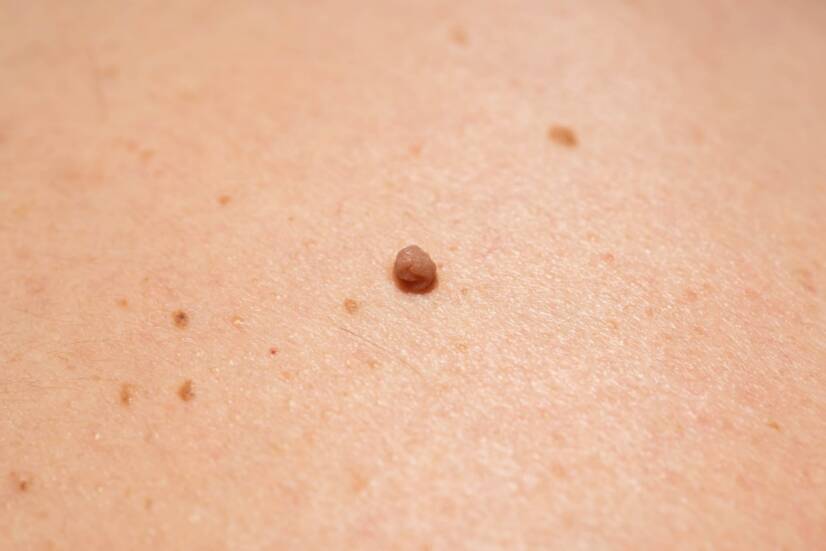
What does a skin fibroma look like?
A cutaneous fibroma looks like a small growth up to approximately 2 centimetres in size. It has a sharply defined shape and does not change size or shape after it has finished growing. It is usually of an overhanging pedunculated nature with a smoother surface.
Fibroma is not a communicable disease. Its growth is individual and spontaneous.
Types of fibromas
There are a large number of types of fibromas due to the vastness of connective tissues in the body.
We recognize angiofibromas (vascular fibroma), lymphatic cystic fibromas, ovarian and uterine fibromas, fibroadenomas of the breast, myxofibromas in subcutaneous tissue and many others.
Hard fibroma (fibroma durum)
A skin fibroma of a hard nature (fibroma durum) contains a minimum of fibroblast cells and a large number of fibers.
An example of a hard fibroma is dermatofibromas - small skin growths that grow at a slow rate. They are usually pink, red or brown in colour. They are most common in the lower legs, trunk and arms.
Another type of hard fibroma is keloids - hard tissue that forms in the area of skin scars. This is an aesthetically unfavourable enlarged scar. The face, shoulders and chest are particularly prone.
A keloid scar can form if the skin scar is not well cared for after surgery.
Hard skin fibroma (fibroma durum). Source of photography: Getty Images
Soft fibroma (fibroma molle)
A soft fibroma (fibroma molle) is a skin growth hanging on a thinner stem. It contains more fibroblast cells and fewer fibres.
Soft skin fibromas are most commonly found in the neck, armpit, breast and groin area around the genitals.
The exact etiology of fibroids is not well understood by the professional community. Genetics, age, hormonal changes and hormonal balance, the state of the immune system and the mechanical factor itself play a major role.
Their growth and formation is primarily determined by genetic factors.
Factors increasing the risk of fibroids:
Genetic factor
Increased age
Weak immune system
Autoimmune diseases
Hormonal changes
Hormonal treatment
Mechanical damage
Skin reaction to irritation
Insulin resistance
Presence of certain forms of human papillomavirus
Older age: The risk of fibroid growth increases with older age. As age increases, the immune system's defence function deteriorates.
Deficient immune system: low immune function and autoimmune diseases increase the possibility of fibroid formation.
Mechanical factor: Regular rubbing and irritation of the skin over a long period of time can lead to the formation of fibromas. This happens especially in specific areas such as the armpits, neck, groin or even the oral cavity.
Skin reaction to injury: after certain injuries such as insect bites, burns, wounds, piercings or surgery, the skin may react by forming fibrous fibres and forming a skin growth - a fibroma.
Hormone treatment: pharmacological treatment with hormones can lower the immune system and increase the risk of fibroid formation.
Examples of this include the use of hormonal contraceptives or immunosuppressive treatment (immune suppressants). In women, physiological hormonal changes may occur, for example, during pregnancy.
Symptoms
Usually fibromas do not show any symptoms or specific manifestations. They appear on the skin as a pale, pink to brown growth. They are usually only an aesthetic problem.
Sometimes fibromas, which grow in the connective tissues of organs, can cause problems.
In some rare cases, a skin fibroma may show signs of itching, redness or burning. This symptom is specific to fibromas located in close proximity to sebaceous glands, blood vessels or nerves.
Fibromas on the skin are very impractical growths as they can catch on clothing.
An itching sensation can be felt when the fibroma is mechanically damaged and irritated.
Diagnostics
Fibromas can be detected at home by inspection (by sight) and palpation (by touch). These are cutaneous fibromas. It is possible to find an excessively soft or hard circular lesion or a small growth in a skin, pink to brown colour.
Diagnosis of fibromas on the skin falls under the care of a dermatologist (skin doctor). Alternatively, an ophthalmologist or ear doctor may be consulted if the fibroma is located in specific areas such as the mouth, ear or eye.
The doctor will diagnose the skin growth by looking at it and then using a dermatoscope (instrumented magnifying glass) to confirm or rule out a fibroid. Sometimes it may be a wart, mole or papilloma.
Depending on the appearance and behaviour of the skin growth, the doctor will perform a biopsy to make sure that the skin growth is not malignant or cancerous.
The final diagnosis will therefore only be made by microscopic examination of the tissue sample taken from the skin growth.
It is advisable to have skin growths examined as they may be malignant.
Internal fibromas are not visible at a glance and can only be detected by instrumental diagnostic tests such as ultrasound, CT (computed tomography) or MRI (magnetic resonance imaging).
Medical examination of skin formations with a dermatoscope. Source: Getty Images
Prevention of fibroid formation
There is virtually no prevention of fibroids due to the genetic factor for their formation. It is therefore not possible to prevent the eventual formation of fibroids.
Health maintenance is recommended, especially in the form of immune system support, immunity and hormonal balance.
It is advisable to have regular check-ups of skin growths, lumps and moles by a dermatologist to rule out the harmfulness of the skin growth.
How it is treated: Skin fibroma - fibroma
Treatment: how to remove fibroids (mold, laser, nitrogen and surgery)
pubmed.ncbi.nlm.nih.gov - Collagenous fibroma (desmoplastic fibroblastoma): report of four cases and review of the literature. T Hasegawa, T Shimoda, S Hirohashi, K Hizawa, T Sano
medicalnewstoday.com - What to know about dermatofibromas. Medical News Today. Jon Johnson
BERGEROVÁ, Yvonne, BRYCHTA, Pavel and Jan J. STANEK, ed. Aesthetic plastic surgery and corrective dermatology. Prague: Grada, 2014. ISBN 978-80-247-0795-2
ŠTORK, Jiří. Dermatovenerology. 2nd ed. Prague: Galén, c2013. ISBN 978-80-7262-898-8
I completed my bachelor's degree in physiotherapy at the Faculty of Biomedical Engineering of the Czech Technical University in Prague. I continued and completed my master's degree in physiotherapy at the Faculty of Health Engineering of TnUAD. I am currently pursuing my PhD rigorosis at the Slovak University of Health Sciences in Bratislava. During my studies I worked as a physiotherapist in the rehabilitation clinic of Vamed Mediterra in Prague and then as a physiotherapist at the Regional Hospital in Liberec in the Department of Neurology. During my employment I was part of the medical team in the Covid19 department. I am mostly interested in human musculoskeletal system, rehabilitation, physiotherapy in gynaecology and natural medicine. My hobbies include exercising, running, writing and managing social media.
. Source of photography: Getty Images")
. Photo source: Getty Images")