Thromboembolic disease: origin and how it relates to pulmonary embolism

Thromboembolic disease: origin and how it relates to pulmonary embolism
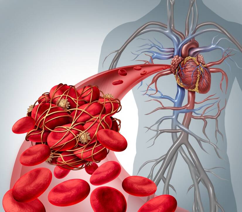
Photo source: Getty images

Thromboembolic disease poses a risk of developing serious complications that threaten a person's life.

Characteristics

Thromboembolic disease - thromboembolism, together with acute coronary syndrome and stroke form a group of diseases that significantly threaten human health and life.

The basis of cardiovascular disease are risk factors that may be uncontrollable and controllable.

What does it mean?

The first group consists of uncontrollable factors, examples of which are increasing age and genetic predisposition. We cannot change these by our actions.

The second part consists of a group of factors that are directly influenced by our actions. Examples are lack of exercise, overweight and obesity, smoking, dietary intake, psychological burden and general lifestyle.

It is reported that...
Thromboembolic disease is one of the most common causes of mortality in industrialized countries.

In the population of healthy people, it is a rare disease.
Approximately 0.01% of people under the age of 40 are affected.
And 0.1 to 0.2% of the population between the ages of 40 and 60.

TED = Thromboembolic disease. 

The risk of TED in a healthy population is significantly increased by various situations that occur during life. Plus, already associated congenital and acquired bleeding disorders contribute a significant amount.

Consequently, multifactorial influence significantly increases the incidence rate.
And... 
The more factors involved...
the higher the rate of developing complications. 

Sufficient prophylaxis has the opposite effect.

Prophylaxis = a set of activities designed to protect against the development of disease = protection from disease.

Effective and timely prophylaxis significantly reduces the risk of complications.

It has been reported to reduce the incidence of death due to thromboembolism by 50-75%.

Thromboembolic disease combines two disease states. These are:

  1. deep vein thrombosis
  2. pulmonary embolism

Thus, the presence of a thrombus, i.e. a blood clot in the veins and embolization into the pulmonary circulation, is behind the onset of the disease.

What is deep vein thrombosis?

It is characterized by the presence of a morbid formation of blood clots - thrombi in the venous system.

The most common thrombus formation occurs in the veins of the lower extremities.

Less commonly, they arise in the pelvic area, in the veins of the kidneys or upper limbs, but also directly in the large hollow veins.

With regard to the site of thrombosis, the following rule applies:

The higher a blood clot forms, the higher the risk of pulmonary embolism.

Thrombosis is a condition of diseased blood clotting in the blood vessels or in the heart. The balance between blood clotting and fibrinolysis is disturbed.

Blood clotting = haemocoagulation to prevent bleeding from injury. 
Fibrinolysis = the process of dissolving blood clots

In intact blood vessels, blood clots do not normally form. Since they are regulated by the process of fibrinolysis.

In the event of a disturbance of the equilibrium, two states can arise:

  1. excessive blood clotting = thrombosis
  2. bleeding = excessive bleeding

The formation of embolism is promoted by the accumulation of blood in the veins of the lower extremities and the change in blood flow in them. The higher the thrombosis occurs, the higher the risk of the blood clot breaking off.

The deep venous system, in short, is made up of large veins that run around the arteries. Blood enters them through the confluence of smaller veins and from the superficial parts of the human body.

In general, changes in hemodynamics, or blood flow, impaired blood coagulation, or clotting, and disruption of the vessel wall are involved in the development of deep venous thrombosis.

Plus the current state of the fibrinolytic system.

Embolism is the embolism are...

In a broader sense, it is a condition where a foreign object becomes lodged in the vasculature. This can be of various origins.

Embolization occurs, for example, due to:

  • blood clot - thrombus = thromboembolism
  • fat = fat embolism
  • air = air embolism
  • amniotic fluid
  • tumour cells
  • foreign body

A thromboembolism is a blood clot that grows on the wall of a vein.
After its release, it is expelled into the bloodstream.
Next, depending on the size, a blood vessel is clogged in another part of the body.
Embolism occurs. 

An example is embolism to the lungs.

Table: thromboembolisation by site of origin

Site of blood clot formation Where blood vessels are clogged
Veins of the lower limbs Pulmonary arteries. The clot does not pass through the smallest vascular plexus in the lungs to the brain,  paradoxically, in congenital heart defects and septal defects, stroke can also occur
Right heart Pulmonary arteries 
Jugulars Pulmonary arteries, often air embolism, alongside surgery or as a result of trauma
Left heart brain, kidneys, spleen, abdominal arteries, arteries of the lower limbs, thrombus formation in arrhythmias and valvular defects 
Aorta brain, kidneys, spleen, abdominal arteries, arteries of the lower limbs  
Pulmonary veins brain, kidneys, spleen, abdominal arteries, arteries of the lower limbs  

Pulmonary embolism occurs when the pulmonary vessels become blocked. The extent of the blockage and the size of the embolus determines the overall course and condition. 

This is an acute condition that has a high risk of death. It is reported to rise to 30% if left untreated.

Massive embolization into the main trunk of the pulmonary artery causes reflex cardiac arrest and death.

Learn more about:
Thromboembolic disease. 
Its causes.
Symptoms and treatment

Thromboembolic disease from a closer look

= A disease process that arises on the basis of two subunits.

The first is deep vein thrombosis and the second is pulmonary embolism.

1. Trombosis

It arises in different places. Most often it is the veins of the lower extremities. Less often it is the upper limbs, pelvis or large veins. 

Embolization from the lower limbs accounts for up to 85% of the proportion. 

Thrombosis in the shin area can proceed asymptomatically or to a degree. These blood clots are mostly dissolved and are not a source of embolization.

A thrombus forms and is subsequently dissolved.
It does not cause discomfort or has only mild manifestations.
It is not the cause of embolization.

The situation changes, and the risk is increased by thrombus in the region of the jugular vein (vena poplitea) and above.

Free-floating thrombi are also dangerous. A free-floating thrombus is attached to the wall of a blood vessel in the lower parts of the limb, however, its end floats around freely in the blood stream at a higher level. There is a risk of its detachment and the formation of an embolus.

Forms of venous thrombosis:

  1. ascendent - thrombus in the veins of the calf, spreading upwards to the thigh and pelvis, over hours, days, but also weeks
  2. transfascial - arising from the superficial veins of the lower limbs, the risk of which is spreading to deep vein thrombosis
  3. descendent - a thrombus develops in the pelvic area and causes a large swelling of the lower limb, pain and discolouration of the limb

Deep vein thrombosis = DVT.

2. Pulmonary embolism

Pulmonary thromboembolism is a condition where a detached thrombus clogs a blood vessel, the pulmonary artery.

Veins going: 
from the heart = arteries
towards the heart = veins

Pulmonary embolisation occurs in cases where the pulmonary duct is mechanically occluded. This closure can occur to varying degrees and at any location. 

The extent of vessel closure may be partial or complete. 

Its form may be:

  1. periferal, subsegmental - mild form
  2. central segmental, lobular - neutral form
  3. central with massive obstruction - severe forma, heart failure to death

Lung embolism = LE.

DVT + LE = XY

Thromboembolic disease = pulmonary embolism resulting from deep vein thrombosis.

These two disease states develop most often as a consequence of another disease, on the basis of another disease state and pathological process.

It is reportedly the third most common disease after coronary syndromes and stroke.

In the case of thromboembolic disease, delayed diagnosis and underestimation of the risks associated with the disease are of particular importance.

The course of the disease can vary, namely from clinically mute, i.e. asymptomatic, to mild, to massive and fatal.

Question: What causes thromboembolic disease?

Causes

The basis is the formation of a thrombus, i.e. a blood clot in the blood vessels. The pathophysiology was already pronounced in 1856 by Rudolf Ludwig Carl Virchow, a German physician.

Virchow's triad includes:

  1. a change in hemodynamics, a term encompassing a disturbance of blood flow in the blood vessels
    • turbulence in blood flow
    • blood stasis
    • may be caused by:
      • aneurysm of a blood vessel
      • heart failure
      • valve defect
      • limited limb mobility
      • long journey
      • mechanical oppression of the blood vessel
  2. coagulation disorder, i.e. increased susceptibility to blood clotting
    • disturbances in the balance of the blood clotting system
    • hereditary factors
    • acquired disease, for inflammatory disease, tumor, pregnancy, smoking
  3. disruption of the vascular wall
    • platelets and other clotting components are seeded onto the broken inner wall of the blood vessel
    • the cause may be:
      • trauma, injury
      • surgery
      • pathological process of blood vessels, but also in the vicinity of blood vessels

 In thromboembolic disease, a multifactorial influence is involved. As already mentioned, in a healthy person the risk of developing it is low.

However, the situation changes as the number of factors increases.

More factors = higher risk of thromboembolic disease.

In general, risk factors include:

Did you know?

In relation to surgery, the increased risk is mainly reported for hip surgery, both for orthopaedic procedures such as hip replacement, but also for fracture.

Joint replacement, artificial joint = total endoprosthesis - TEP.

In this context, it has also been reported that the risk increases from 0.7 % to 30 % during hip TEP surgery.

This compares with a risk of 1.8-7% for knee TEP.

Any surgical procedure increases the risk of thromboembolic disease.

However, laparoscopy reduces the level of intervention in the body , while the person is also mobilised more quickly. Which reduces the risk rate from 0.06 to 0.9%.

Aggravating risk factors:

  • age, especially above 40 years of age
  • obesity
  • smoking can increase the risk up to 1.5 times, after 6 months it drops to the level of a non-smoker
  • hormone therapy - 2-fold risk
  • hormonal contraception - 3-fold risk
  • pregnancy and the postnatal period up to 5-fold risk along with other factors
  • oncological disease is a major factor in the development of TECH, with the adverse effects of chemotherapy and patient immobilization contributing to this

Table: risk factors for the development of TECH

Groups of factors Factors
Congenital risk
  • coagulation disorders
    • V Leiden mutation 
    • antithrombin III deficiency
    • protein C and S deficiency
Acquired uncotrollable
  • age
  • familial predisposition
  • acquired coagulation disorder
  • antiphospholipid antibodies
Acquired cotrollable
  • varicose veins of the lower limbs
  • obesity
  • acute illness
  • chronic disease
  • kidney disease
  • pregnancy and postnatal period
Environmental influence
  • trauma, injury
  • fixation of the limb with plaster, splint, brace
  • immobilisation for illness and injury
  • surgery
  • certain medications such as hormonal contraceptives, corticosteroids, chemotherapy
  • long journey
  • lack of exercise
  • excessive physical exertion
  • smoking
  • cannulation of a blood vessel - central venous catheter

Thromboembolic disease in children and adolescents

Blood clots also develop during childhood and adolescence. Acquired risk factors are a major component, with a reported ratio of 95 %, compared to 60 % in the adult population.

Among all, these are mainly risk factors such as:

  • insertion of a central venous catheter, i.e. cannulation of a large vein, either into the superior and inferior vena cava and nearby tributaries (jugularis, subclavian or femoralis veins)
    • alebo umbilikálny katéter a iný cievny katéter
  • injuries and polytraumas
  • after major surgery
  • in cases of heart defects
  • artificial valve replacements
  • autoimmune diseases
  • Kawasaki disease
  • after organ transplantation
  • hormonal contraception in adolescent girls
  • smoking
  • congenital conditions of excessive blood coagulation

The most common sites of thrombosis:

  • the site of insertion of the cannula into the vein
  • renal veins
  • portal vein
  • the right atrium
  • veins of the lower limbs in the adolescent population

Symptoms

Symptoms depend on several factors.

In most cases, thrombi in the veins of the calf completely dissolve, even without treatment. However, approximately 20 percent will expand in an upward direction.

Calf thrombi are low risk. The risk increases with the location of the thrombus.

The risk increases especially for thrombi in the jugular vein area and upwards. But there are also studies that claim that even thrombosis in the calves is a possibility for the development of massive pulmonary embolism.

Thrombosis is also serious in terms of damage to the valves in the veins of the lower limbs

Did you know?
The valves in the venous system of the lower limbs help the blood to flow properly andupward progression against gravity.
In addition to the valves, blood flow is also assisted by the muscles of the lower limbs and pulsation of the arteries along which the deep veins run.

In thrombosis, the possibility of a number of symptoms such as:

  • swelling of one of the lower limbs
    • characteristic asymmetric unilateral swelling
    • or the swelling of both limbs is aggravated, with more pronounced swelling of only one limb
  • lower limb pain
    • aggravated by movement
    • feeling of tension in the limb
    • feeling of heavy legs
    • tactile pain
  • limitation of lower limb mobility
  • increased filling of superficial veins
  • the limb may feel warmer to the touch

Pulmonary embolisation can manifest as::

  • sudden onset of breathing difficulties - dyspnoea
  • accelerated breathing - tachypnea
  • accelerated heartbeat - tachycardia
  • cyanosis - bluing of the skin
  • chest pain
  • cough
  • coughing up blood
  • increase in body temperature
  • during massive embolization also occurs:
    • feeling of anxiety
    • fear of death
    • collapse
    • petechiae - tiny spots of bleeding under the skin, like the head of a needle
    • failure of right heart function
    • state of shock - cardiogenic shock
    • death

It is reported that pulmonary embolism manifests when 30-50% of the pulmonary vasculature is occluded.

Chronic complications include post-thrombotic syndrome and chronic thromboembolic pulmonary hypertension or cor pulmonale.

Post-thrombotic syndrome is a condition where there has been damage to the veins. These are narrowed at the site of thrombosis and have damaged valves. This causes stagnation of blood, or the accumulation of blood in the lower limb, as the function of pushing the blood upwards towards the heart is impaired.

These changes subsequently again contribute to the formation of thrombosis.

In addition to swelling, it is also manifested by pain in the limb, skin changes (colour and quality of the skin) and even ulcer formation. These are also known as venous ulcers, also called leg ulcers

Diagnostics

Diagnosis is focused on history taking. In the personal history, it is important to find out about previous thrombotic conditions, in women, an indication of repeated miscarriages in the past is important.

Thrombosis and hypercoagulable states are high risk factors for miscarriage. 

Furthermore, the family history reveals the occurrence of diseases such as thrombosis, but also heart attack, or stroke

Next...

An important part is the physical examination and clinical picture of the disease.

In this case, both lower extremities are examined, their circumference, swelling, color, skin temperature, the presence of pulsations and the filling of superficial veins are compared. Special examinations such as Homans and plantar signs are complemented.

Blood pressure, pulse, regularity of heartbeat, oxygenation of blood with oxygen, respiratory rate, blood supply to the periphery, as well as to the lips and mucous membranes for possible cyanosis, and listening with the aid of a phonendoscope to the actual breathing and heart sounds are examined.

Blood collection for laboratory examination is also important. Here, blood parameters are monitored in the blood count, the values of various coagulation factors and general coagulation tests, D-dimer, plus biochemistry, liver tests, genetic testing in the case of genetic testing.

Important examination methods include:

  • ECG
  • echocardiogram
  • Duplex sonography - examination of blood vessels, showing the condition of blood vessels and blood flow throughout the limb
  • venography - invasive method and administration of contrast agent
  • CT, including angiogram
  • MRI,
  • including angiogram

In the differential diagnosis it is necessary to think about other diseases. Examples are cardiac decompensation, heart attack, respiratory failure - respiratory failure, respiratory tract infection, lung disease, liver disease (cirrhosis, cancer, impaired function) and kidney disease, but also other conditions after trauma, in the case of tumour, abscess or lymphoma, or thrombophlebitis.

Various scoring systems are also available to assess the likelihood of pulmonary embolisation, such as the Geneva or Wells's scores.

Table: Wells's score of pulmonary embolism

Predisposing factors
  • medical history shows DVT or PE 
  • surgical procedure or immobilisation
  • oncological disease
  • 1,5 points
  • 1,5 points
  • 1 point
Symptoms
  • haemoptysis - coughing up blood
  • 1 point
Clinical signs
  • heart rate more than 100/minute
  • DVT symptoms
  • 1,5 points
  • 3 points
Clinical condition
  • other diagnosis is less likely than PE
  • 3 points
Assessment Clinical probability of PE Points total
Low 0 - 1
Moderate 2 - 6
High more than 7
2-tier classification
PE is not likely 0 - 4
PE is likely more than 4

Course

The course of the disease - thromboembolism directly depends on the area of the location of thrombosis and its extent.

The presence of small blood clots in the veins of the calf may not manifest itself. This is a clinically mute, or asymptomatic course of the disease.

These small blood clots mostly dissolve on their own, without necessary treatment, and are not the cause of embolization.

Alternatively, there is only mild discomfort. These may be in the form of a feeling of heavy legs, pain in the calf area, or a slight swelling.

In this way, it is a possibility that the disease progresses for several days or weeks.

As well as swelling, thrombosis can also subsequently progress, i.e. progress.

The reason for the worsening of the swelling is blood stasis, that is, its accumulation in the lower limb. It is caused by impaired function of the venous valves and insufficient blood flow against gravity.

At this point, the risk of complication and thus embolization increases.

After detachment, the thrombus is referred to as an embolus.

And now, within seconds, embolization occurs. If the embolus enters the right heart, it is ejected into the pulmonary circulation.

Depending on its size and the location of the obstacle created, health problems subsequently begin to be associated.

An example is sudden onset shortness of breath, or the feeling of not having enough air. There is also coughing, even coughing up blood, chest pain, fear of death and, in the case of massive pulmonary embolism and closure of the pulmonary trunk, even death.

Read also: 
Thrombosis is not a disease of the elderly. Young people are at risk, too!
Atherosclerosis and arterial thrombosis.

Dôležitá je prevencia a profylaxia...

Prevention and targeted prophylaxis before the onset of health complications is of great importance. 

Pre-existing predispositions must be detected and treated early. Anticoagulants are used. Wearing elastic stockings is recommended.

In the case of hospitalization, early mobilization is important so that the patient is not immobilized for a long time.

Emphasis must also be maintained when working with prolonged sitting or avoiding prolonged monotonous sitting and standing postures. Sufficient physical activity is therefore necessary.

Putting your lower limbs into an elevated position helps. This improves the return of blood to the upper body and reduces the pressure in the veins of the lower limbs.

Get enough exercise and limit sedentary lifestyle!

Preventive is regular and sports activity. Only a health problem can be limiting. However, a sufficiently long walk every day is sufficient.

The other side consists of a rational diet and sufficient drinking. It is necessary to maintain a suitable body weight.

Important: you should completely give up smoking.

Will you be travelling by car or plane and for a long time?

Consult your doctor/GP. They will help determine the appropriate form of prevention.

When travelling or working at your desk, the following will help:

  • sufficient and regular breaks
  • changing position and walking around
  • stretching the muscles of the lower limbs
    • moving your toes up and down
    • flexing your calf muscles, thigh and gluteal muscles
  • do not drink any alcohol and do not to smoke
  • drink plenty of fluids
  • do not take sleeping pills and do not sleep - then a person is not able to change position and exercise muscles
  • wear elastic compression stockings

What is traveller's thrombosis

How it is treated: Thromboembolic disease

Treatment of thromboembolic disease: medications and invasive methods

Show more
fshare on Facebook

Interesting resources

  • Ortel, TL; Neumann, I; Ageno, W; et al. (13 October 2020). "American Society of Hematology 2020 guidelines for management of venous thromboembolism: treatment of deep vein thrombosis and pulmonary embolism". Blood Advances4 (19): 4693–4738. doi:10.1182/bloodadvances.2020001830. PMC 7556153. PMID 33007077.
  • Heit JA, Spencer FA, White RH (January 2016). "The epidemiology of venous thromboembolism". Journal of Thrombosis and Thrombolysis41 (1): 3–14. doi:10.1007/s11239-015-1311-6. PMC 4715842. PMID 26780736.
  • Abbattista M, Capecchi M, Martinelli I (January 2020). "Treatment of unusual thrombotic manifestations". Blood135 (5): 326–334. doi:10.1182/blood.2019000918. PMID 31917405.
  • National Clinical Guideline Centre – Acute and Chronic Conditions (UK) (2010). "Venous Thromboembolism: Reducing the Risk of Venous Thromboembolism (Deep Vein Thrombosis and Pulmonary Embolism) in Patients Admitted to Hospital". PMID 23346611.
  • Shatzel, Joseph J.; O'Donnell, Matthew; Olson, Sven R.; Kearney, Matthew R.; Daughety, Molly M.; Hum, Justine; Nguyen, Khanh P.; DeLoughery, Thomas G. (January 2019). "Venous thrombosis in unusual sites: A practical review for the hematologist". European Journal of Haematology102 (1): 53–62. doi:10.1111/ejh.13177. ISSN 0902-4441. PMID 30267448.
  • Windecker, Stephan; Stortecky, Stefan; Meier, Bernhard (July 2014). "Paradoxical Embolism". Journal of the American College of Cardiology64 (4): 403–415. doi:10.1016/j.jacc.2014.04.063. PMID 25060377.
  • Martinelli I, Bucciarelli P, Mannucci PM (2010). "Thrombotic risk factors: basic pathophysiology". Crit Care Med38 (suppl 2): S3–9. doi:10.1097/CCM.0b013e3181c9cbd9. PMID 20083911. S2CID 34486553.
  • Bovill EG, van der Vliet A (2011). "Venous valvular stasis-associated hypoxia and thrombosis: what is the link?". Annu Rev Physiol73: 527–45. doi:10.1146/annurev-physiol-012110-142305. PMID 21034220.
  • Rosendaal FR, Reitsma PH (2009). "Genetics of venous thrombosis". J. Thromb. Haemost7 (suppl 1): 301–4. doi:10.1111/j.1538-7836.2009.03394.x. PMID 19630821. S2CID 27104496.
  • Khan NR, Patel PG, Sharpe JP, Lee SL, Sorenson J (2018). "Chemical venous thromboembolism prophylaxis in neurosurgical patients: an updated systematic review and meta-analysis". Journal of Neurosurgery129 (4): 906–915. doi:10.3171/2017.2.JNS162040. PMID 29192859. S2CID 37464528. Patients requiring cranial and spinal surgery present a unique situation of elevated risk for VTE but also high risk for disastrous outcomes should bleeding complications occur in eloquent areas of the brain or spinal cord.open access
  • Stein PD, Beemath A, Meyers FA, et al. (2006). "Incidence of venous thromboembolism in patients hospitalized with cancer". Am J Med119 (1): 60–8. doi:10.1016/j.amjmed.2005.06.058. PMID 16431186.
  • Jackson E, Curtis KM, Gaffield ME (2011). "Risk of venous thromboembolism during the postpartum period: a systematic review". Obstet Gynecol117 (3): 691–703. doi:10.1097/AOG.0b013e31820ce2db. PMID 21343773. S2CID 12561.
  • Varga EA, Kujovich JL (2012). "Management of inherited thrombophilia: guide for genetics professionals". Clin Genet81 (1): 7–17. doi:10.1111/j.1399-0004.2011.01746.x. PMID 21707594. S2CID 9305488.
  • Turpie AGG (March 2008). "Deep Venous Thrombosis". The Merck's Manuals Online Medical Library. Merck.
  • Beyer-Westendorf J, Bauersachs R, Hach-Wunderle V, Zotz RB, Rott H. Sex hormones and venous thromboembolism - from contraception to hormone replacement therapy. Vasa. 2018