Pulmonary embolism: causes, symptoms, probability testing, and treatment
Most common symptoms
- Malaise
- Chest pain
- Painful Breathing
- Spirituality
- Fever
- Increased body temperature
- Blue leather
- Sweating
- Low blood pressure
- Swelling of the limbs
- Disorders of consciousness
- Dry cough
- Pressure on the chest
- Fatigue
- Anxiety
- Damp cough
- Vomiting
- Coughing up blood
- Confusion
- Accelerated heart rate
- Heart enlargement
Causes
It represents up to 85% of embolisations. However, there are other obstructions that may be behind the occlusion of the vessel: adipose tissue, air, amniotic fluid or tumour cells.

In the case of thrombosis, and therefore in the case of lung embolism, there are predisposing factors that cause problems, especially when combined. They are also referred to as the Virchow's Triad. Detection of the presence of risk factors is crucial in determining subsequent preventive measures.
In addition, there are common risk factors for lung embolism. They can be innate or acquired.
Thrombosis occurs as a combination of several factors, also referred to as the Virchow's Triad:
- change in the coagulation mechanism, i.e. blood clotting
- disruption of the internal vascular mucosa, i.e. the endothelium
- slower blood flow
- vascular change as in varicose veins, inflammation
- in cardiac arrhythmia
- the current state of the fibrinolytic system is also important
The most common risk factors to cause a thromboembolic disease:
- advanced age
- genetic and familial predisposition
- increased levels of coagulation factors
- decreased levels of proteins C, S, or antithrombin III
- antiphospholipid syndrome
- dehydration
- vascular inflammation
- Lower limb varicose veins
- blood stasis in the lower limbs, i.e. affected backflow
- long flight
- standing for a long time
- sitting for a long time
- long drive
- more than 4 hours
- smoking
- obesity
- lack of movement, immobilisation
- complete immobilisation for more than 3 days
- partial immobilisation of the lower limb with a splint, plaster
- surgery
- especially major surgeries
- in the abdominal cavity
- joint replacements of the lumbar or knee joint, i.e. total endoprosthesis (TEP)
- artificial valve prostheses, stents
- central venous catheter
- hormonal changes
- hormonal contraceptives
- pregnancy, especially in the 2nd and 3rd trimesters
- postpartum (or postnatal) period
- tumourous disease
- ulcerative colitis
- infection and sepsis
- injuries, burns, polytrauma
- past recovery from embolism, thrombosis, stroke or heart attack
- drug abuse
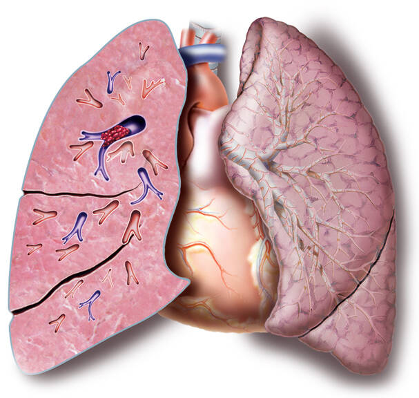
A blood clot, or a thromboembolus, or other material travels through the blood vessels until it creates a barrier to blood flow, an obstruction in the pulmonary stream. The onset of difficulties and the image of a massive pulmonary embolism in a healthy person are formed by the closure of 50% of the pulmonary vessels.
The embolus creates an obstacle in branches of the pulmonary artery. Depending on the extent and restriction of the blood flow, there is a disruption of the pulmonary circulation. Impaired pulmonary circulation is due to a reduction in venous return to the left heart.
The blood returns to a lesser extent from the right side of the bloodstream to the left heart, which causes reduced pressure in the major circulatory system and increased pressure in the pulmonary artery (pulmonary hypertension) and in the right heart. This is the cause of overloading the right heart.
If the extent of embolisation is large, such as when obstructing a pulmonary artery strain, the patient may suddenly die. If the embolisations are recurrent but small, chronic right heart failure, pulmonary hypertension, and later right ventricular dilatation occur.
Learn more: Enlargement and dilation of the heart
Embolisation occurs mainly due to deep venous thrombosis of the lower extremities. There is a proximal and a distal form. The proximal form is located above the knee, mainly from femoral (44.1%) and iliofemoral thrombosis (13.5%). Overall, the risk for embolisation us up to 50% if left untreated.
For example, take a distal vessel from the lower leg with thrombosis in the calf area. The risk is 5-25% in case of non-treatment. In about 5.1% of cases, the source of embolization is the right heart.
As many as 33.9% of postmortem examinations showed unknown sources of embolisation.
The blood clot adheres to the vessel wall and some thrombi hold on to it better. Others are unstable and in most cases are released into the bloodstream after mechanical pressure, for example due to coughing, during defecation, vomiting, or standing up too quickly.
Symptoms
When the vessel is clogged, the blood flow in the affected part of the lungs is disrupted. Moreover, a pulmonary tissue infarction can occur if the vascular supply to the lungs is impaired (less than 10% of cases).

In some cases, it can also be asymptomatic. The overall clinical picture depends on several factors:
- extent of the obstruction
- size of the embolus
- duration of onset
- condition of the cardiovascular system
Typical symptoms of pulmonary embolism are difficulty breathing and rapid breathing. Chest pain may be an associated problem. Tje heart rate is elevated. Other associated symptoms that can lead to a diagnosis include cough, coughing up blood, and collapsing.
Table: percentage of typical symptoms in pulmonary embolism
| Symptom | Percentage | Description |
| dyspnoea | 82 | difficulty in breathing abrupt onset it also arises at rest, without prior overload the percentage is indicated in up to 95 cases |
| tachypnoea | 60 | unusually rapid breathing over 20 breaths per minute in all its forms |
| chest pain | 49 |
may resemble a myocardial infarction or has a pleural character
|
| increased heart rate | 40 | tachycardia accelerated pulse / heart rate above 100 per minute |
| cough | 20 | |
| collapse | 14 | syncope, or fainting, temporary loss of consciousness may be the first manifestation of the disease |
| hemoptysis | 7 | coughing up blood |
In case of massive pulmonary embolism, the hemodynamics are impaired, and blood pressure in the lungs increases. He is a burden to the true heart. The right heart is failing. As a result of the reduced backflow of blood to the left heart, the circulating blood pressure drops. The result is cardiogenic shock. The risk of a damaged blood supply to the heart is a heart attack.
Learn more: First aid in heart attack
The massive form of pulmonary embolism presents with symptoms such as:
- increased sweating
- pallor
- low blood pressure
- tachycardia
- rhythm disorders - gallop rhythm
- pulmonary hypertension
- acute dilatation of the right ventricle
- increased central venous return, which can be seen in the increased filling of the jugular veins
- disorder of consciousness, collapse
Untreated massive pulmonary embolism has a high risk of death.
Treated embolism has a 20 percent risk of death.
Sudden death occurs in approximately 10% of cases.
Other symptoms may cause the skin to turn bluish, i.e. cyanosis. It can be present at the periphery first, but also centrally later on. The sufferer may be show signs of fear of impending death.
Hiccups, also known as singultus, can also be associated. The affected person may have an upset stomach or vomit. In the chronic type, the patient may present with swelling of the lower limbs, increased fatigue and general weakness. Collapse may be the first sign of illness. Ther emay be bladder and bowel incontinence.
Submassive embolism presents with less severe problems. But it is also characterised by shortness of breath or pain behind the sternum. There are no signs of shock in this type. Vascular occlusion is less than 50%.
Another form is successive pulmonary embolism characterised by recurrent embolisation of smaller clots. It is also referred to as the relapsing form. Its incidence is reported to be about 29%. It is often mistaken for other lung or heart disease.
Symptoms of pulmonary embolism:
- sudden shortness of breath
- chest pain
- cough
- coughing up blood
- palor
- sweating
- fear of impending death
- fainting
- rapid breathing
- increased pulse
- low blood pressure
- cardiogenic shock
- cyanosis
- dizziness
- deep vein thrombosis in the legs
- temperature above 38,5 °C
Pulmonary embolism is treated by
Pulmonary embolism is examined by
Other names


