- ncbi.nlm.nih.gov - Pseudotumor cerebri - Mondragon J, Klovenski V.
- mayoclinic.org - Pseudotumor cerebri (idiopathic intracranial hypertension)
- hopkinsmedicine.org - Pseudotumor Cerebri
- pubmed.ncbi.nlm.nih.gov - Update on idiopathic intracranial hypertension in adults: a look at pathophysiology, diagnostic approach and treatment
- solen.sk - Idiopathic intracranial hypertension - pseudotumor cerebri from the perspective of an ophthalmologist, Petr Sklenka, MD, Pavel Kuthan, MD, Department of Ophthalmology, 1st Faculty of Medicine, Charles University in Prague.
Pseudotumor cerebri: What is idiopathic intracranial hypertension?
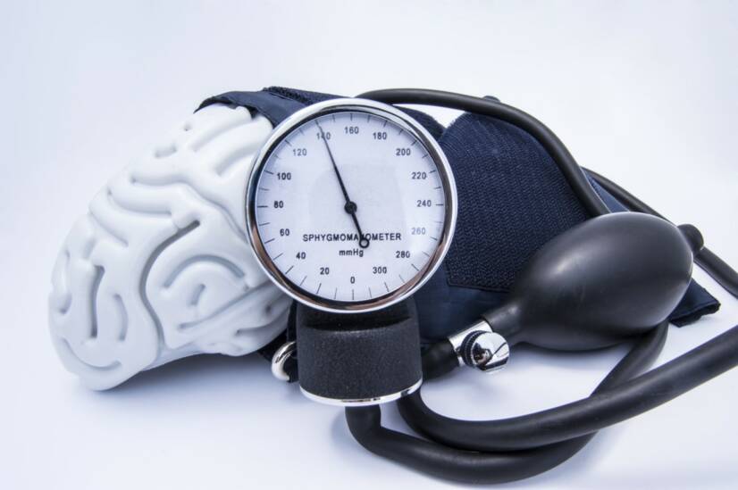
Photo source: Getty images
Pseudotumor cerebri is a disease also known as idiopathic intracranial hypertension.
Most common symptoms
- Headache
- Double vision
- Tinnitus
- Loss of sense of smell
- Loss of field of vision
- Deterioration of vision
Show more symptoms ᐯ
Characteristics
Pseudotumor cerebri, idiopathic intracranial hypertension, occurs when the pressure inside the skull (intracranial pressure) increases excessively without apparent cause. Hence the name "idiopathic", meaning from unexplained causes.
Symptoms mimic those of a brain tumour, but imaging tests show no expansive process in the brain.
Pseudotumor cerebri can occur in children and adults, but most commonly in obese women of childbearing age.
The disease is manifested by headache, swelling of the papilla of the optic nerve (with associated visual disturbance) or pulsatile tinnitus.
Pseudotumor cerebri is the older name for a neurological disease called idiopathic intracranial hypertension (IIH).
It is a clinical condition characterized by an increase in intracranial pressure in the absence of any identifiable causative factor.
This means that any medical condition, venous abnormality or use of or exposure to drugs that could cause the secondary development of intracranial hypertension should be excluded by appropriate investigations.
We can then make a diagnosis of pseudotumor cerebri.
The incidence of this disease in the population of Western countries is approximately 0.9/100 000 inhabitants per year. If we consider only the female population aged 15 to 44 years, the incidence rises to 3.5 cases per 100 000 inhabitants per year.
An even higher figure is obtained if we consider only those women aged 20-44 who exceed their ideal weight by more than 20%. There are up to 19 such women with intracranial hypertension per 100 000 population per year.
In Asian countries, the prevalence is lower (0.03/100 000/year). This is due to the different prevalence of obesity worldwide. For example, there are ten times more people with obesity in the USA than in Asian countries.
These data show that female sex and obesity are strongly associated with morbidity from pseudotumor cerebri.
Men account for only 9% of diagnosed cases, suggesting a possible role for sex hormones as a cause of disease in women of reproductive age.
Like women, men suffering from IIH are usually obese. They develop the disease later, at an average age of 37 years. For women, it is as early as 28 years.
Very rarely, the disease is diagnosed in the elderly and in children under 3 years of age.
Causes
Dysregulation of cerebrospinal fluid (CSF) dynamics is involved in the development of this disease, but the exact mechanism is still unknown.
Overproduction of CSF in the brain, decreased reabsorption and abnormal pressure gradients in the cerebral veins are involved.
With regard to obesity, increasing body mass index (BMI) values are directly correlated with the risk of intracranial hypertension and a more severe course.
Even an increase in BMI after cure of IIH is a risk factor for recurrence.
There are many hypotheses as to how obesity contributes to the development of pseudotumor cerebri. One is the central distribution of body fat around the abdomen, which increases venous pressure, which in turn inhibits fluid reabsorption in the brain.
Another theory takes into account the increased formation of microthrombi in blood vessels, especially in obese patients, which interrupts venous circulation, again resulting in impaired recirculation of fluid.
In addition, elevated levels of fbrinogen, D-dimer, clotting factors and leptin, which regulate the production, secretion and reabsorption of liquor in the brain by various complex responses of the body, have been found in obese patients with IIH.
Since IIH is predominantly diagnosed in obese women of childbearing age, the influence of female steroid hormones can be assumed.
Another disease that is a risk factor in the development of pseudotumor cerebri is the so-called obstructive sleep apnoea. It occurs very often in obese people, especially in men.
Vitamin A and retinoids are factors that are also associated with an increased risk of this disease. This is mainly due to excessive intake or, conversely, long-term low doses of vitamin A.
The reabsorption of liquor is also impeded by altered venous outflow caused by venous obstruction. Reabsorption of liquor is ensured by the so-called arachnoid granulations, which protrude into the cerebral venous sinus.
Their function is impaired if there is a narrowing in the venous sinus, i.e. stenosis.
Other diseases that are associated with secondary intracranial hypertension:
- Addison's disease
- Anemia
- Blood clotting disorders
- Kidney disease
- Lupus
- Polycystic ovary syndrome
- Sleep apnea
- Parathyroid insufficiency
There was no confirmed association between IIH and pregnancy, thyroid disease, iron deficiency anaemia and antibiotic use.
Regarding hormonal contraception, the effect on the development of increased IH is not yet known.
Symptoms
Headaches
Up to 84% of patients with this condition describe a headache. It is the most common first symptom that brings the patient to the examination.
It is characterised by a progressively worsening course with daily occurrence. It is a constant, non-pulsating pain that worsens with coughing or the Valsalva manoeuvre (exhalation with the nose plugged and mouth closed).
It is most often bilateral, felt mainly behind the forehead and eyes. It is similar to tension headaches or migraine attacks.
If experienced as a migraine, unilateral pain is present with intolerance to light, noise, nausea and vomiting.
Visual disturbances
The second most common symptom is visual disturbances.
Patients suffer from visual field disturbances to loss of vision. The difficulties may not be present all the time but are variable in nature. However, as the disease progresses, the symptoms worsen.
Most commonly, patients have loss of peripheral visual field on the outer lateral sides. Less commonly, visual loss begins to manifest on the sides of the visual field towards the nose.
These are transient "visual obscurations", transient episodes of unilateral or bilateral vision loss due to partial ischemia of the optic nerve target (the point where the optic nerve exits the retina). The blurring is caused by increased pressure on the tissues.
Seizures usually last less than a minute and are often triggered by a change in position. Over time, there is a complete restoration of vision.
Visual acuity is normal in up to 2/3 of cases, except in patients with severe vision loss or visual impairment in the papillomacular region, which is the site of sharpest vision in the eye.
About a fifth of patients suffer from double vision, called diplopia, caused by paralysis of the sixth cranial nerve, which innervates the eye muscles.
Papiloedema
Papiloedema is the most specific symptom of intracranial hypertension. It can be observed on examination of the ocular background.
It is a swelling of the papilla of the optic nerve (the part of the nerve curled on the retina) due to increased pressure acting on the optic nerve.
Papiloedema can have four grades:
- early
- full-blown
- chronic
- atrophic papiloedema
In clinical practice, the so-called Frisén scale is used to assess papiloedema.
The swelling is usually bilateral. However, a certain proportion of patients have asymmetric swelling or worse findings in one eye compared to the contralateral eye according to the Friesen scale.
Papiloedema usually develops rapidly and up to 10% of cases result in permanent loss of vision.
Hearing impairment
Tinnitus is the perception of unpleasant whistling, humming, ringing, and other sounds without external stimulus. It is most often bilateral, pulsatile, and synchronous with the heart rate. It can have a variable frequency, ranging from daily to monthly.
Olfactory disturbance
Some patients also describe changes in the sense of smell or even a complete loss of the sense of smell, called anosmia.
Otolikvorea or rinolikvorea
This is a leakage of liquor from the cranial space, where the liquor flows out through the ear or nose. This is a relatively rare symptom.
It is caused by chronically increased intracranial pressure, which leads to remodelling of the skull base and the formation of communications between the internal environment and the sinuses.
Pseudotumor cerebri may also have symptoms of intracranial hypotension, i.e. symptoms of reduced intracranial pressure, which is the opposite of this disease.
Neuropsychiatric condition
In pseudotumour cerebri, the patient's mental status is usually normal. However, neuropsychological functions such as speed of thought, attention and visuospatial processing may be adversely affected.
Diagnostics
Diagnosis of patients with signs and symptoms of intracranial hypertension includes neuroimaging, lumbar puncture with assessment of pressure and biochemical analysis of liquor, ophthalmoscopy, visual acuity and perimeter examination, and complete blood count.
Neuroimaging
Magnetic resonance imaging (MRI) with venography (MRV) is the preferred imaging modality. It is used to exclude other secondary causes of intracranial hypertension.
The examination shows the brain parenchyma and ventricles well.
Other findings on MRI that may suggest a diagnosis of pseudotumor cerebri (but are not 100% diagnostic) include stenosis of the transverse sinus (narrowing of the venous outflow), posterior scleral plexus, distension of the perioptic subarachnoid space, empty sella (missing pituitary gland), and optic nerve abnormalities.
Computed tomography (CT) is performed if acute exclusion of cerebral ischemia or tumor is necessary or if the patient has contraindications to MRI. However, this method is less sensitive and specific than MRI.
Lumbar puncture
A useful investigation is the examination of the lymph fluid and its pressure during lumbar puncture.
A pressure greater than 25 cm H2O in adults and greater than 28 cm H2O in children aged 1 to 18 years with negative neuroimaging findings supports the diagnosis of pseudotumour cerebri.
Laboratory analysis of the liquor includes cell count and differential analysis, glucose and protein content, Gram staining, and microbiological culture.
Ophthalmological examination
Ophthalmoscopy detects the presence of opacity of the optic disc, papiloedema.
Visual acuity examination evaluates the degree of visual loss due to the disease.
Perimeter examination will provide information on the deterioration of peripheral vision.
Complete blood count
Performed to rule out anemia or lymphoproliferative disease as the cause of optic nerve papilla edema.
Course
The course of the disease depends on several factors. The first is the speed of onset of symptoms. The faster the onset, the more aggressive the treatment the disease requires.
The extent of vision loss at first presentation and the grade of papiloedema are also important. Significant vision loss and a worse grade of papiloedema at the time of diagnosis means a higher risk of permanent vision loss.
Patients with pseudotumour cerebri suffer from symptoms of this disease that significantly complicate their lives for months or years, even if treatment is started early.
Some patients suffer permanent consequences such as persistent papillary edema, increased intracranial pressure and permanent loss of visual field.
How it is treated: Pseudotumor cerebri
Treatment of pseudotumor cerebri: drugs to relieve discomfort and surgery
Show morePseudotumor cerebri is treated by
Other names
Idiopathic intracranial hypertension
Interesting resources

MUDr. Andrea Bullová
Physician
After graduating from an eight-year high school, I knew exactly who I wanted to become in life. Studying at the Faculty of Medicine at Comenius University in Bratislava fulfilled a long-held dream and I was able to start fulfilling my mission as a doctor. After obtaining my diploma and M.D. degree, my first professional steps led to the St. Elizabeth Cancer Institute in Bratislava. At the Stereotactic Radiosurgery Clinic I worked with patients with brain tumours and eye tumours. After a year of working with oncology patients, in 2020 I moved to the Neurology Clinic of SZU St. Michael's University Hospital in Bratislava, where I am still working today. I like to spend my free time outdoors, whether cycling, hiking or just walking in the nearby forest.
View all articles by the same author


