Keratoconus is a degenerative disease of the cornea. It is not clear why it arises. Early diagnosis and treatment are important, however, a complete cure is not possible.
Keratoconus is a non-infectious degenerative disease of the cornea. The cornea arches asymmetrically and conically, resulting in impaired vision to blindness.
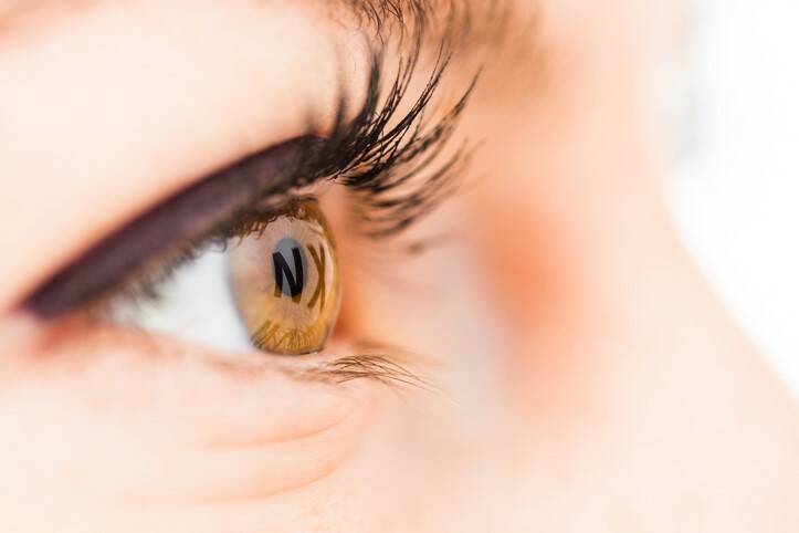
In most cases, this disease is confused with myopia or astigmatism. Because in the beginning, he has the same symptoms. It can develop completely unnoticed for many years, which worsens subsequent treatment.
It most often affects the cornea of both eyes, in up to 85% of cases.
It is said that the cause is congenital inferiority of corneal tissue, the exact reason of which has not yet been clarified. A genetic and partially hereditary basis is assumed.
It often occurs together with other diseases, such as allergies, atopic eczema, Down's, Turner's, or Marfan's syndrome.
Excessive eye contact in children also increases the risk of development. It arises in childhood and during puberty, 10 - 20 years of age. The sooner he starts, the faster he progresses - he progresses. After the age of 40, the overall progress is more modest.
The disease is rare and affects approximately 1 in 1,500 people. It is more often diagnosed in young men. Therefore, it is often referred to as a disease of young people.
The cornea is an important part of the optical system of the eye. When it is disturbed, a visual impairment occurs. It has a regular domed curved shape, while the keratoconus causes its asymmetrical conical arching and thinning of the thickness.
Vision gradually deteriorates during keratoconus, and the disorder is very poorly corrected. In the worst cases, blurred vision, swelling, and rupture of the cornea occur. In this case, the only solution is a corneal transplant.
What is the cornea?
The cornea, professionally the cornea (cornea), is, together with the whites of the eyes, the sclera, the tight envelope of the eye. The cornea has differently arranged fibers than white, which guarantees its transparency.
The cornea makes up about 20% of the surface of the eye.
It is thus a transparent, important, and first element of the optical system of the eye. Its total optical power is up to two-thirds, which is 43 diopters.
A healthy eye has an optical power of about 60 diopters.
It forms the first mechanical as well as chemical barrier between the outside world and the inside of the eye. From the eye, it is the most stressed part that resists external influences and is exposed to the risk of injury.
Corneal injury or injury causes sharp stabbing pain in the eye and increased tearing.
Inside the eye, it is surrounded by the ventricular fluid of the eye, and on the outside, a tear film urges it. For more information on the tear film, see the article on dry eye.
It does not contain any vessels, but a number of nerve endings. These give it sensitivity to touch, changes in temperature, and chemical properties. The eyelids close reflexively when in contact with the cornea.
The cornea is not even a millimeter thick. Its outer part (towards the circumference) has a thickness of 0.8 and the inner (towards the center) 0.6 mm. Its diameter is 12 mm.
Despite its small width, it consists of five layers.
5 layers of cornea :
epithelium
Bowman's membrane
tree
Descemet's membrane
endothelium
The table describes the 5 layers of the cornea
Layer
Description
Epithelium
outer layer
formed by another 4 - 6 layers
about 10% of the corneal thickness
a tear film adheres to its surface
prevents the infection from entering the cornea
the basement membrane belongs to this layer and also has a protective function
Bowman's membrane
formed by collagen
thickness 8 - 12 .m
non-cellular division of epithelium and stroma
it does not regenerate, it heals with a scar in case of injury
Corneal tree
the largest part of the cornea - 90% of the thickness
the arrangement of the collagen fibers ensures transparency
it consists of about 300-500 fibers
between the fibers are keratocytes - corneal cells
Descemet's membrane
strong and resistant to infection
prevents the passage of leukocytes into the cornea
it grows in width with age
at birth about 3 and in adulthood about 8 - 12 µm
collagen type is similar to Bowman's membrane
Corneal endothelium
one type of cell that is about 5000 / mm 2 at birth
overtime, their number decreases to 2000 cells/mm 2
the cells are not regenerated
layer thickness 4 - 6 .m
provides nutrient transfer and hydration
separates the cornea from the eye chamber
The cornea does not contain blood vessels. Blood supply is provided by the marginal layer. The rim is interwoven with blood vessels and is referred to as a limbus.
Furthermore, nutrients are introduced into the cornea via ventricular fluid and tears.
Oxygen supply is provided from the outside environment. The most important role is played by the tear film, which urges the cornea. Its quality is therefore very important and dry eye syndrome is risky for eyesight.
The tear film and tears also have a defensive, cleansing function, preventing clouding and drying of the cornea. They also affect the optical properties.
The cornea is the first part that communicates with the external environment. Photo: Thinkstock
The cornea is sensitive to injury and degenerative changes in terms of optical properties. In addition to keratoconus, it can be affected by other problems, such as:
keratoglobus
pleucidal marginal degeneration
crocodile skin degeneration
Cornea guttata
lipid keratopathy
zonal keratopathy
epithelial and Bowman's membrane dystrophy
and other
Keratoconus, keratoglobus and marginal pleucidal degeneration are classified as ectatic corneal degenerations.
Causes
The exact cause is not revealed even today. Keratoconus is a degenerative disease of the cornea and is characterized by arching but also thinning thickness. Which causes vision problems.
Both eyes are affected, but asymmetrically, ie one more.
Familial occurrence, and therefore hereditary, is assumed. The genetic factor plays a role.
It has been found that in most cases it occurs together with allergic diseases, such as atopic eczema, hay fever, but also with diseases such as Down's, Turner's, or Marfan's syndrome.
Civilization factors, such as stress and poor lifestyle, are also said to be involved in the outbreak. Mechanical stress, and thus poor wearing of contact lenses and frequent eye wiping in children, also have a negative effect.
Symptoms
As the onset of the disease is at a relatively young age, from the 10th to the 30th year of life, the symptoms also start early. The rule is that early occurrence is also characterized by a faster progression of difficulties, ie progression.
The initial symptoms appear mostly from the 13th to the 27th year.
The second group of people at risk is between the ages of 40 and 60. In this case, however, it does not have such a significant course.
Blurred vision is one of the first and most common symptoms. This is primarily compensated by diopters, ie glasses or lenses.
It is therefore very often confused with myopia or astigmatism.
This is a mistake, as delaying appropriate treatment complicates the overall condition.
An alarming indicator is if the eyesight deteriorates by one cylindrical diopter per year.
The manifestation is therefore a faster deterioration of vision. Myopia and astigmatism deepen.
Manifestations of keratoconus are difficulties such as:
visual acuity impairment
exacerbation of myopia (minus diopters)
and astigmatism (cylindrical diopters)
bad correction of visual impairment
rapid progression and deterioration even with diopters per year
image distortion
blur or distort the image
double vision
driving or reading is also a problem
duplication, ie duplication of the observed object
reduction of tolerance for wearing contact lenses
unnatural tilting of the head to the side
advanced corneal opacity
and thus a reduction in transparency
up to the loss of vision, ie blindness
lightness
hydrops, swelling, corneas
it causes pain
Diagnostics
Keratoconus is a chronic disease. The anamnesis is important, but especially a special professional eye examination.
The eye examination also includes the evaluation of the visa, ie visual acuity using an optotype. An objective refraction is evaluated with an autorefractor. Subjective refraction is also evaluated.
Examination of the anterior segment of the eye includes an examination of the eyelids, conjunctiva, tear film
The examination also takes place with the help of a slit lamp. Corneal topography is performed, a photograph of the anterior segment of the eye as aberrometry.
Examination with a slit lamp. Photo: Thinkstock
The most modern investigative devices include Galilei, Pentacam, and Orbscan.
The examination looks for signs such as:
Bowman's shadow - the cause is the scars of the cornea, the cornea gradually arches
Fleischer ring - yellow, brown, or gray coloration of the cornea is observed
Munson's sign - or even Munson's sign
at an advanced stage
when viewed below, the lower lid arches into a V-shape
Axenfeld's symptom when the arched part of the cornea is characterized by a loss of sensitivity to touch
Vogt striae - cracks of the Descemet membrane
keratoconus scarring - scars in the area of the corneal arch
hydrops, ie swelling in the acute stage
as a result of rupture of the Descemet's membrane
sudden deterioration in visual acuity
Course
The course of the disease is long, ie chronic. Typically, the sooner it starts, the faster it progresses, that is, progress and worsening difficulties.
The first symptoms are mild and often overlooked. Many people do not even know he has keratoconus. The most common is decreased visual acuity when the symptoms are confused with myopia or astigmatism.
This is a complication of subsequent and delayed treatment. Gradually, the difficulties develop and cause a significant deterioration in vision.
Neglected treatment can lead to corneal clouding, rupture, and blindness of the damaged eye.
In most cases, keratoconus is present in both eyes, with asymmetric progression. Simply described that one eye is affected more.
The disease can be alleviated with the help of state-of-the-art methods, but it still cannot be completely cured.
How it is treated: Keratoconus
Keratoconus and its treatment: By stage, correction and surgery
"Keratoconus". NORD (National Organization for Rare Disorders).
"Keratoconus" (PDF). The University of Texas Health Science Center at San Antonio.
"Facts About the Cornea and Corneal Disease". NEI.
"Cornea Research Foundation of America - Keratoconus". www.cornea.org.
Kandel, Himal; Pesudovs, Konrad; Watson, Stephanie L. (March 2020). "Measurement of Quality of Life in Keratoconus". Cornea. 39 (3): 386–393.
Romero-Jiménez, Miguel; Santodomingo-Rubido, Jacinto; Wolffsohn, James S. (1 August 2010). "Keratoconus: a review". Contact Lens & Anterior Eye: The Journal of the British Contact Lens Association. 33 (4): 157–166, quiz 205.
Fecarotta, Christopher; Huang, Wendy (27 July 2015). "Pediatric genetic disease of the cornea". Journal of Pediatric Genetics. 03 (4): 195–207.
Wang, Y.; Rabinowitz, Y.S.; Rotter, J.I.; Yang, H. (2000). <403::aid-ajmg11>3.0.co;2-a "Genetic epidemiological study of keratoconus: Evidence for major gene determination". American Journal of Medical Genetics. 93 (5): 403–409.
"FDA approves first corneal cross-linking system for treatment".
"Keratoconus - NORD".
"Keratoconus Origin". dictionary.com.
Feder R, Kshettry P (2005). "Non-inflammatory Ectactic Disorders, Chapter 78". In Krachmer J (ed.). Cornea. Mosby. ISBN 978-0-323-02315-3.
Epstein A (2000). "Keratoconus and related disorders" (PDF). North Shore Contact Lens.
Pantanelli S, MacRae S, Jeong TM, Yoon G (November 2007). "Characterizing the wave aberration in eyes with keratoconus or penetrating keratoplasty using a high-dynamic range wavefront sensor". Ophthalmology. 114 (11): 2013–21
The secondary medical school in Nitra gave me the basis for my career in the field of health and diseases. Thanks to it, I worked for 2 years in the traumatology clinic and outpatient clinic at the Nitra Hospital. Since 2006 I was employed in the emergency medical service, where I stayed until 2017.
I completed my bachelor's degree at the University of Constantine the Philosopher in Nitra in the field of emergency health care. The bachelor's degree allowed me to continue my mission as a paramedic. In the meantime, I got a job at the emergency line 155. I have been working in pre-hospital health care until today.
I had an interest in people, health and even diseases in my childhood, which gave me the prerequisite to pursue this topic in adulthood. Studying and acquiring new information in practice provided me with a great basis for writing professional texts, in the form of articles that can be understood by ordinary people. Thus, my interest in the Health Portal has a solid foundation in years of practice and personal interest. Similarly, I am also interested in healthy eating, nutrition and overall healthy lifestyle. I fill my free time with family and sports.