Chronic venous disease, insufficiency: What are its causes and symptoms?

Chronic venous disease, insufficiency: What are its causes and symptoms?
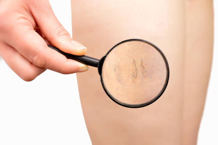
Photo source: Getty images

Chronic venous disease is a common problem, the development of which is also supported by a modern lifestyle. It negatively affects the quality of life, it worsens over time.

Most common symptoms

Show more symptoms

Characteristics

Chronic venous disease includes a wide range of diseases that affect the veins and last for a long time. These are mainly diseases of the veins of the lower extremities.

You may also come across: chronic venous disease of the lower extremities.

Today, it is one of the most common problems, also because its development supports a modern lifestyle.

The fact is, however, that a relatively large percentage of people underestimate it and neglect its treatment. Alternatively, instead of professional treatment, it treats itself.

Chronic venous disease has a long history and is progressive. 

What does that mean?

It worsens over time, and can result in chronic venous insufficiency. In addition to the aesthetic problem in the form of an ulcer, it also groups other difficulties.

Chronic venous insufficiency  ≠ chronic venous disease .

About chronic venous insufficiency speak of, abbreviated CHVI, when it meets one condition.

The condition for the evaluation of CHVI is:

Presence of increased venous pressure

Chronic venous disease, abbreviated as CHVO, combines all diseases or anomalies that affect the veins in the long term.

The disease is said to affect: 
half  to two-thirds of the population of developed countries.

Women are affected in a 2: 1 ratio compared to the male population.

Men neglect this disease moreWomen are more careful about the aesthetic aspect. But in many cases, they self-heal, which has a negative impact.

Progressive nature means that CHVO worsens over time and can go as far as CHVI with all its negative and serious manifestations. 

Therefore, early detection of vein problems and earlyprofessional treatment is important.

Brief information about veins in brief

The vessels that carry blood back to the heart are called the veins. 

The veins collect deoxygenated blood from the whole body. This also applies to the lower limbs. However, gravity impairs the return of blood from this place of the human body.

Auxiliary mechanisms have been developed for better blood flow from the legs, such as a muscle pump or a valve system.

The muscle pump is actually the muscles of the lower limbs, such as those in the forelegs - the muscles of the calf. Their movement compresses the veins, which helps to push the blood towards the higher parts.

Plus ...

The veins of the lower limbs contain valves. Like heart valves, their job is to direct blood flow. Closing the valve does not return blood to the lower part of the vascular bed.

In the lower limbs we recognize three systems of veins, namely:

  1. superficial veins
  2. deep venous system
  3. connecting cores

The superficial veins  (1.) are placed closer to the body surface. They collect blood from tissues. 

The connecting cores (3.) connect the cores placed on the surface and those in-depth.

The system of deep veins (2.) includes larger veins, which run together with the arteries and are in their immediate vicinity.

This placement of deep veins is also important.

Arteries, unlike veins, contain a strong muscle layer. These muscles in the arterial wall are involved in the flow of blood. 

The contraction of the arterial wall also helps in the flow of blood in deeply laid veins.

Want to know more  about chronic venous disease
What causes it?
How does it manifest itself?
And what is important in its prevention and treatment?
Read on  to us .

What is chronic venous disease

The definition defines it as a broad term for all diseases and anomalies of the venous system that occur chronically. It is also one of the long-term inflammatory diseases of the veins.

But what is it all about?

The basis of the problem must be sought at the microscopic level.

It is a leukocyte-endothelial interaction. Thus, the action between white blood cells (leukocytes) and the inner lining of the vessel wall (endothelium).

White blood bodies exaggerately attach to the inner wall of the veins, which results in its gradual weakening or even damage.

The vascular wall destroyed in this way does not fulfill its function sufficiently, which gradually begins to manifest itself. The veins lose their strength, shape and are more easily permeable to fluid.

As a result of the whole cascade of disease changes, characteristic difficulties are associated. These do not end with swelling or pain in the legs.

Causes

The cause is a long-term progressive process that takes place at the microscopic level. It involves several complex mechanisms. 

The development of CHVO and CHVI is caused by disease processes:

  1. blood reflux - venous reflux, in case of impaired venous valve function
    • disturbed blood flow - backflow
      • the blood is not sufficiently pushed higher, but is pushed by gravity back, into the lower parts of the vessel
    • accumulation of blood in the legs
  2. vascular obstruction - an obstruction in the flow of blood, such as thrombosis
  3. muscle pump disorder - dysfunction of calf muscles in neurological diseases
  4. combination of several

Among all the mechanisms, it is necessary to mention, for example:

  • increased pressure in the veins
  • venous valve damage
  • decreased venous tone 
  • increased permeability of capillaries - the smallest vessels 
  • increased blood density
  • swelling of the tissue
  • excessive leukocyte action on the vein wall
  • thrombosis, ie the formation of blood clots

By joint action occurs:

Venous blood
flow deteriorates,  ie the backflow of blood from the legs 
+
slows down the flow of blood in the veins and capillaries 

increases the pressure in the veins

blood accumulates in the lower limbs 

leukocytes are deposited on the vascular wall  

an inflammatory reaction develops 

thrombosis progresses 

the vascular wall is damaged 

the venous valves are destroyed 

the blood circulation deteriorates
+ the
difficulties progress. 

A number of disease changes follow each other, which worsens the condition of veins and tissues.

How does Prof. MUDr. Viera Stvrtinova, CSc. compare in her presentation CHVO: 
What was before?
An egg or a hen? 

In chronic venous disease, the division into primary and secondary form is given.

Primary disease accounts for 70 to 90 % of all cases. 
The exact cause of this form of PGI is unknown.

A ...

The secondary form arises as a result of another pathological event. Examples are injury,  post-thrombotic syndrome, phlebothrombosis, cancer, vascular malformations and others.

Plus, there is an innate form among the causes.

Thus, the cause and initial factor of chronic venous disease may not be known. 
But ... 
Several factors are involved in its development.

The SPA has a multifactorial origin.

Different factors support each other. 

You ask: Who is at risk of chronic venous disease?

Risk factors in the development of protected areas are:

  • age, with an increased risk, especially in people over 65 years of age
  • hereditary and genetic predisposition, such as diseases in relatives
    • Varicose veins
    • venous thrombosis, thromboembolism
    • forearm ulcer
    • blood clotting disorders
  • female gender
    • hormonal changes mainly during the period of menopause - transition
    • pregnancy and multiple births
    • hormonal contraceptives
    • hormonal treatment
  • venous thrombosis in the past and its insufficient treatment
  • obesity and overweight
  • injury
  • greater body height
  • lack of exercise
    • sedentary lifestyle
    • sedentary work
    • work standing without movement
    • long sitting on the toilet
  • disproportionate muscle load
  • tight clothing and high heels, unsuitable shoes
  • traffic jam
  • lack of fiber in the diet and poor eating habits
  • hot environment
  • smoking
  • long travel
  • walking on city pavement

PGI is associated with other disease states. If it is neglected, there is a risk of deteriorating health and the risk of developing complications.

Read about therefore: Diseases of 
chronic venous insufficiency
Deep venous thrombosis
Thrombophlebitis
Thromboembolic disease

These risk factors are also divided into uncontrollable and influenceable from a practical point of view.

To a group of uncontrollable know their efforts work. This includes, for example, age, gender or hereditary and genetic factors.

Vice versa.

We can change influenceable risk factors through our efforts. Examples are overweight and obesity, lack of exercise, smoking or a generally unsuitable lifestyle.

When preventing or exacerbating the disease, it is important to evaluate the risk factors present.
Their elimination follows.
This effort is the basis for success in averting the progress of difficulties.

Symptoms

Manifestations of the disease are divided into subjective and objective.

Subjective are those felt by the affected person. He describes them to the doctor.

They are objectively visible from the outside and can be observed. The doctor can examine them.

The table shows the subjective and objective manifestations of the PLA

Subjective Objectively
  • lower limb pain (DK)
  • or it may be a vague feeling
    • feeling discomfort
  • dilated veins
    • telangiectasia - vascular whisks, threads
    • corona phlebectatica - enlarged red,
      blue to purple veins
      on the inside of the ankle
  • feeling heavy legs
  • swelling of the lower limbs - edema
    • unilateral swelling -
      swelling of one affected limb
    • in other diseases
      with present swelling, DK
      may worsen in one limb
    • worsens during the day
    • it is tempered by the position
      with the DK unloaded
  • tired legs
  • skin changes - trophic skin changes
    • hyperpigmentation
    • subcutaneous fibrosis
    • dermatosclerosis
    • lipodermatosclerosis
    • white atrophy
    • to forearm ulcer - ulcus cruris
  • feeling hot in the legs
    • burning 
    • tingling
    • tension - the feeling that the leg breaks
    • feeling pulsating - rarely
In the evaluation of the clinical picture in CHVO
, a division
into three basic types is used : 
  1. telangiectasia
  2. reticular veins
  3. Varicose veins
  • calf cramps - muscle cramps
1. Teleangiectasia Permanently dilated small skin coils
smaller than 1 mm - clumps.  They are clearly visible when illuminated
from a distance of 2 meters. Known names
as whisks, threads,
professionally also spider veins. Corona phlebectatica - ankle glow.
  • excessive sweating of the feet
2. Reticular veins  Widened cores with a diameter of 1 to 3 mm 
  • itchy skin DK
These two types of
dilated veins are sometimes
referred to as microvarices
aggravating factors :
  • in women, the hormonal cycle
    • deterioration during menstruation
    • during pregnancy
    • in hormonal treatment
  • summer season - high temperatures
  • position - long sitting and standing without changing position
  • night time - cramps in DK occur at night
    • the difficulties are more intense at night
    • worsening procedure even during the day 
    • causes  insomnia
    • restless legs syndrome
3. DK varicose veins - varicose veins of the lower limbs Vacuum vein extensions, zigzag
surface veins
with a diameter greater than 3 mm.  Varicose
veins can affect all three types of veins,
namely superficial, connective or deep.

Diagnostics

Diagnosis takes place on several levels. In doing so, it combines several investigative methods.

Investigation methods:

  • medical history - the patient tells the doctor his subjective difficulties, which he feels
    • the doctor detects previous diseases such as thrombosis, inflammation of the veins, the presence of other diseases
    • family occurrence or genetic predisposition is determined
  • clinical examination - a doctor examines a person
    • the condition of his lower limbs
    • standing or lying examination
    • clinical trials - Schwartz test, Trendelenburg test
  • non-invasive methods
    • ultrasound - SONO examination, duplex sonography, Doppler USG
    • phlebography, CT, MRI
    • plethysmography
    • VeinViewer - projection of the venous system on the skin with an infrared camera
  • invasive approach
    • measurement of venous blood pressure
    • contrast X-ray phlebography
    • CT venography
    • isotopic examination
  • microcirculation assessment

The investigation methods also proceed in that order.

During diagnosis, the condition is evaluated according to the CEAP classification.

CEAP stands for Clinical Ethiology Anatomy Pathophysiology, meaning that it is an evaluation of a CHVO according to clinical symptoms, cause, anatomical location and pathophysiology.

The classification was established in 1994 in the Hawaiian Islands by the American Venous Forum.

CEAP classification in the table

C -
Clinical
  • C0  = no finding on visual and tactile examination
  • C1  = telangiectasia or reticular veins
    • dilated small blood capillaries
    • so-called red threads in the subcutaneous tissue
  • C2  = varicose veins - varicose veins
  • C3  = edema - swelling
  • C4  = skin hyperpigmentation or swelling
    • hyperpigmentation is a change in skin color
  • C5  = healed forearm ulcer
  • C6  = active ongoing foreleg ulcer
Plus value is added according to the course:
  • + S  = symptomatic form of the disease
  • + A  = asymptomatic form of the disease
    • without the presence of manifestations
E -
Ehtiology
  • Ec  - congenital = congenital origin
  • Ep  - primary form with unknown cause
  • Es  - a secondary form for a known cause,
    such as post-thrombotic syndrome, after injury
  • En - unidentified form
A - Anatomical
  • As  - anatomic superficial = superficial veins - value 1 - 5
  • Ad  - anatomic deep = deep venous system - value 6 - 16
  • Ap  - anatomic preforating - connecting veins - value 17 and 18
  • depending on the vein involvement, it is accurately evaluated by a 
    number from 1 to 18
P -
Pathology
  • Pr  - blood reflux 
  • After  - obstruction
  • Pr ,  o  = combination
  • Pn  - not detected

Course

The course of the disease may be unrecognized in the initial stage and the disease is asymptomatic.

Chronic venous disease is long-term and progressive. This means that it develops over time and the manifestations of the disease increase. 

Alternatively, slight dilation of the skin vessels is present. And in the form of blue-violet spider-shaped, filamentous, whiting veins. 

The opposite is the presence of large dilated varicose veins, or varices.

However, the presence of small or large vasodilation may not indicate the severity of the condition of the deep vein system.

The aesthetic problem is only one side of the disease.

Of the subjective difficulties, it is possible that increased fatigue of the lower limbs or a feeling of heavy legs is the first to appear. 

Later, nocturnal pain or muscle cramps develop. Increasing the intensity of difficulties both at night and during the day suggests the progression of the disease.

Therefore, a professional examination is required.

The return of blood from the lower limbs gradually deteriorates, in addition to macrocirculation, microcirculation is also disrupted at the level of the smallest vessels - capillaries, where metabolism occurs.

Blood circulation is limited at the level of the vessels themselves, the surrounding tissue and skin. 

Skin changes are also associated, which, in addition to changes in skin pigmentation and color, can lead to skin defects. This is called a forearm ulcer.

What is a forearm ulcer?

It is a skin defect that reaches deep, has an irregular shape and the shape of a circular wound.

A chronic wound that is difficult to heal and heal.

Its symptoms are:

  • deep defect
  • extensive skin damage
  • circular shape
  • irregular edges
  • the presence of exudation, i.e. inflammatory effusion
    • so-called wound wetting
  • coated defect base
  • swelling of the limb

People often come to the doctor with an ulcer present, which can be healed, but also in the active stage.

It most often arises due to chronic venous insufficiency.

How it is treated: Chronic venous

Treatment of chronic venous disease: medication? Movement and lifestyle are a must

Show more
fshare on Facebook

Interesting resources

  • "Chronic Venous Insufficiency". Society for Vascular Surgery. December 1, 2009.
  • Whiteley MS (2011). "Understanding Venous Reflux - the cause of varicose veins and venous leg ulcers". Whiteley Publishing. Archived from the original on October 25, 2017. 
  • Patel S, Surowiec S. "Venous Insufficiency". National Center for Biotechnology Information, U.S. National Library of Medicine
  • Evans CJ, Fowkes FG, Ruckley CV, Lee AJ (1999). "Prevalence of varicose veins and chronic venous insufficiency in men and women in the general population: Edinburgh Vein Study". Journal of Epidemiology doi:10.1136/jech.53.3.149. PMC 1756838. PMID 10396491.
  • Robertson LA, Evans CJ, Lee AJ, Allan PL, Ruckley CV, Fowkes FG (May 2014). "Incidence and risk factors for venous reflux in the general population: Edinburgh Vein Study. Eur J Vasc Endovasc Surg. 2014 Aug;48(2):208-14. 
  • Patel, Shivik K.; Surowiec, Scott M. (2020). "Venous Insufficiency". StatPearls. StatPearls Publishing. 
  • Avril, Stéphane; Badel, Pierre; Dubuis, Laura; Rohan, Pierre-Yves; Debayle, Johan; Couzan, Serge; Pouget, Jean-Fraçois (25 January 2012). Gefen, Amit (ed.). Patient-Specific Modeling in Tomorrow's Medicine. Springer. p. 218, 220. ISBN 9783642246180
  • Barron GS, Jacob SE, Kirsner RS (Sep 2007). "Dermatologic complications of chronic venous disease: medical management and beyond". Ann Vasc Surg21 (5): 652–62. doi:10.1016/j.avsg.2007.07.002. PMID 17823046.
  • "Chronic Venous Insufficiency". The Lecturio Medical Concept Library
  • Patrick C Alguire, Barbara M Mathes. "Diagnostic evaluation of lower extremity chronic venous insufficiency". Retrieved 2017-09-12. Topic last updated: Dec 04, 2017.
  • Antignani, P. L. (August 2001). "Classification of chronic venous insufficiency: a review". Angiology. 52 Suppl 1: S17–26. 
  • Hafner, Jürg (1999-01-01). Management of Leg Ulcers. Karger Medical and Scientific Publishers. p. 81. ISBN 9783805566544.
  • Maryam Azhdari, Marzie Zilaee, Majid Karandish, Seyed Ahmad Hosseini, Anahita Mansoori, Mohadeseh Zendehdel, Sara Khodarahpour (2020). "Red Vine Leaf Extract (AS 195) Can Improve Some Signs and Symptoms of Chronic Venous Insufficiency, a Systematic Review". Phytotherapy Research34 (10): 2577–2585. doi:10.1002/ptr.6705. PMID 32314844. S2CID 216047649.
  • Frick, RW (March 2000). "Three treatments for chronic venous insufficiency: escin, hydroxyethylrutoside, and Daflon". Angiology51 (3): 197–205. doi:10.1177/000331970005100303. PMID 10744007. S2CID 19421726.
  • "Chronic Venous Insufficiency". hopkinsmedicine.org.
  • Mowatt-Larssen, Eric; Shortell, Cynthia (2010). "CHIVA". Seminars in Vascular Surgery23 (2): 118–22.