Osteoporosis, osteogenesis in children? Why does it arise, how is it manifested, treated

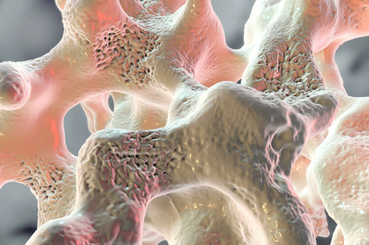
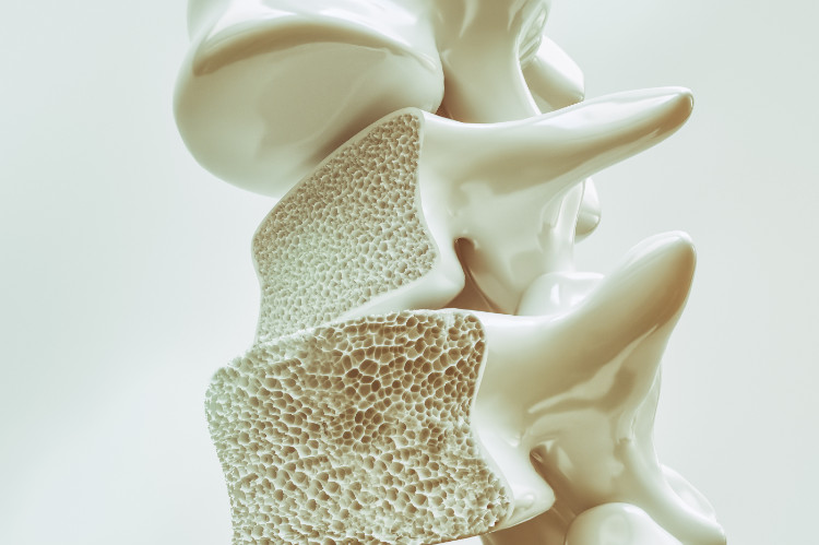
Osteoporosis is a disease that causes thinning of the bones by loss of bone mass. Its increase year by year is related to the increasing age of the population. This shows that it is a disease affecting mainly the older generation. It is rare in children.
Article content
The so-called silent bone thief affects about 5 to 7% of the total number of people.
A slight increase in prevalence has also been noted in our offspring. Nevertheless, this disease is still spoken of very cautiously in relation to the very young, especially in the case of repeated bone fractures with an inappropriate mechanism of injury.
These can also occur in osteogenesis imperfecta, a disease that is almost unknown to the public. It is often diagnosed during pregnancy or shortly after birth. What should we know about these two serious diseases?
What does bone tissue consist of?
The skeleton is the basic pillar of the musculoskeletal system. It forms a kind of support for the soft parts (bone skeleton) of our body and protects them from damage. It consists of bones that act as levers and, with the help of muscles and nerves, allow movement.
Bone (Lat. os) is made up of bone cells, which we call osteocytes. The spaces between them are filled by the organic intercellular component osteoid, collagen and elastic fibres. Bone tissue (matrix) belongs to the connective tissue and is highly mineralised (compounds of calcium, phosphorus, magnesium and sodium). The architectural units of bone are the bone lamellae and the bone beams.
The basic structure of bone
- The periosteum - The uppermost part of the bone. It can also be called a kind of tough connective tissue. It is found on the surface of every bone except the ends of joints. It contains abundant vascular supply that provides the nourishing function of the bone. It also contains numerous sensory nerves that are responsible for the excruciating pain of a bone fracture. The bone itself has no nerve endings and does not hurt.
- Bone tissue (compaction, spongiosis) - Forms the bone itself. On the surface, the compaction is rough and plate-like. Inside, in addition to the cavities, there is spongy bone tissue (spongiosis).
- Bone marrow (medulla ossium) - It consists of connective cells and fibres and the vasculature. It fills the marrow cavities. We know red and yellow bone marrow. Red bone marrow plays an important role, especially in childhood, as it is an important blood-forming organ.
Peculiarities of bone structure in childhood
The bone structure of a child and an adult differs considerably on several levels. In the newborn, the bone is still immature. The lamellar structure of the bone, which is the basic building unit, is not formed. Similarly, the tubular parts of long bones are composed only of ligaments, between which osteocytes are irregularly scattered. Also, the bone marrow is located between the fibers and the rafters, and the marrow cavities are almost absent. Therefore, the bone is protected by the periosteum, which is extremely strong.
However, a strong and firm periosteum is not only typical for the neonatal period. Despite the advanced remodelling of the bone tissue, its strength does not change. For this reason, we can see bone fractures in children with an intact periosteum, which also forms a kind of support for the fracture.
As the child grows, the immature bone tissue gradually rebuilds. The architectural structure of the bones (lamellae, rafters) is formed. Around the 5th month of life, Haversian lamellae are formed and the remaining lamellae are formed by one year of age.
At about one year of age, the marrow cavities enlarge and complete and blood cells form.
The final remodelling of the bone structure ends around the second year of life. The bone takes on its final form and in essence differs only minimally from the structure of adult bone. By the age of 12, only the end portions, tendon attachments and joint heads and capsules have changed.
What are the basic differences between osteoporosis and osteogenesis imperfecta?

Osteoporosis is a systemic metabolic disease of the bones. The metabolism in the bone tissue is disturbed. Consequently, bone loss and thinning of the bones (a decrease in bone density - bone density) occurs. The thinning of the bones results in their fragility and increased brittleness. Osteoporosis in children is called juvenile. Children are more likely than their peers (even if there has been no traumatic event) to complain of back and limb pain. People mistakenly think that osteoporosis is bone softening. This is a misconception.
Primary osteoporosis due to mineral deficiency in bone tissue does not occur in children. It is typical of older age. The causes of osteoporosis in adulthood have their basis in childhood. In the literature, osteogenesis imperfecta is often classified as a secondary type of osteoporosis (resulting from a primary disease). It could be argued that this is a subcategory of osteoporosis.
Osteogenesis imperfecta is a genetic disease of the connective tissue. It is characterised by the fragility of the bones as in osteoporosis. They are deformed, fragile, easily broken, and hernias are common. Hearing impairment due to deformities of the auditory ossicles and brittle teeth, which in this disease have a typical bluish tinge along with the sclerae of the eyes, are no exceptions.
It is not uncommon in this disease for fractures to occur during intrauterine development and even more frequently during birth. In severe forms of the disease, it can be detected sonographically as early as around the 16th week of pregnancy. With DNA (chorionic villus) testing and subsequent analysis, diagnosis can be accelerated to the 13th to 14th week of pregnancy.
Causes, course and known risks
- Osteoporosis is caused by disturbances in the metabolism of minerals in the bone. Because bone is metabolically very active, such disturbances are uncommon. When they do occur, they can significantly impair the density and thus the structure of the bones, which continues throughout life (it slows down in adulthood but does not disappear). They become thinner, brittle and weak and often deform, hurt and break. Osteoporosis significantly affects the quality of life of the patient.
Its incidence increases with age - the so-called senile osteoporosis. Primary osteoporosis has its main cause in a mineral deficiency. In secondary osteoporosis, the cause of its occurrence is some primary disease (inflammation, tumours, endocrine diseases, genetics, the action of certain drugs, alcohol, cigarettes). Juvenile or even childhood type of osteoporosis is very rare. This is because the formation of new bone mass until puberty predominates over its breakdown.
Osteoporosis is often asymptomatic. The first time it is thought of is when a bone is broken by a low-intensity trauma mechanism. The long bones of the forearm, which cause intense pain, and the vertebrae (lower thoracic vertebrae and upper sacral vertebrae) are most commonly broken. Their damage may not be detected because of the lower intensity of the pain and failure to seek medical attention.
Undiagnosed vertebral fractures and deformities cause a gradual reduction in the child's height, or a hump develops. Mobility, balance or breathing difficulties are impaired. In some children, osteoporosis manifests itself only in minor back pain, pain between the shoulder blades or in the limbs, which are rarely given much importance by parents.
- Osteogenesis imperfecta is exclusively a genetic disease caused by a mutation in the gene that codes for type 1 collagen (more than 90% of cases). Collagen can form in sufficient quantity but of poor quality. The second possibility is a reduced quantity and good quality. The third possibility is a small quantity and also poor quality. This results in a defect in the connective tissue or a complete inability of the child patient to form this connective tissue.
In this disease, fractures are very common already in the intrauterine period of fetal development. There is a risk of a severe course of labour with multiple bone fractures during the expulsion phase of the fetus. Therefore, sectioning is preferred. With early diagnosis of this disease, some women opt for abortion.
Children born with this insidious diagnosis also have severe bone deformities, increased joint mobility, hearing impairment due to deformity of the auditory bones, and respiratory disturbances due to the difficulty in expanding the lung tissue due to the deformities. The milk teeth and whites of the eyes are typically colored blue.
The risks of this disease are multiple fractures sometimes with fatal consequences, respiratory problems, poor quality of life and early death. With a combination of insufficient quantity and quality of collagen, children do not live beyond one year.
What are the treatment options?
In osteoporosis, the progression of the disease is crucial. Regular exercise, rehabilitation, and an appropriate diet with sufficient calcium and vitamin D, which is important for proper bone function, play an important role in treatment.
Salmon calcitonin, alendronate, bisphosphonate II generation, calcitriol, fluoride salts are used.
Treatment of osteogenesis imperfecta is long-term (several years) and requires multiple specialists. Parental awareness of the disease, risks, and adverse prognosis is required. It is important that parents handle the child carefully and avoid pulling. The child requires frequent positioning so as not to stress certain bones more. Dietary modification (minerals, vitamin D) and rehabilitation are required.
Of the drug therapy, bisphosphonates or pamidronate have been the most successful.
Both diseases in children require increased attention and caution, as there is a risk of fractures. The risk is increased by children being restless and always trying something new. Treatment of osteoporosis is more favourable than treatment of osteogenesis imperfecta. However, much depends on the doctor, the parents' attitude, patience and, above all, the stage at which the disease is.
Even osteogenesis imperfecta in the lower stages allows the child to lead an almost normal life with some limitations.


