Tooth Decay: Causes and Manifestations

Tooth Decay: Causes and Manifestations
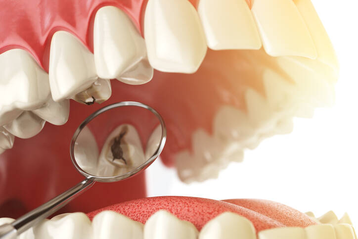
Photo source: Getty images

Tooth decay is a common dental disease. It is an acquired defect of the tooth tissue. This disease has been known for a long time. However, its incidence has been increasing especially during the last hundred years. This is due to the changes in the dietary regimen of people. The emergence of caries is conditioned by many factors, prevention, the composition of the diet, but especially by poor oral hygiene and dental care.

Most common symptoms

Show more symptoms

Characteristics

Tooth decay is defined as the progressive demineralization and subsequent damage to the hard tooth tissue. This disease affects both the deciduous and permanent dentition. It is the most common infectious disease in children and adults alike and is often neglected.

FAQs: 
What causes tooth decay and what symptoms does it have?
What does tooth decay look like and how to detect it?
Can there be deep/interdental/under filling tooth decay?
What is the treatment and can it be treated at home?

The causes are multifactorial. However, the mechanism is conditioned by the presence of dental plaque, which is colonized by a variety of bacteria. These microbes ferment sugar, resulting in a decrease in pH.

If the supply of simple sugars is reduced, the pH rises again.

After the intake of simple sugar, the pH drops sharply, within minutes. However, the return to higher values takes longer, even tens of minutes. The pH value on the tooth surface and in the saliva is approximately the same. Complex mechanisms to maintain its equilibrium respond to the reduced pH by releasing calcium and phosphate from the tooth enamel. Demineralisation of the tooth enamel occurs.

At a higher pH, on the tooth surface, minerals are redeposited in the tooth enamel. If the state of demineralisation lasts long enough, damage occurs on top of the enamel. This can be observed as stains on the tooth, which can be white in colour and, in long-term demineralisation, are also coloured yellow to brown.

This stage is still reversible. It may not progress to deep damage to the tooth. A sufficient supply of minerals and the removal of negative factors is necessary. If the disruption of the tooth tissue goes into a deeper layer, the tooth must be treated with drilling and a dental filling, i.e. a filling.

Tooth decay is a risk for the development of complications that can lead to inflammation of the tooth pulp, gums, and even periodontitis. Premature tooth loss is serious. A risky complication is the spread of bacteria from the inflamed tooth through the bloodstream into the body. This can lead to various diseases, such as heart disease. This is why consistent plaque removal and prevention of tooth decay is very important.

TIP: Learn more in How to have healthy teeth.
Read also Periodontal Disease

What we need to remember:

  • tooth decay is caused by infectious disruption of the tooth enamel
  • damage to hard tooth tissue
  • various bacteria are found on tooth enamel
  • if they have enough carbohydrates, there is a decrease in pH
  • this subsequently demineralises the tooth enamel
  • the tooth's sensitivity to cold, hot or sweet foods increases
  • which is also usually the first sign of tooth decay
  • if it is only a superficial caries at an early stage, treatment without special dental intervention is also possible
  • if the patient improves their dental hygiene
  • increases fluoridation and calcium content
  • if the pH is low for a long time, tooth decay penetrates through the tooth enamel into the dentin
  • inflammation of the pulp, the root and even suppurative inflammation can occur
  • the risk of periodontal disease
  • a serious complication is the spread of bacteria from the inflamed tooth to the whole body

Where is tooth decay most common?

The most common is the decay of the tooth crown on the surface used for chewing or in the interdental spaces. However, decay can also occur in the cervical region or around the edges of crowns or fillings. Thus, it is not rare for tooth decay to form under a filling, after treatment. The cause may be a lack of hygiene.

Tooth decay thrives in places such as various grooves, notches, pits, depressions, but also in the area of the exposed tooth root and between the teeth.

Table: different types of caries distribution

Type of dental decay Description
hard tissue damage enamel
dentin
cementum
most commonly, a combination of the above
the rate of caries formation acute, yellow discoloration and soft areas
chronic, harder darker areas
stopped, after removing the negative influence, when tooth decay no longer continues, the area remineralizes
in relation to the dental pulp superficial
central
closer to the pulp
penetrating into the marrow
according to place of origin primary, on an undamaged tooth
secondary, already on the treated tooth, at the filling
recurrent, arising under the filling

Causes

Tooth decay is the most common infectious disease. This means that infection is behind its occurrence. And therefore the spread of microorganisms. These thrive especially in the area where the plaque covers the tooth. This place is suitable for the attachment and multiplication of bacteria.

Dental plaque is colonised by various microorganisms. Food debris is also deposited on this biofilm. When sufficient sugars are added, the pH drops, which is the cause of demineralisation. If it persists, it is the cause of tooth decay. It is therefore important to remove plaque sufficiently.

Mechanical cleaning is necessary to remove biofilm, i.e. plaque. Rinsing with mouthwash is beneficial but not sufficient. Likewise, excessive consumption of sugars is inappropriate, as is prolonged sipping of sweetened drinks or juices.

Microbes in plaque causes do the following:

  1. they ferment sugars
  2. produce organic acid
  3. which lowers the pH
  4. which causes demineralisation of tooth enamel
  5. surface damage occurs
  6. after lowering the concentration of sugars the pH rises, however, slowly
  7. with prolonged exposure to acids, and therefore bacteria, the damage penetrates deeper
  8. tooth decay develops
  9. inflammation progresses deeper
  10. risk of complications
    • tooth decay spreads to the gum
    • bloody gums
    • loose teeth
    • development of periodontitis
    • spread of infection into the body

Table: effects on tooth decay

Risk factor Description
Microorganisms and bacteria also referred to as cariogenic microorganisms
  • Streptococcus (mutans, sobrinus)
  • Lactobacilli
  • Actinomycetes
  • Fusobacteria
  • Bacilus subtilis
Sugar increased intake of simple sugars such as mono- and disaccharides
bacteria convert sugars in food into acids
sweet drinks, sour juices, cola, sour fruit and other sweets the time and duration of exposure are also important 
Dental plaque a place for the successful capture and multiplication of micro-organisms
Quality of dental tissue is to some extent also genetically determined
the composition of saliva or mineral levels change during pregnancy, increasing the risk of tooth decay
Oral hygiene regular but sufficient oral hygiene is important
mechanical cleaning of the teeth should take place at least once every 24 hours
Reduced mineral content low fluoride exposure and low calcium
especially in childhood

You need to pick a suitable toothbrush for mechanical brushing.
See how to do that by reading How to pick the right tooth brush.

Symptoms

In the early stage, the symptoms of tooth decay are not noticeable. At first it proceeds asymptomatically, i.e. asymptomatically. The white soft layer on the tooth is characterised by the presence of dental plaque. Subsequently, stains are formed when exposed to an acidic environment.

In the acute course, white spots on the teeth are present. Long-term erosion and demineralization cause stains that may be yellow or brown in colour. Typically, tooth decay only begins to show when the tooth enamel is eroded

A person first has teeth sensitive to cold or, on the contrary, too hot food. External exposure to cold or heat also has a negative effect. Similarly, the intake of acidic or sweet food, sweets and drinks also affects the teeth.

Tooth sensitivity is thus usually the initial symptom of tooth decay. Later, at an advanced stage, tooth pain also sets in. This is especially when stressed, such as when chewing, but subsequently also at rest. If tooth decay penetrates into the tooth pulp, inflammation occurs.

If tooth pulp gets inflamed, the tooth will start to hurt. However, this may subside later, which is a symptom of nerve endings dying. Purulent inflammation can penetrate into the nearby bone. When the tooth hurts after a bite, it is a manifestation of bone involvement, so dental or stomatosurgical intervention is required.

From a visual point of view, there is dark discoloration of the tooth as well as a depth defect. When caries is not treated properly, it happens that the decay spreads even under the filling. As a consequence, the filling may fall out. The greatest risk is the spread of the infection to the entire hanging apparatus of the tooth and further into the body.

A short list of symptoms of tooth decay:

  1. initial silent (asymptomatic) stage
  2. plaque, or biofilm on teeth
  3. white, later yellow to dark spots on the enamel
  4. sensitivity to hot, cold, sweet or sour
  5. visual manifestations of decay, dark areas and defects in the enamel
  6. pain persists when tooth decay penetrates deeper
  7. inflammation of the bone marrow
  8. temporary disappearance of pain in nerve damage
  9. painful biting when the inflammation extends to the bone
  10. damage to the hanging apparatus, bleeding gums, gingivitis
  11. risk of periodontitis, tooth is getting loose
  12. the spread of bacteria into the body, for example into the heart, and the development of endocarditis is dangerous

Diagnostics

The early stage is asymptomatic. However, the presence of dental plaque is visible. Subsequently, stains on the tooth can be noticed. The direct evidence and diagnosis of caries is the presence of dark defects in the tooth enamel. Increased sensitivity and later tooth pain are mentioned in the patient's medical history and clinical course.

Other methods for diagnosis include X-ray examination of the teeth. This reveals the depth of damage to the tooth. A specialist examination is carried out by a dentist. A special method is endoscopy, measuring the electrical conductivity of the tooth tissues, and a method using a laser.

Dental X-ray, which will show the condition of the teeth, tooth decay and fillings
Dental X-rays reveal the condition of the teeth, tooth decay and existing fillings. Photo source: Getty images

The dentist diagnoses decay:

  • by visually inspecting the teeth
  • with a cold spray
  • with warm wax
  • doing an X-ray image

Table: area and depth assessment of tooth decay

Phase Description
Initial phase the first stage of demineralisation and surface damage
  • white, flat spots on the enamel that are opaque
  • yellow or dark stains of the enamel when the pH is lowered for a long time
Existing damage i.e. a lesion penetrating deeper under the enamel
assessed as follows:
  • caries superficialis, i.e. damaged tooth enamel
  • caries media, a defect affecting the dentin, or tooth pulp
  • caries profunda, advances to the vicinity of the nerve
  • caries sicca, which is actually an inactive tooth decay

Course

The course, and therefore the development, of tooth decay depends on a number of factors. It has been suggested that four factors are necessary for caries to develop, like genetic make-up or sufficient hygiene.

Other factors involved in tooth decay are:

  • tooth
  • bacteria
  • diet
  • time

The tooth is the place where plaque is deposited and bacteria take up residence. Predisposing sites are especially those where the tooth has more notches, unevenness. Decay thrives mainly where there is poor access. And this is also the place between two teeth, that is, the interdental spaces. But also the place on the wall surfaces, the notches of the teeth and the area by the gums or the smooth surfaces of the incisors.

Bacteria are microorganisms that inhabit different parts of the human body under normal conditions. The skin or the digestive tract. Probably the best known are the intestinal bacteria, i.e. the microflora. Similarly in the oral cavity. They form a plaque on the surface of the teeth. They produce acids and therefore create a low pH.

A diet high in simple sugars feeds the carious bacteria. These produce more acids. Examples are sweetened drinks, cola, acidic liquids such as juices, but also acidic fruits and various sweets. The amount or frequency of intake of risky foods is therefore influential.

The time it takes for plaque to mature is approximately 24 hours, after which it becomes aggressive to tooth enamel. The pH of the oral cavity is sharply reduced after eating food. This condition persists for approximately 30 minutes. After about 40 minutes, the pH increases due to saliva.

It is therefore a good idea to divide the food intake into approximately 5 portions. This results in a pause between meals of 2-3 hours. Sweets and even sugary drinks are preferable to be taken after the main meal. It is inappropriate and risky to drink sweetened liquids or juices over a prolonged period of time. This maintains a low pH in the oral cavity for a long time.

How does the caries progress?

  1. tooth decay develops over a long period of time due to a bacterial infection
  2. the latter is the cause of demineralisation, which is caused by the conversion of carbohydrates into organic acids
  3. which gradually erodes tooth enamel
  4. increased sensitivity of the tooth to heat, cold, sweet or sour
  5. decay can occur anywhere on the tooth, near the neck of the tooth or under a filling or crown
  6. as the infection gradually spreads, there are certain initial symptoms, e.g. pain
  7. it is a long-term process when the infection penetrates from the surface of the tooth to its pulp and root
  8. untreated tooth decay causes decay and death of the tooth and damage to the mandible
  9. is the result of other diseases and defects at the level of the teeth and other parts of the body

Tooth decay prevention is very important

Dental care and overall oral hygiene is necessary from the first teeth. Even young children should have preventive check-ups. Therefore, parental awareness is also an important factor for the future condition of the child's teeth. Of course, thorough dental care is necessary throughout life. In the same way, regular preventive checkups and timely treatment of dental problems are of great importance.

Factors affecting tooth quality:

  • how much the individual, parents are informed/educated about the relevant topics
  • parental attitudes towards oral hygiene that children notice
  • family brushing habits
  • quality of oral hygiene
    • mechanical teeth cleaning at least once every 24 hours
    • interdental cleaning (interdental brushes, dental floss)
  • a person's diet to be balanced
    • a harder diet, such as fresh vegetables or fruit, is also suitable
  • lower consumption of sugary foods, sweets, sweetened drinks
  • negative effects of prolonged pacifier use in children
  • eating habits, break between meals between 2 - 3 hours
  • prolonged sipping of sweet or acidic liquids is non-watery
    • sweetened drink in a bottle for children also has a negative effect
    • early learning to drink liquids from a glass
  • replacement of simple sugars by natural sweeteners (stevia)
  • if a person cannot brush his or her teeth after eating, the use of sugar-free chewing gum is advisable
  • the right toothbrush and toothpaste with quality ingredients that the tooth needs

How it is treated: Tooth Decay

Treatment: How to treat tooth decay? You can't do it at home, you need a specialist!

Show more

Dental Health & Information : How Do You Know You Have a Cavity?

fshare on Facebook

Interesting resources

  • Laudenbach, JM; Simon, Z (November 2014). "Common Dental and Periodontal Diseases: Evaluation and Management". The Medical Clinics of North America98 (6): 1239–1260. doi:10.1016/j.mcna.2014.08.002. PMID 25443675.
  • "Oral health Fact sheet N°318". who.int. April 2012. Archived from the original on 8 December 2014. Retrieved 10 December 2014.
  • Taber's cyclopedic medical dictionary (Ed. 22, illustrated in full color ed.). Philadelphia: F.A. Davis Co. 2013. p. 401. ISBN 9780803639096. Archived from the original on 2015-07-13.
  • SECTION ON ORAL, HEALTH; SECTION ON ORAL, HEALTH (December 2014). "Maintaining and improving the oral health of young children". Pediatrics134 (6): 1224–9. doi:10.1542/peds.2014-2984. PMID 25422016. S2CID 32580232.
  • de Oliveira, KMH; Nemezio, MA; Romualdo, PC; da Silva, RAB; de Paula E Silva, FWG; Küchler, EC (2017). "Dental Flossing and Proximal Caries in the Primary Dentition: A Systematic Review". Oral Health & Preventive Dentistry15 (5): 427–434. doi:10.3290/j.ohpd.a38780. PMID 28785751.
  • Silk, H (March 2014). "Diseases of the mouth". Primary Care: Clinics in Office Practice41 (1): 75–90. doi:10.1016/j.pop.2013.10.011. PMID 24439882. S2CID 9127595.
  • "Oral health". www.who.int. Retrieved 2019-09-14
  • Schwendicke, F; Dörfer, CE; Schlattmann, P; Page, LF; Thomson, WM; Paris, S (January 2015). "Socioeconomic Inequality and Caries: A Systematic Review and Meta-Analysis". Journal of Dental Research94 (1): 10–18. doi:10.1177/0022034514557546. PMID 25394849. S2CID 24227334.
  • Otsu, K; Kumakami-Sakano, M; Fujiwara, N; Kikuchi, K; Keller, L; Lesot, H; Harada, H (2014). "Stem cell sources for tooth regeneration: current status and future prospects". Frontiers in Physiology5: 36. doi:10.3389/fphys.2014.00036. PMC 3912331. PMID 24550845.
  • Vos, T (Dec 15, 2012). "Years lived with disability (YLDs) for 1160 sequelae of 289 diseases and injuries 1990–2010: a systematic analysis for the Global Burden of Disease Study 2010". The Lancet380 (9859): 2163–96. doi:10.1016/S0140-6736(12)61729-2. PMC 6350784. PMID 23245607.
  • Bagramian, RA; Garcia-Godoy, F; Volpe, AR (February 2009). "The global increase in dental caries. A pending public health crisis". American Journal of Dentistry22 (1): 3–8. PMID 19281105.
  • Health Promotion Board: Dental Caries Archived 2010-09-01 at the Wayback Machine, affiliated with the Singapore government. Page accessed August 14, 2006.
  • Richie S. King (November 28, 2011). "A Closer Look at Teeth May Mean More Fillings". The New York Times. Archived from the original on November 29, 2011. Retrieved November 30, 2011. An incipient carious lesion is the initial stage of structural damage to the enamel, usually caused by a bacterial infection that produces tooth-dissolving acid.
  • Johnson, Clarke. "Biology of the Human Dentition