Otosclerosis is a progressive disease that affects the bones of the inner ear. This disease leads to gradual hearing loss, in some cases in both ears.
Otosclerosis most commonly affects the pinna, the last of a series of three tiny bones located in the middle ear.
Currently, there are several treatment options for hearing loss. The most preferred is surgical replacement of the stirrup or implantation of a hearing aid.
The term otosclerosis is derived from the words "oto", meaning ear, and "sclerosis", meaning hardening of the tissues.
Otosclerosis is a disease caused by abnormal bone remodeling in the middle ear.
All bone remodeling occurs naturally during life. This remodeling is a dynamic process in which bone tissue is rebuilt by replacing old tissue with newly formed bone. In otosclerosis, this process is pathologically altered. Its proper function, namely the transmission of sound from the middle ear to the inner ear, is impaired.
Otosclerosis is most common in Caucasians, the so-called Caucasian population. It is slightly less common in the Asian population.
It affects women up to 2 times more often than men.
The first signs of the disease appear in the second or third decade of life. Rarely, otosclerosis can affect children and adolescents.
Causes
Hearing is one of the five senses by which humans perceive sound sensations. The organs of hearing are the pinna, the outer ear, the middle ear and the inner ear. The correct perception of sound depends on a series of events in which sound waves in the air are converted into electrochemical signals in the ear.
The auditory nerve then transmits these signals to the auditory centres in the brain.
In the first step, sound waves enter the outer ear and pass through a narrow passage called the ear canal. At the end of the ear canal is a thin, white, translucent membrane - the eardrum.
The incoming sound waves vibrate the eardrum. From the eardrum, these vibrations are transmitted to three small bones in the middle ear called the hammer, anvil and stirrup.
These small bones act as an amplifier. They amplify the sound vibrations and "send" them to the cochlea. The cochlea is a spiral-shaped organ in the inner ear that is filled with fluid.
The incoming sound vibrations are transmitted from the hammer to the anvil and then to the cochlea. The cochlea is supported by what's called the oval window of the cochlea. The amplified vibrations agitate the fluid inside the cochlea. A wave is created. It causes the tiny hair cells to move up and down.
The movement of these cells creates an electrical signal that is then transmitted by the auditory nerve to the brain.
The result is the recognition of sensation as sound.
The hair cells located at the bottom of the cochlea recognize high-pitched sounds, and the hair cells closer to the middle recognize lower-pitched sounds, such as the barking of a large dog.
Otosclerosis causes the intermaxillary joints to "harden" and one of the bones to become stuck.
In 80% of cases, the last of the row of bones, the stirrup, is affected by otosclerosis.
Because it is not mobile, it is no longer able to vibrate and transmit sound vibrations. Such a signal deteriorates and hearing becomes progressively worse.
Remodelling occurs in all bones during life. It is a natural phenomenon that is important, for example, in the healing of bone fractures.
Normal bone remodelling of large bones occurs at a rate of approximately 10% per year. In small ear bones, bone remodelling is very slow, only 0.13% per year.
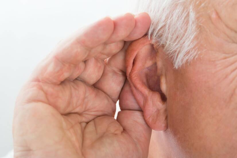
Hearing loss is caused by a malfunction of the tiny bones of the ear. Source: Getty Images
Patients with otosclerosis have abnormally increased bone remodeling. Pathologically rapid bone remodeling leads to the accumulation of rapidly formed and redundant bone tissue.
Abnormal bone remodeling in otosclerosis occurs in three stages:
Otospongiosis phase - there is increased activity of osteoclasts (cells that engulf old bone) and the formation of new blood vessels, leading to bone resorption and spongiosis formation
Transitional phase - osteoblasts migrate to new spongioses
Otosclerotic phase - formation of sclerotic deposits that put pressure on blood vessels and impair microcirculation
The cause of this increased bone remodeling is not yet known. Some researchers believe it could be related to the immune system and mediators of inflammation called cytokines. A proper balance of these substances is essential for healthy bone remodeling.
Some studies suggest that otosclerosis is a hereditary disease. Up to 60% of patients with otosclerosis report a family history of the disease. The remaining 40% have a sporadic history of otosclerosis.
Hormonal fluctuations may be another risk factor. Examples include puberty, pregnancy and menopause, which are also associated with rapid hearing loss in patients with previously diagnosed otosclerosis. Scientists have even identified estrogen receptors on otosclerotic cells, supporting the theory that female sex hormones influence the progression of the disease.
Abnormal parathyroid function with abnormal calcium and phosphate levels may also be a potential cause of otosclerosis. However, otosclerosis only affects the temporal bone, of which the stapes is a part.
Otosclerosis is more common in people who have had the infectious disease measles in childhood. The exact reason for measles affecting the onset or progression of otosclerosis is not yet known.
Symptoms
The most common and main symptom of otosclerosis is hearing loss.
It usually starts in one ear and progresses to the other ear as the disease progresses. Up to 80% of patients in the more advanced stages of otosclerosis have bilateral hearing loss.
The first symptoms include the inability to recognise low tones or whispers. Patients have difficulty hearing male voices or vowels in words. Hearing loss in one ear is noticeable, for example, when making telephone calls.
Patients begin to put the phone to the other ear, which they can hear better.
Other symptoms include uncomfortable dizziness, balance problems or tinnitus. Tinnitus is a constant whistling, knocking, ringing, buzzing or hissing in the ears. Up to half of otosclerosis patients suffer from this symptom.
Vertigo, i.e. dizziness and vertigo, occurs in connection with an affection of the semicircular canals. The semicircular canals are located near the cochlea and together with it form the auditory-balancing organ. The semicircular canals are responsible for the balance of the body.
Summary of possible manifestations of the disease:
Gradual hearing loss, first in one ear and in 80% of cases eventually in both ears.
initially low tones and whispers
hearing impairment - hearing loss can also be found under the term hypoacusis
dizziness, vertigo
difficulty maintaining balance
humming, whistling in the ears - tinnitus
Diagnostics
Otosclerosis is one of the diseases of the hearing. Therefore, its diagnosis is carried out by a doctor specialised in otorhinolaryngology (ENT). He has instruments and equipment that can diagnose impairments in all sections of the organ of hearing. The examination is performed by a specialist in ENT - otorhinolaryngology. Source: Getty Images
Otoscopic examination
The first step is a classic otoscopic examination. The doctor examines the outer ear area with an otoscope - a device with a thin funnel and light. Since otosclerosis takes place in the deep structures of the ear, an otoscopic examination is usually normal.
An exception is the presence of redness along the eardrum, which is called Schwartz's sign. This sign may not be present in all patients with otosclerosis and is therefore not a major diagnostic condition.
Audiometry
Audiometry is traditionally used to diagnose hearing disorders, which include otosclerosis.
The result of an audiometric examination is an audiogram. An audiogram is a curve that shows the intensity of sound (loudness) and the frequency of tones that are needed for the patient to hear them. It is examined in a quiet room, with headphones on. Each ear is examined separately, as the disorder may occur in only one ear.
An audiogram can distinguish air conduction disorder from perceptual hearing loss.
If the bone conduction is fine and the disorder is in the air conduction, it is a conduction disorder. This means that sound is not being transmitted from the middle ear to the inner ear. Therefore, the problem is in the middle ear and the inner ear is intact.
If the patient hears equally poorly through the bone and air channels, it is a perceptual disturbance. This means that the disturbance is in the inner ear.
Otosclerosis is typically manifested by low-frequency transductive hearing loss. On an audiogram, a bone conduction defect is present in the frequency regions around 2,000 Hz. This diagnostic feature is called Carhart's notch.
The curve of the audiogram can be used to determine the degree of hearing loss.
The World Health Organization (WHO) has developed an International Classification of Hearing Loss:
mild hearing loss - 26-40 dB
moderate hearing loss - 41-55 dB
moderate hearing loss - 56-70 dB
severe hearing loss - 71-90 dB
severe hearing loss - more than 91 dB
The progression of otosclerosis can also be monitored by means of an audiogram. The progression of the disease is directly proportional to the degree of hearing loss and the change in the frequency of the sound heard. Initially, there is an impairment in the perception of low-frequency tones. In the stages when the conductivity deteriorates due to hardening of the joints between the bones, the impairment manifests itself in the perception of all and high frequencies.
Over time, 10 % of otosclerotic patients also develop cochlear lesions (damage to the inner ear - cochlea). Extensive cochlear involvement is characterised by mixed hearing loss in all frequencies on the audiogram.
Tympanometry
Tympanometry investigates the condition of the middle ear and in particular the transmission of acoustic energy. It focuses on the mobility of the eardrum and middle ear bones. The measurement works on the principle of changes in air pressure in the ear canal.
The pathological result is a flattening of the tympanogram curve. This occurs only in the high stages of otosclerosis. Therefore, tympanograms in patients with early otosclerosis are often normal.
Imaging examination
A useful examination is high-resolution CT scan, which has a high diagnostic sensitivity and specificity. This examination can reveal abnormalities in the patient's anatomy and also the degree of severity of the disease.
CT findings in otosclerosis include various anatomical abnormalities in the thickness of the ossicles and the size of the middle ear and eardrum structures.
High-resolution CT can also reveal a disorder of the inner ear, i.e., the cochlea. Affection of the cochlea is visible as a demineralized area around the cochlea - the double ring sign.
High-resolution CT is also used in planning surgical treatment for otosclerosis.
Course
Hearing loss starts suddenly and gradually worsens.
Many patients with otosclerosis may not even notice it in the early stages. Later, they feel they can no longer hear low tones or whispers.
As the disease progresses, hearing deteriorates even when high-frequency tones are perceived.
Secondary damage in otosclerosis is the involvement of the structures of the inner ear - the cochlea. The resulting hearing damage in the cochlea is called mixed, as both sensorineural (perceptual) and conductive hearing loss is present. It affects 1 in 10 patients.
The prognosis of the disease is unfavourable.
Unfortunately, there is no known treatment to slow or stop the progression of the disease. Some patients suffer from progressive hearing loss that extends to the other ear.
Even with surgical treatment, otosclerosis is not cured. In 90% of cases, hearing improves and in half of the patients the tinnitus disappears.
How it is treated: Otosclerosis
Treatment of otosclerosis: no cure, will surgery help?
health.gov.sk - pdf download Otosclerosis - Ministry of Health, doc. MUDr. MUDr. Zuzana Kabátová, CSc.
MUDr. Andrea Bullová
Physician
After graduating from an eight-year high school, I knew exactly who I wanted to become in life. Studying at the Faculty of Medicine at Comenius University in Bratislava fulfilled a long-held dream and I was able to start fulfilling my mission as a doctor. After obtaining my diploma and M.D. degree, my first professional steps led to the St. Elizabeth Cancer Institute in Bratislava. At the Stereotactic Radiosurgery Clinic I worked with patients with brain tumours and eye tumours. After a year of working with oncology patients, in 2020 I moved to the Neurology Clinic of SZU St. Michael's University Hospital in Bratislava, where I am still working today. I like to spend my free time outdoors, whether cycling, hiking or just walking in the nearby forest.