Cerebral oedema is divided into vasogenic, cellular, osmotic and interstitial based on the causes of its occurrence.
It can arise from a variety of causes, including head trauma, vascular ischemia, intracranial lesions, or obstructive hydrocephalus leading to interstitial edema.
The consequences of cerebral edema can be devastating. If left untreated, it can even be fatal.
Edema is the technical name for swelling.
Edema is the body's natural response to some kind of injury. It occurs, for example, with trauma, burns, shock, inflammation and infection, or an allergic reaction.
It is the accumulation of free fluid and its pouring into the site of injury. The injured area swells, increases in volume and expands.
Most of the body's organs are held loosely in body cavities. They are surrounded by soft tissue, muscle and skin that are elastic. This gives these organs the ability to expand freely.
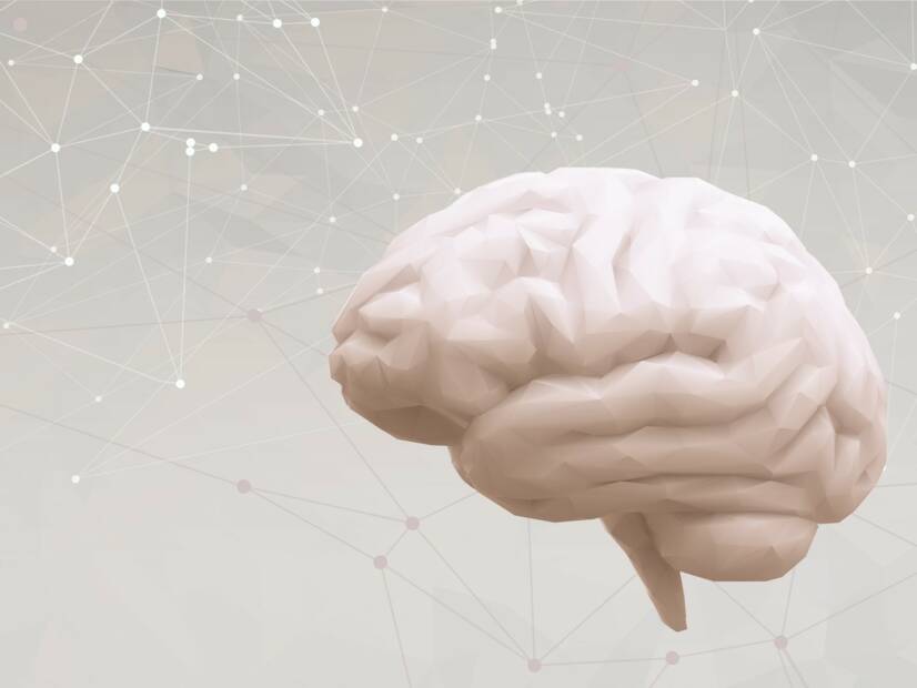
The brain is fairly tightly surrounded over its entire surface by the skull or hard bone.
It is because of this fact that the brain has a limited ability to increase its volume. When swelling occurs, the pressure inside the skull begins to increase rapidly and important brain centres are compressed.
Cerebral oedema can affect all ages, genders and ethnic groups. The true frequency of cerebral oedema may be underestimated because of its often non-specific symptoms.
It can arise from a variety of causes, including head trauma, vascular ischemia with inadequate blood supply, intracranial lesions, or obstructive hydrocephalus leading to so-called interstitial edema.
The consequences of cerebral oedema can be devastating, even fatal if left untreated.
Causes
The explanation of the mechanism of brain damage in cerebral edema is based on the so-called Monroe-Kellie doctrine.
The Monroe-Kellie doctrine states that the cranial cavity is volume invariant and contains incompressible components: a solid component consisting of brain matter (approximately 1400 ml) and a liquid component consisting of blood (approximately 150 ml) and cerebrospinal fluid (approximately 150 ml).
Because of this solid and constant volume, an increase in the volume of one of these components must automatically lead to a loss and compression of the other component in the same amount.
In cerebral oedema, the brain 'swells'. Its relative volume increases.
This increased brain volume reduces blood flow to the brain. Increased pressure can also cause further structural damage to both the edematous and non-edematous parts of the brain.
Cerebral edema can result from a variety of disorders.
The main types of damage are divided based on the cause into:
vasogenic
cellular
osmotic
interstitial
By these mechanisms, brain edema occurs in diseases such as brain tumor, trauma, hypoxia, i.e., non-oxygenation of the brain, infection, metabolic disorders, or acute hypertension.
Neurological causes may include hepatitis (inflammation of the liver), Reye's syndrome, carbon monoxide poisoning, lead poisoning and brain swelling at high altitude. A rare cause of cerebral oedema is the so-called pseudotumour cerebri.
1. Vasogenic cerebral oedema
This is the most common form of oedema. It results from disruption of the blood-brain barrier.
Through the broken blood-brain barrier, ions and proteins flow more freely and pass into the space outside the blood vessels. This causes fluid to be drawn into the brain tissue.
For example, in brain tumours, chemicals such as vascular endothelial growth factor (VEGF), glutamate and leukotrienes are produced. These chemicals increase the permeability of fluid through the arterial vessels around the tumours.
This leads to infiltration of fluid into the brain tissue, especially the white matter. This so-called peritumoral oedema leads to the typical clinical picture in patients with brain tumours.
In 65 % of patients, there are disturbances of thought, speech and consciousness.
2. Cellular or cytotoxic oedema
Occurs within minutes after brain injury.
It affects cells in the brain. Three types are distinguished:
Glial cells - Another name for neuroglia, these are the supporting nerve cells. It mainly has a supportive, protective, nourishing and regenerative function for the nerve cells themselves.
Neurons - Nerve cells
Endothelial cells - Endothelium is a thin layer of cells that make up the inner lining of blood vessels.
Cytotoxic edema results from a failure of ion transport across cell membranes. Sodium enters the cell freely. The mechanism to exclude it in excess is non-functional.
Subsequently, other anions begin to flow into the cell, trying to return the cell to a neutral state. This leads to intracellular oedema, i.e. 'swelling' of the cell itself.
This form of edema is caused, for example, by traumatic brain injury or sudden stroke.
3. Interstitial brain oedema
Interstitial brain oedema is caused by the displacement of cerebrospinal fluid (liquor) from the space of the brain ventricles, into the space of brain tissue.
The increased intracranial pressure pushes the fluid between the brain cells. The fluid thus accumulates in the extracellular space, i.e. around the cells, mainly in the white matter of the brain.
Patients with hydrocephalus or meningitis suffer from this type of swelling.
4. Osmotic oedema
Generally arises in disorders affecting osmolarity, such as hyponatremia, diabetic ketoacidosis, or other similar metabolic diseases.
The brain cells in these cases withdraw water from the plasma, leading to extensive edema.
The histopathology of cerebral edema may show both underlying pathology (tumor, infection, anoxic changes) and diffuse edema of either cell bodies in cytotoxic edema or interstitial spaces.
Symptoms
Cerebral oedema can be asymptomatic and only visible on imaging scans, but it can also cause life-threatening complications.
A detailed history provides valuable information that will point to the possible cause of the oedema.
Patients may have a history of, for example, head trauma, stroke, cancer, metabolic disease or other factors.
The clinical picture of cerebral edema varies from asymptomatic to severe impairment of vital signs, coma and death. Symptoms appear in most patients when intracranial pressure (ICP) rises above 20 cm H2O.
Symptoms can vary widely depending on the location and extent of cerebral edema.
Localized cerebral edema can cause:
weakness
visual disturbances
seizures
sensory changes
diplopia - double vision
other neurological disorders
The following symptoms may be present in diffuse cerebral edema:
Symptoms vary depending on the location and extent of the swelling. Source: Getty Images
Diagnostics
Early diagnosis of cerebral oedema is extremely important. It can prevent brain herniation, permanent sequelae or death.
A detailed neurological examination will provide valuable information. There is usually a change in mental state, for example, slowness, drowsiness or, on the contrary, agitation and uncontrollable behaviour.
An important feature is the development of fixed and dilated pupils.
Patients should undergo a computed tomography (CT) scan of the brain. A CT scan may show oedema, which is visible as an area of low density and loss of differentiation between grey and white matter.
In addition, there may also be narrowing and flattening of the cerebral ventricles, smoothing of the cerebral convolutions and disappearance of the spaces between them. Such a finding is indicative of an increase in intracranial pressure.
The cause of the oedema may be revealed during the first CT scan, especially in cases such as brain tumour, cerebral ischaemia or hydrocephalus.
CT scanning is also used as a follow-up examination to monitor progression or to correct therapy for edema.
CT/MRI scanning is of great importance. Source: Getty Images
Magnetic resonance imaging (MRI) is also beneficial. In specific T2 weighting, bright large foci are seen that show fluid.
Elevated intracerebral pressure is monitored with a special monitor. A probe is inserted into the brain or into a brain ventricle via a so-called ventriculostomy.
Course
The course and development of cerebral oedema depends on the cause at which it arose.
If the cause of the oedema is acute, such as cerebral ischaemia or infection, the development will be rapid and dramatic.
In chronic (for example, metabolic) diseases, oedema develops more slowly. The onset of symptoms is creeping and initially unnoticeable.
Edema may manifest itself, for example, by slowing of the psychomotor pace, general inefficiency, forgetfulness, difficulty in walking, etc.
The swelling in brain tumours depends on the malignancy of the tumour. If it is a highly malignant and aggressive tumour, the oedema will develop more rapidly. The first symptoms may therefore be loss of consciousness or an epileptic seizure.
The prognosis of cerebral oedema is highly variable. It depends on the degree of brain involvement, the severity and the cause of the oedema.
If the patient falls into a coma after being diagnosed with oedema, the prognosis may be poor.
If treatment is not started in time, the patient may die or suffer permanent effects of irreversible brain damage.
Cerebral oedema as a consequence of reversible and treatable diagnoses, such as diabetic ketoacidosis or untreated hypertension or mild head injury, has a relatively good prognosis and the patient may recover completely.
solen.sk - Current perspectives on the issue of mild brain damage, MUDr. Štefan Sivák, PhD., MUDr. Vladimír Nosáľ, PhD., prof. MUDr. Egon Kurča, PhD. from the Department of Neurology, 1st Faculty of Medicine, Charles University and UNM in Martina.
solen.cz - Pathophysiology and therapeutic options of intracranial hypertension, Miroslav Kalina, MD, Department of Neurology, Na Homolce Hospital, Prague.
ncbi.nlm.nih.gov - Cerebral edema, Sara M. Nehring; Prasanna Tadi; Steven Tenny.
ncbi.nlm.nih.gov Pathophysiology and treatment of cerebral edema in traumatic brain injury, Ruchira M. Jha; Patrick M. Kochanek, J. Marc Simard
pubmed.ncbi.nlm.nih.gov - Cerebral edema and elevated intracranial pressure, Matthew A Koenig
MUDr. Andrea Bullová
Physician
After graduating from an eight-year high school, I knew exactly who I wanted to become in life. Studying at the Faculty of Medicine at Comenius University in Bratislava fulfilled a long-held dream and I was able to start fulfilling my mission as a doctor. After obtaining my diploma and M.D. degree, my first professional steps led to the St. Elizabeth Cancer Institute in Bratislava. At the Stereotactic Radiosurgery Clinic I worked with patients with brain tumours and eye tumours. After a year of working with oncology patients, in 2020 I moved to the Neurology Clinic of SZU St. Michael's University Hospital in Bratislava, where I am still working today. I like to spend my free time outdoors, whether cycling, hiking or just walking in the nearby forest.