Angina pectoris: What it is and what are the symptoms of a stable or unstable form of chest pain?

Angina pectoris: What it is and what are the symptoms of a stable or unstable form of chest pain?
Photo source: Getty images

Angina pectoris, or angina pectoris, is one form of ischemic heart disease. It arises as a result of a mismatch between the need and the supply of blood, and therefore also oxygen and nutrients, to the heart. It typically occurs with increased physical or mental stress. Attacks of angina pectoris recur and are manifested by pressure or animal pain in the chest.

Characteristics

Angina pectoris or angina pectoris is known by the abbreviation AP , but also as thoracic or cardiac angina. The name originated from the Latin angera , which means animal, and the words pectoris , which means chest .

Literally a  typical symptom of chest tightness
Chest pain / chest tightness. 

Most often you are interested in: 
What is angina pectoris, why is it divided into a stable and unstable form? 
How does it manifest itself? 
How to prevent and treat?

Angina pectoris is one of the forms of ischemic heart disease . Its incidence increases with age, especially in men. In older age, the representation is approximately the same for both sexes. However, it also affects young people.

AP is divided into several forms.

These can be acute or chronic . The acute form is also called unstable angina pectoris , abbreviated NAP . We can also find it as an unstable AP. The chronic has been going on for a long time and is represented by stable angina pectoris , abbreviated SAP

There is another species, and that is vasospastic or Prinzmetal's angina pectoris .

Angina pectoris is classified as ischemic heart disease and its main cause is the disparity between the need and the influx of oxygenated blood into the cells of the heart muscle, ie cardiomyocytes. It most often occurs with increased need , and this arises as a result of excessive load . Exercise can be physical or mental .

The main reason is atherosclerosis-damaged coronary (heart) arteries , which fail to meet the requirement of increased blood flow to the muscle. The inner diameter of the vessel is narrowed, which causes a reduction in blood flow at the time of increased heart rate. Similarly, such a disparity can cause aortic narrowing , cardiac hypertrophy, or hyperthyroidism .

Also read the article on coronary heart disease  at the Medical Center.

The classification of angina pectoris is summarized in the table below

Form AP Description
Stable angina pectoris
  • chronic form
  • repeats
  • arises from increased exertion
  • the reason is 70% stenosis of the coronary artery
  • in the room recedes within 15 - 20 minutes
  • ST segment depressions are present on the ECG
Unstable angina pectoris
  • also referred to as an unstable AP
  • subunit of acute coronary syndrome 
  • newly created AP
  • impaired stable AP
  • it also arises in peace
  • takes longer
  • the reason is the rupture of the atherosclerotic plaque with subsequent thrombosis
  • the vessel is not completely occluded
  • there is no myocardial necrosis, ie heart attack
  • on the ECG are ST depression
Vasospastic angina pectoris
  • also referred to as Prinzmetal AP
  • arises in young people
  • without atherosclerotic changes
  • most often it starts at 4 o'clock
  • the reason is spasm, ie the contraction of the coronary artery
  • complete occlusion of the vessel, so-called occlusion, can occur
  • The ECG has a picture of STEMI (myocardial infarction with ST elevations)
  • there is no necrosis of the heart muscle

Causes

The cause of angina pectoris is the disparity between the need for blood flow , ie oxygenation, and the supply of blood to the heart muscle . Cardiac activity, or the work of the heart muscle, requires a constant supply of oxygen and nutrients. The disposal of metabolic products and wastes is also important.

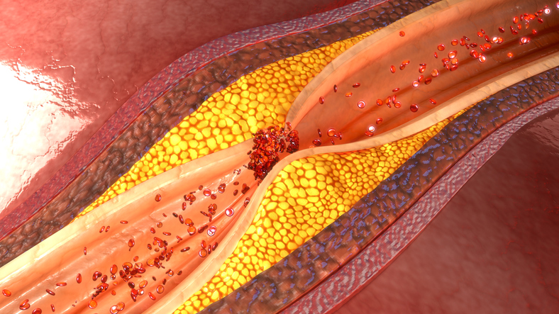
The most common cause is atherosclerosis of the coronary arteries . The inner diameter, ie the lumen of the vessel, is reduced by the atherosclerotic process. In a room, this narrowing can take up to 70% of the artery space and cause no problems. However, during exercise, oxygen consumption increases .

Angina pectoris and atherosclerosis and atherosclerotic plaque in the coronary artery
AP for coronary artery atherosclerosis. Photo source: Getty images

The heart performs more activity, tachycardia occurs , which can be perceived as a fast pulse and a fast heartbeat. This physiological process requires increased blood flow  and such a  damaged vessel cannot provide it . This results in anginal chest pain and other problems.

The cause is stable angina pectoris

If the difficulties arise from increased exertion , in the form of physical or mental stress, it is a stable angina pectoris . SAP is classified as a chronic form of coronary heart disease. It is a transient condition that does not cause irreversible damage to the heart, but only temporary non-blood supply to the subendocardial layer of the heart wall.

Increased effort can be, for example, harder workrunninglonger walking , walking up stairs  or having an argument , or stress . Interruption of overload will cause relief and complete relief of difficulties. And at most within 15 - 20 minutes. Even with this form of AP, the vessel can be narrowed to 70% of its internal diameter and difficulties occur depending on the extent and duration of the load.

In a person with AP, the onset of difficulties can also be provoked by a cold environment, the transition from heat to cold air in winter.

Or unstable angina pectoris

The opposite is unstable angina pectoris . The reason is the rupture of the atherosclerotic plaque with subsequent placement of platelets on the damaged layer. A thrombus is formed, thrombosis . However, this condition does not completely block the lumen of the vessel and at least partial congestion of the heart muscle is maintained.

Although ischemia develops with the onset of difficulties, it does not progress to necrosis , ie myocardial infarction. Difficulties arise in peace , without prior effort. They last more than 20 minutes. Newly occurring angina pectoris or deteriorating stable angina pectoris is also referred to as NAP .

A special type is vasospastic angina pectoris

This form is also referred to as Prinzmetal AP . The reason is not the atherosclerotic plaque and coronary artery damage, but its spasm , ie contraction . The main reason for the narrowing is unknown and is not the result of an increased burden . However, it may be due to cocaine use.

Usually, coronary artery spasm is a transient condition and resolves within 30 minutes. It also occurs at a young age and is rarely the cause of complete withdrawal to myocardial infarction.

What other causes are behind this disease?

Other causes of AP include a mixed form that combines atherosclerotic narrowing and vascular spasm. But also coronary syndrome X . In this case, difficulties arise due to increased effort. However, neither spasm nor coronary atherosclerosis is demonstrated. The cause is probably microvascular changes, ie at the level of the smallest coils of the heart.

Cardiac ischemia can also occur for other reasons , such as:

  • narrowing of the aortic valve
  • congenital heart defects
  • enlargement of the heart
  • hypertension
  • hyperthyroidism
  • vascular inflammation such as Kawasaki disease
  • injury
  • embolism
  • lack of oxygen in the inhaled air
  • anemia
  • fever
  • tachycardia

TIP: other causes of fast heartbeat in the article on the Medical Center.

Risk factors for the development of ischemia of the heart, and therefore also AP:

  • atherosclerosis
  • high blood pressure
  • diabetes
  • overweight and obesity
  • thrombosis
  • disorder of fat metabolism
    • high blood cholesterol 
    • excess fat in the diet
  • a diet poor in fruit and vegetables and overall poor eating habits
  • insufficient physical activity and sedentary lifestyle
  • excess salt
  • smoking
  • alcohol
  • drugs
  • excessive mental stress
  • family occurrence and genetic burden
  • male
  • older age

Symptoms

Angina pectoris is clinically manifested as a typical chest tightness . Thus, the pain is animal or pressure , but it can also have the  character of burning or a vague negative perception. This condition is also referred to as chest discomfort . Patients often describe it as having a stone on their chest or as someone sitting on their chest.

The pain is located in the middle of the chest, behind the sternum. It often radiates to the shoulders, upper limb, fingers, but also to the neck, sledge, jaw, between the shoulder blades or to the upper abdomen. The difficulties with SAP are provoked by effort . In the case of NAP, they start without a previous load .

Difficulties with AP arise after:

  • physical exertion
    • harder work
    • beh
    • fast walking
    • walking longer distance
    • walking up the stairs
    • in case of more severe vascular involvement, walking for a shorter distance
  • mental stress, such as stress, quarrel, emotional state
  • when switching to a cold environment
  • even after a meal, in case of eating a larger portion of food
  • with NAP it is also at rest

Symptoms that may occur with AP:

  • chest pain , ie angina (angina)
    • chest tightness , as a typical manifestation of AP
    • pressure
    • burning
    • discomfort
  • radiation of pain
    • shoulders
    • upper limb to toes
    • neck
    • sledge and upper jaw
    • between the blades
    • upper abdomen
  • shortness of breath , difficulty breathing, feeling short of air
  • nausea
  • feeling sick (vomiting)
  • sweating
  • pallor
  • dizziness
  • fainting
  • short-term loss of consciousness, collapse, syncope
  • anxiety
  • fear of death, the so-called horror mortis

Diagnostics

The diagnosis of angina pectoris is based on the anamnesis and the clinical picture . The doctor finds out the conditions of the difficulties, their nature and the accompanying problems. Furthermore, laboratory blood tests are performed, which also serve to identify risk factors, such as high cholesterol or blood sugar.

The basic examinations include an ECG , followed by a 24-hour ECG, the so-called Holter ECG . but also exercise ECG and bicycle  ergometry  (or running). They are used also challenge tests a combination of drugs with the imaging method. Subsequently, ECHO is performed , which is an ultrasonographic examination of the heart and its activity. Other methods include CT, MRI or SPECT (tomographic scintigraphy, ie single photon emission computed tomography).

A special examination method is coronarography (angiography). It is a contrast X-ray examination of the coronary (heart, coronary) arteries . Catheterization, ie the insertion of a catheter into the larger peripheral veins up into the coronary arteries and the subsequent injection of a contrast agent. During continuous X-ray examination, a restriction or closure of the bloodstream is found.

This method is diagnostic and therapeutic , as the enlargement of the narrowed artery, ie  angioplasty , can also be performed during catheterization . The concept of ballooning and subsequent introduction of STENT is well known . It is a minimally invasive procedure, also referred to as PTCA , PKI (percutaneous transluminal coronary angioplasty / intervention). Recovery after coronarography, resp. Angioplasty is fast.

AP is classified by the Canadian Cardiovascular Society - CCS method into:

  • Class I  - effort tolerance, if AP at high effort intensity
  • II. class  - tolerates walking on the plane for 200 meters
    • climb the stairs to the 2nd floor
    • AP for emotional burden
    • after a heavier meal
    • in a cold environment
    • in the morning after waking up
  • III. class  - tolerates only minimal effort
    • with a significant reduction in normal physical activity
    • AP while walking on the plane for a few meters
  • IV. class  - chest pain and at rest
    • inability to perform any activity without manifestations of AP

Of course, in the case of chest pain, it is important to differentiate the cause, ie the differential diagnosis . As for the pain in this locality there are various diseases, such as:

  • aortic aneurysm
  • pulmonary embolism
  • peptic ulcer
  • oesophagitis
  • GERD, or gastroesophageal reflux
  • spine ache
  • tumors in the chest
  • panic disorder and anxiety
  • herpetic infection

Course

The course of angina pectoris depends on several circumstances . And how serious is the coronary artery disease to what extent. At what point that vases or is it a more vascular disability. What mechanism it evokes, and thus whether it is SAP or NAP. 

Usually, animal chest pain is a typical symptom . Other difficulties can also be associated. However, with SAP, effort is given as a trigger . Following the end of the load, the problems subside. It is usually within a few minutes, but no longer than 15-20 minutes.

With NAP, the onset of difficulties is not conditioned by activity or stress. It starts in peace and lasts longer . The unstable form is also classified as the first episode of angina pectrois, but also such a stable AP, the course and intensity of which deteriorated. Important early professional examination and treatment, regardless of the form of AP.

Other symptoms mentioned may also be associated, such as radiation, pain , lack of air (shortness of breath, dyspnoea). In some cases of ischemia of the heart, chest pain may not work out. A person is weak, tired, easily exhausted and may have a feeling of mild shortness of breath.

How it is treated: Angina pectoris

How is angina treated and can it be cured?

Show more

What causes angina pectoris?

fshare on Facebook

Interesting resources

  • White PD (1931). Heart Disease (1st ed.). Macmillan.
  • Boden WE, O'Rourke RA, Teo KK, Hartigan PM, Maron DJ, Kostuk WJ, et al. (April 2007). "Optimal medical therapy with or without PCI for stable coronary disease". The New England Journal of Medicine356 (15): 1503–16. doi:10.1056/NEJMoa070829. PMID 17387127.
  • Tobin KJ (July 2010). "Stable angina pectoris: what does the current clinical evidence tell us?". The Journal of the American Osteopathic Association110 (7): 364–70. PMID 20693568.
  • "MerckMedicus: Dorland's Medical Dictionary". Retrieved 2009-01-09.
  • Hombach V, Höher M, Kochs M, Eggeling T, Schmidt A, Höpp HW, Hilger HH (December 1988). "Pathophysiology of unstable angina pectoris--correlations with coronary angioscopic imaging". European Heart Journal. 9 Suppl N: 40–5. 
  • Simons M (March 8, 2000). "Pathophysiology of unstable angina". Archived from the original on March 30, 2010. Retrieved April 28, 2010.
  • "What Is Angina?". National Heart Lung and Blood Institute. Retrieved April 28, 2010.
  • Kaski JC, ed. (1999). Chest pain with normal coronary angiograms: pathogenesis, diagnosis and management. Boston: Kluwer. pp. 5–6. ISBN 978-0792384212.
  • Guyton, Arthur. "Textbook of Medical Physiology" 11th edition. Philadelphia; Elsevier, 2006.
  • "Heart Attack and Angina Statistics". Archived from the original on 2010-04-13. Retrieved 2010-04-13.
  • "Angina". Texas Heart Institute. October 2012. Archived from the original on 2014-08-17. Retrieved 2010-05-04.
  • Gulati M, Shaw LJ, Bairey Merz CN (March 2012). "Myocardial ischemia in women: lessons from the NHLBI WISE study". Clinical Cardiology35 (3): 141–8.